4414
Accelerated whole-brain Bloch-Siegert B1-mapping using segmented 3D EPI1Institute of Medical Engineering, Graz University of Technology, Graz, Austria, Graz, Austria
Synopsis
To exploit acceleration potential of Bloch-Siegert shift based $$$B_1^+$$$-mapping, we implemented a 3D segmented EPI sequence with variable number of interleaves. Additionally, sampling of the k-space center with variable block-sizes was investigated. Compared to a fully sampled gradient-echo sequence, we show that acceleration by a factor up to 50 is feasible. Depending on the desired accuracy, whole-brain 3D $$$B_1^+$$$-mapping is possible in 5 to 10 seconds.
Introduction
Estimating the $$$B_1^+$$$-field is essential for many quantitative MRI methods and with the advent of ultra high-field MRI systems the demand of fast an accurate methods has become even more crucial.In Bloch-Siegert shift based $$$B_1^+$$$-mapping an off-resonance RF pulses is applied which induces a phase shift proportional to the peek amplitude $$$B_{1p}$$$ of the RF pulse 1. Because the shift is proportional to the RF power deposition, the method is SAR demanding, requiring long acquisition times. To increase the efficiency, the frequency of the RF-pulse can be decreased2. Another possibility is a dense sampling of the k-space center in combination with variational image reconstruction3. Further, acquisition can be accelerated with 2D spiral and EPI readouts 4.
Here, we investigate the potential of faster image acquisition with a 3D segmented EPI sequence. To exploit errors associated with faster acquisition, the number of interleaves and the block-size of the k-space center were varied. This work shows that with an adequate choice of interleaves and block-size, whole-brain 3D $$$B_1^+$$$-maps can be acquired in 5 to 10 seconds.
Methods
Bloch-Siegert shift: Assuming that the off-resonance frequency $$$\omega_{RF}$$$ of the RF pulse with peak amplitude $$$B_{1p}$$$ is much larger than the field inhomgoneities $$$\Delta \omega_{B0}$$$ ( $$$\omega_{RF} >> \Delta \omega_{B0}$$$) and $$$|\gamma B_{1p}| << \omega_{RF}$$$, the Bloch-Siegert phase shift $$$\phi_{2BS} $$$ is given by1:\begin{equation} \phi_{2BS} =B_{1p}^2 \int_0^T \frac{(\gamma B_{1n}(t))^2}{\omega_{RF}} dt = B_{1p}^2 K_{BS}\end{equation}
wher $$$B_{1n}(t)$$$ is the normalized pulse envelope and the constant $$$K_{BS}$$$describes the pulse properties.
Sequence: We implemented a 3D segmented EPI with a variable number of interleaves $$$N_{Intlv}$$$ in phase encoding direction $$$k_y$$$ 5. For each shot, one partition $$$k_z$$$is encoded. In addition, a navigator echo prior and after the echo train and after was integrated to measure time-dependent $$$\Delta \omega_{B0}$$$ fluctuations. Before the imaging-part, one shot was acquired without phase encoding gradients to correct for Nyquist ghosts. Furthermore, 20 preparation pulses were executed to approach faster a steady-state. Because of the variable implementation of $$$N_{Intlv}$$$, the sequence can be scaled to a standard RF-spoiled gradient-echo sequence, and up to the acquisition of one partition in a single shot. In addition, a variable block-size in the k-space center can be selected to measure $$$n_y$$$ time $$$n_z$$$ out of $$$N_y$$$ times $$$N_z$$$ fully sampled k-space lines.
In-vivo measurements: Brain MRI of a healthy subject were carried out on 3T MR system (Magnetom Vida, Siemens Healthcare, Erlangen, Germany) with the following sequence parameters: binominal -1 1 water excitation pulse with $$$\alpha = 20^\circ$$$ and RF-spoiling increment of $$$90^\circ$$$ 6, $$$4ms$$$ Gaussian saturation pulse with nominal $$$\phi_{BS,nominal} = 46^\circ$$$, $$$TR = 45ms$$$, $$$BW=2000Hz/px$$$,$$$FOV=256x256x192mm^3$$$ and matrix size$$$N_x=64$$$, $$$N_y=64$$$ and $$$N_z=48)$$$.
First, fully sampled k-space ($$$n_y=64, n_z=48$$$) images were acquired with different $$$N_{Intlv}$$$. Second, the block-size was varied ($$$n_y= [32,16,12]$$$ and $$$n_z=[24,12,8,6]$$$), each with variable $$$N_{Intlv}$$$.
Image reconstruction: For all EPI data-sets ($$$n_y \neq N_{intlv}$$$), Nyquist-correction prior image reconstruction was applied. To combine the individual coil images and to estimate $$$\phi_{2BS}$$$, each coil image for the positive $$$\omega_{RF}$$$ was multiplied by the complex conjugate of the negative $$$\omega_{RF}$$$ coil image and summed up.
Results and Discussion
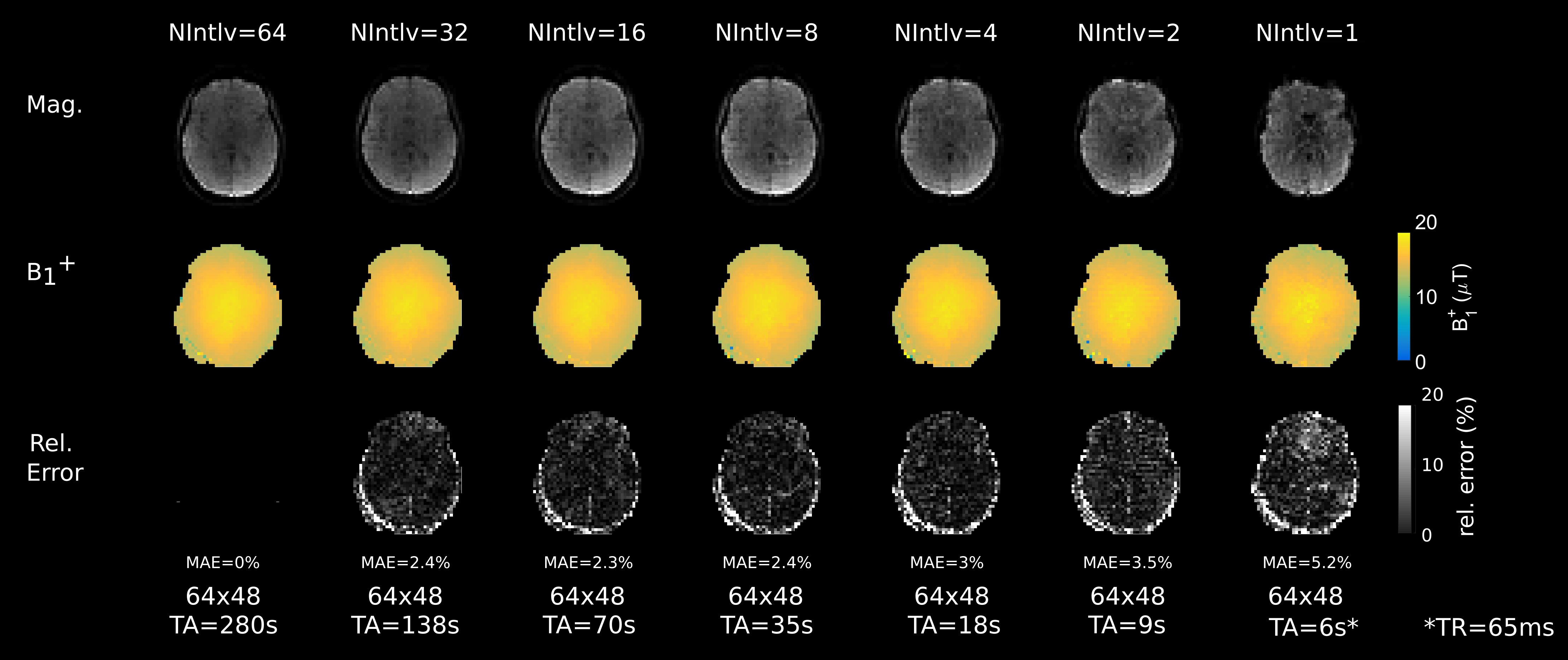
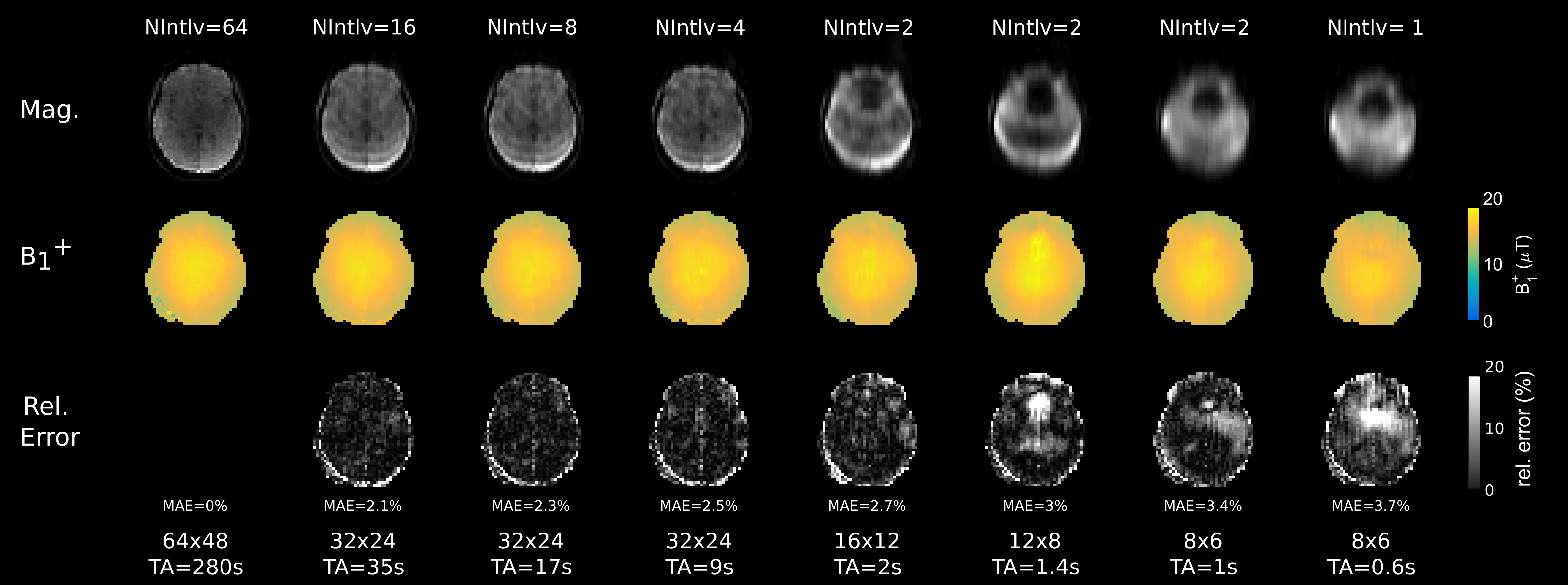
Figure 1 shows the reconstructed images for different $$$N_{intlv}$$$ (top), the estimated $$$B_1^+$$$ maps (middle), and the absolute error with respect to the gradient-echo images ($$$N_{intlv} = 64$$$). For the fully sampled k-space, visually a good correspondence between $$$B_1^+$$$-maps from $$$N_{intlv}=64$$$ with an acquisition time $$$TA=280s$$$ to $$$N_{intlv}=4$$$ ($$$TA=18s$$$) can be observed. Further reduction of $$$N_{intlv}$$$ leads to an increase of the mean absolute error (MAE). For $$$N_{intlv}=1$$$, distortions become more severe, which is also reflected in the $$$MAE$$$ by $$$5.2\%$$$.The results for different block-patterns for varying $$$N_{intlv}=1$$$ are depicted in Figure 2. When comparing the quality of $$$B_1^+$$$-maps and the $$$MAE$$$ with the fully sampled results (Figure 1), it can be observe that the $$$MAE$$$ tends to be smaller for faster $$$TA$$$.
Conclusion
We have demonstrated that with segmented 3D EPI highly accelerated whole-brain $$$B_1^+$$$-maps can be acquired. To improve image quality, and potential further acceleration, variational reconstruction methods will be implemented and the navigator signals will be incorporated to compensate temporal $$$\Delta \omega_{B0}$$$ fluctuations7.Acknowledgements
No acknowledgement found.References
[1] Sacolick, Laura I. and Wiesinger, Florian and Hancu, Ileana and Vogel, Mika W. B1 mapping by Bloch-Siegert shift. Magn Reson Med 2010;63:0740-3194.
[2] Duan, Qi and van Gelderen, Peter and Duyn, Jeff. Improved Bloch-Siegert based B1 mapping by reducing off-resonance shift. NMR in Biomedicine,vol. 26, pp.1070-1078, 2013.
[3] Lesch, Andreas and Schloegl, Matthias and Holler, Martin and Bredies, Kristian and Stollberger,Rudolf. Ultrafast 3D Bloch-Siegert B1 mapping. Magn Reson Med 2019;81:0740-3194.
[4] Saranathan, Manojkumar and Khalighi, Mohammad Mehdi and Glover, Gary H. and Pandit,Prachi and Rutt, Brian K. Efficient Bloch-Siegert B1 (+) mapping using spiral and echo-planar readouts. Magn Reson Med 2013;70:0740-3194.
[5] Kim Butts Ph.D., Stephen J. Riederer, Richard L. Ehman, Richard M. Thompson, Clifford R. Jack. Interleaved echo planar imaging on a standard MRI system. MagnReson Med 1994;31:67-7.
[6] Corbin, Nadège and Acosta-Cabronero, Julio and Malik, Shaihan J. and Callaghan, Martina F. Robust 3D Bloch-Siegert based B1+ mapping using multi-echo general linear modeling. MagnReson Med 2019;82:0740-3194.
[7] Oran OF, Klassen LM, Serrai H, Menon RS. Demonstration and suppression of respiration-related artifacts in Bloch-Siegert shift-based B1+ maps of the human brain. NMR Biomed.2020 Jul;33(7):e4299
Figures