4390
Three-dimensional echo-shifted echo planar imaging with simultaneous blip-up and blip-down acquisitions for correcting geometric distortion1Center for MR Research, University of Illinois at Chicago, Chicago, IL, United States, 2Athinoula A. Martinos Center for Biomedical Imaging, Massachusetts General Hospital, Charlestown, MA, United States, 3Department of Radiology, Harvard Medical School, Charlestown, MA, United States, 4Department of Biomedical Engineering, University of Illinois at Chicago, Chicago, IL, United States, 5USC Stevens Neuroimaging and Informatics Institute, Keck School of Medicine, University of Southern California, Los Angeles, CA, United States, 6Department of Neurology, Keck School of Medicine, University of Southern California, Los Angeles, CA, United States, 7Departments of Radiology and Neurosurgery, University of Illinois at Chicago, Chicago, IL, United States
Synopsis
Geometric distortion is a prevalent image artifact in echo planar imaging (EPI). In a method known as BUDA (blip-up/down acquisition), k-space model-based reconstruction of two EPI datasets acquired separately with opposing phase-encoding polarities has been shown to reduce image distortions. A main disadvantage is that the two-shot acquisition strategy doubles the scan time and increases vulnerability to motion. We herein introduce a novel sequence – 3D echo-shifted EPI with BUDA (esEPI-BUDA) – to address the aforementioned issues by integrating the two acquisitions into one shot. We have successfully applied the 3D esEPI-BUDA technique to functional MRI.
Introduction:
Geometric distortion is a prevalent and obstinate artifact in echo planar imaging (EPI). Among many correction techniques, the approach that acquires two EPI datasets with opposite phase-encoding directions (i.e., blip-up and blip-down) 1–3 is gaining popularity and has been adopted in several large-scale neuroimaging studies 4,5. Distortion correction can be performed in the image domain 1–3. However, image registration errors between the two EPI datasets can be problematic, particularly when the signal-to-noise ratio (SNR) is poor. In a new technique known as blip-up/down acquisition (BUDA) 6–8, Berkin et. al., proposed to transform corrupted k-space data to a distortion-corrected image using a specialized reconstruction model. Similar to the image-based methods, BUDA acquires the two EPI datasets in two shots, which doubles the scan time and is subject to inter-shot subject motion. Herein, we report a novel technique – echo-shifted EPI with BUDA (esEPI-BUDA) – to address the aforementioned problems by integrating the blip-up and blip-down acquisitions into a single shot. We demonstrate this technique in 3D fMRI with reduced image distortion.Methods:
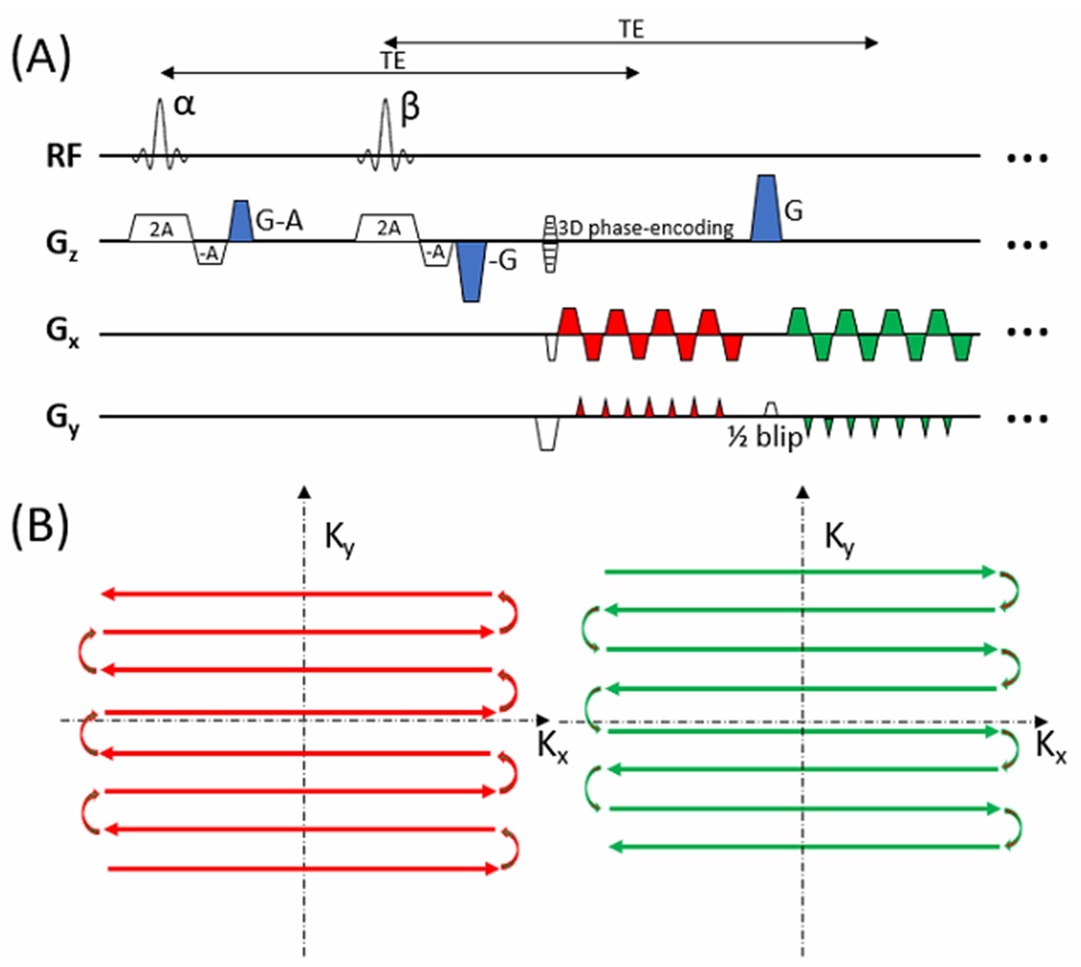
3D esEPI-BUDA:Figure 1 shows the principle of 3D esEPI-BUDA pulse sequence and its corresponding k-space trajectories. 3D esEPI-BUDA uses two RF pulses to acquire the blip-up and blip-down datasets in a single TR (or shot). The two signals from the two RF pulses are time-shifted and individually selected by three dephasing/rephasing gradients (or echo-shifting gradients; shaded in blue) applied along the slab-selection direction (Gz). The first echo-shifting gradient with an area of (G-A) acts as a spoiler to dephase the transverse magnetization shortly after the first RF pulse α. After the transverse magnetization is dephased, the second RF pulse β is applied to excite the stored longitudinal magnetization, followed by the second echo-shifting gradient with an area of (-G). This gradient dephases the transverse magnetization from RF pulse β, while rephasing the transverse magnetization produced by RF pulse α. The rephased signal is acquired by the first EPI echo-train with blip-up phase-encoding (red). The final echo-shifting gradient with an area of G is placed after the first readout echo-train to rephase the signal produced by RF pulse β, while dephasing the signal from RF pulse α. The rephased signal is acquired by the second echo-train with blip-down phase-encoding (green). k-Space data from both echo-trains are under-sampled (e.g., by two-fold) to shorten the echo-train length, thus enabling short and consistent TEs (e.g., 30 ms for fMRI at 3T) for both acquisitions. To equalize the signals for the two echo-trains, the flip angles α and β need to satisfy the following condition:$$\sin\alpha\cdot\cos^{2}\left(\frac{\beta }{2}\right )=\cos\alpha\cdot\sin\beta$$Our esEPI-BUDA sequence also contains a small gradient with ½ phase-encoding blip area (Gy) prior to the second echo-train so that the two k-space trajectories are interleaved (Figure 1B) for effective joint reconstruction.
BUDA reconstruction 6–8:
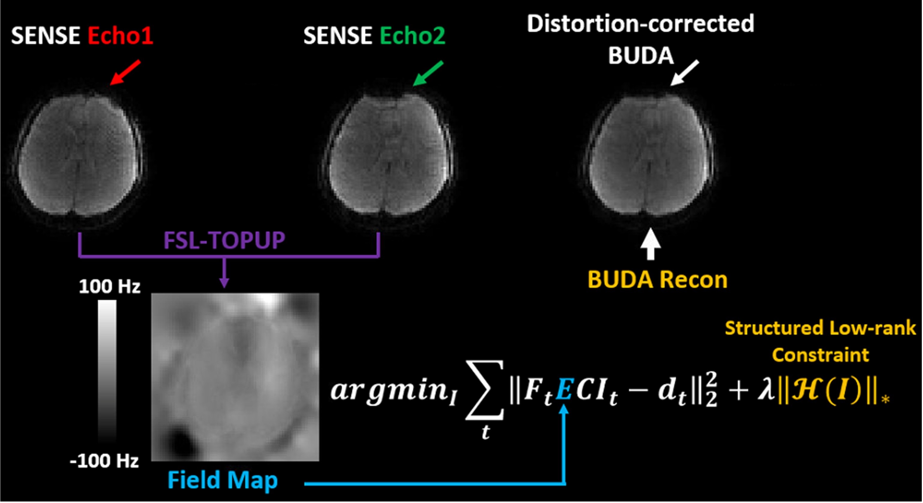
Figure 2 shows the steps involved in 3D esEPI-BUDA image reconstruction. Each echo-train dataset first underwent SENSE reconstruction using TOPUP in FSL 1,9 to estimate a field map E, which was subsequently incorporated to jointly reconstruct the data from both echo-trains according to: $$\arg\min_{I}\sum_{t}\left \|F_{t}ECI_{t}-d_{t}\right\|_{2}^{2}+\lambda\left\|H\left(I\right)\right\|_{\ast }$$where t is the echo-train index (1 or 2), Ft is the Fourier operator, C is the coil sensitivity, and It and dt are the targeted distortion-corrected image and the k-space data for the tth echo-train, respectively. The constraint $$$\left\|H\left(I\right)\right\|_{\ast }$$$ enforces low-rank prior on the block-Hankel representation of the two datasets, and λ is the parameter to tune the weight of structured low rank regularization.
Experiments:
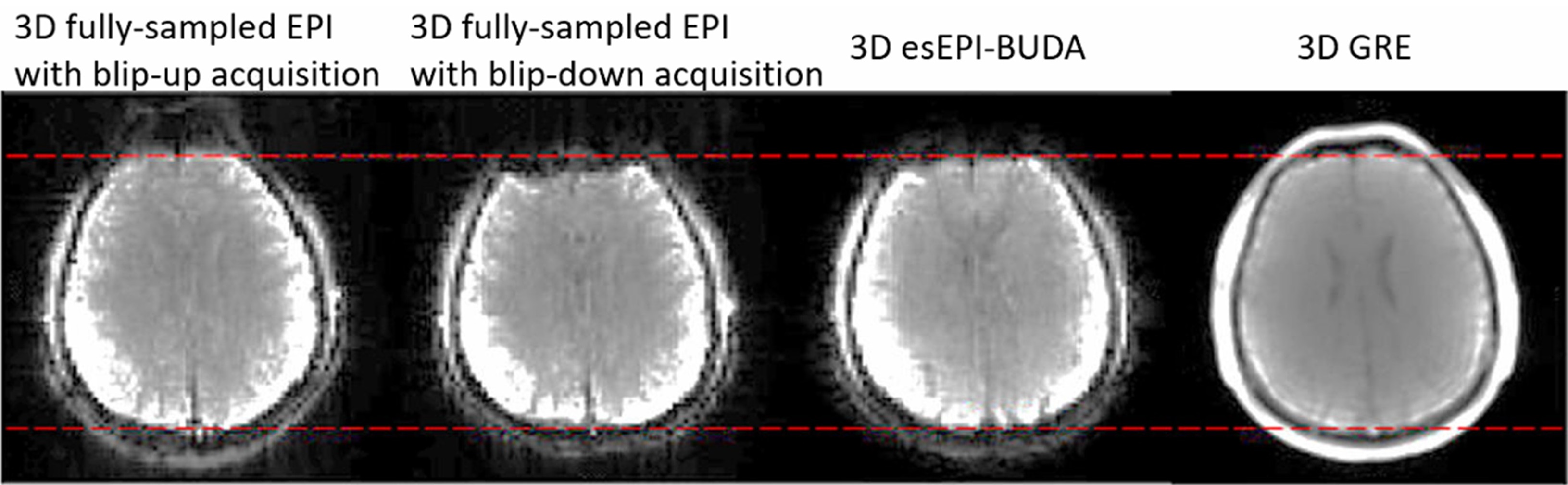
The 3D esEPI-BUDA sequence was implemented on a GE MR750 3T scanner. 3D fMRI experiments were performed on healthy human brains using a 32-channel head coil with a visual stimulation paradigm. The paradigm contained six 48-s blocks with 24-s stimulus followed by 24-s rest. The imaging parameters were: TR/TE=75/30ms, volume TR=2.4s, α≈β=15° (Ernst angle), FOV=220×220×128mm3, acquisition matrix=72×72×32, spatial resolution=3.1×3.1×4.0mm3, and under-sampling factor along the in-slab phase-encoding direction = 2 (i.e., the length of each echo-train = 36; total length of two echo-trains = 72). For comparison, images over the same volume were also acquired using 3D fully-sampled EPI (flip angle=15°) with blip-up and blip-down acquisitions separately.
Results:
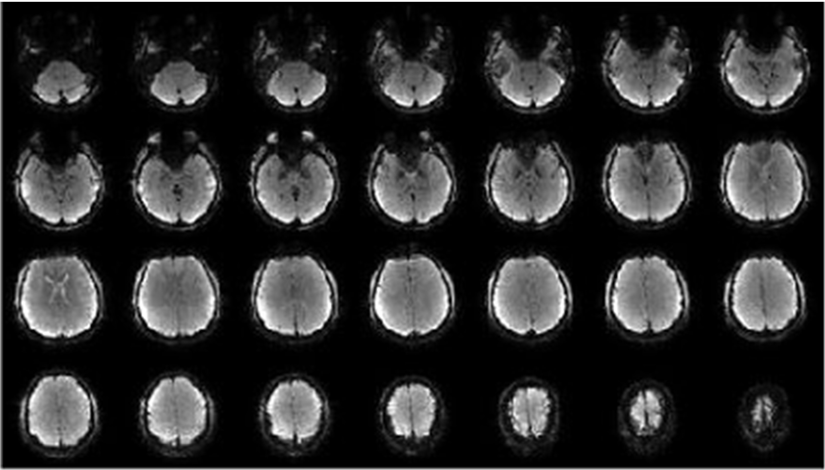
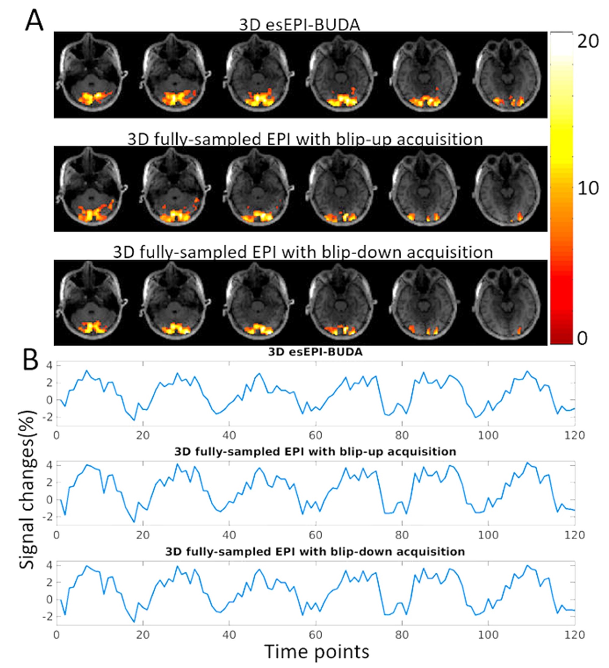
Figure 3 displays a set of representative whole-brain images obtained using 3D esEPI-BUDA, illustrating excellent image quality. The BUDA reconstruction produced images with noticeably reduced geometric distortion, especially at the frontal lobe, when compared to the 3D fully-sampled EPI images (Figure 4). Results from the fMRI experiment are illustrated in Figure 5A where six contiguous activation maps from 3D esEPI-BUDA are overlaid on the corresponding T1-weighted images. fMRI activations were observed in the visual cortex, as expected. Figure 5B shows the time evolution of the BOLD signal change (~4.0%), which is comparable with that of 3D fully-sampled EPI.Discussion and Conclusion:
We have demonstrated a novel pulse sequence – 3D esEPI-BUDA, which accomplished distortion correction with 3D whole-brain coverage by acquiring the blip-up and blip-down datasets in a single shot without increasing the scan time and without being subject to inter-shot motion. Although our demonstration was for 3D fMRI, the same strategy can be extended to other EPI-based applications such as 2D fMRI, 2D/3D diffusion, and 2D/3D perfusion imaging.Acknowledgements
This work was supported in part by the National Institutes of Health (NIH; 5R01EB026716-01 and 1S10RR028898-01). ZC and LY are supported by NIH Grants K25 AG056594, and R01NS118019. The content is solely the responsibility of the authors and does not necessarily represent the official views of the NIH.References
1. Andersson JLR, Skare S, Ashburner J. How to correct susceptibility distortions in spin-echo echo-planar images: Application to diffusion tensor imaging. Neuroimage. 2003;20(2):870-888.
2. Holland D, Kuperman JM, Dale AM. Efficient correction of inhomogeneous static magnetic field-induced distortion in Echo Planar Imaging. Neuroimage. 2010;50(1):175-183.
3. In MH, Posnansky O, Beall EB, et al. Distortion correction in EPI using an extended PSF method with a reversed phase gradient approach. PLoS One. 2015;10(2).
4. Miller KL, Alfaro-Almagro F, Bangerter NK, et al. Multimodal population brain imaging in the UK Biobank prospective epidemiological study. Nat Neurosci. 2016;19(11):1523-1536.
5. Casey BJ, Cannonier T, Conley MI, et al. The Adolescent Brain Cognitive Development (ABCD) study: Imaging acquisition across 21 sites. Dev Cogn Neurosci. 2018;32:43-54.
6. Bilgic B, Poser BA, Langkammer C, et al. 3D-BUDA Enables Rapid Distortion-Free QSM Acquisition. ISMRM. 2020; p0596.
7. Liao C, Bilgic B, Tian Q, et al. Distortion-free, high-isotropic-resolution diffusion MRI with gSlider BUDA-EPI and multicoil dynamic B0 shimming. Magn Reson Med. 2021;86(2):791-803.
8. Cao X, Wang K, Liao C, et al. Efficient T2 mapping with blip-up/down EPI and gSlider-SMS (T2-BUDA-gSlider). Magn Reson Med. 2021;86(4):2064-2075.
9. Smith SM, Jenkinson M, Woolrich MW, et al. Advances in functional and structural MR image analysis and implementation as FSL. Neuroimage. 2004;23:208-219.
Figures