4372
Exposure of infants in baby gradient coils1School of Information Technology and Electrical Engineering, The University of Queensland, Brisbane, Australia, 2Department of Energy, Politecnico di Torino, Torino, Italy
Synopsis
In pediatric magnetic resonance imaging, infants are exposed to rapid, time-varying gradient magnetic fields, leading to electric fields induced in the body of infants and potential safety risks (e.g., peripheral nerve stimulation). In this work, the induced electric fields by the small x, y, z gradient coils in an infant model were numerically evaluated at different model positions. It was found that the induced electric fields in most tissues exceeded the basic restrictions of the ICNIRP 2010 guidelines.
Introduction
Magnetic Resonance Imaging is a non-invasive imaging technology for clinical diagnosis and disease treatment monitoring. It employs electromagnetic (EM) fields to produce high-resolution anatomical images. However, the interaction between these EM fields and living tissue can sometimes lead to detrimental physiological effects. For example, the rapid time-varying gradient magnetic fields can induce electrical fields (E-fields) inside the patient, triggering physiological reactions such as stimulation of peripheral nerves (PNS). PNS ranges from poking of the skin to burning sensations and even intolerable pain [1]. Several international guidelines and standards have been devised to protect patients against these possible EM effects [2-4]. However, these exposure guidelines are usually developed based on numerical analysis of EM induction inside the adult male/female models due to gradient pulsing [5-7]. At present, it lacks appropriate EM safety evaluation of pediatric MRI, and detailed E-fields induced in infants during imaging have not been thoroughly investigated; thus, no guidelines have been made for infant imaging in MRI yet. In this work, the field exposure in an infant model by a set of baby gradient coils (BGC) is numerically evaluated.Methods
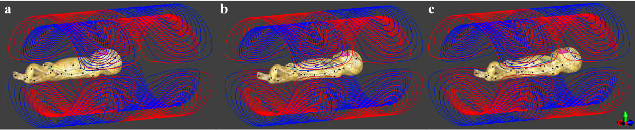
As shown in Figure 1, actively-shielded BGCs were designed [8, 9] to investigate the induced E-fields in the infant's model. The lengths of the primary and secondary coils are 70 cm and 80 cm, respectively. The radii are 19.2 cm, 20.0 cm and 20.8 cm for the primary x, y and z coil, and are 33.9 cm, 34.5 cm, and 35.2 cm for the secondary coils. The BGCs were designed to produce a Go = 30 mT/m gradient strength in a 30 cm diameter of spherical volume (DSV). The gradient field linearity was -5% to 5% in the DSV. The simulation was performed using SIM4LIFE (ZMT, Zurich, Switzerland), integrated with an anatomical infant model, Charlie. The tissue conductivities of the infant model are based on the database developed by the IT'IS Foundation [10]. In the simulations, the coils were driven by sinusoidal currents at 1 kHz frequency to produce the gradient strength Go. In the simulations, the infant model was oriented along an axial direction and facing the positive y-axis of the scanner. Three positions were investigated; the DSV of the gradient coil was located at positions I (head area, Figure 1.a), II (chest area, Figure 1.b), III (body centre, Figure 1.c).Results
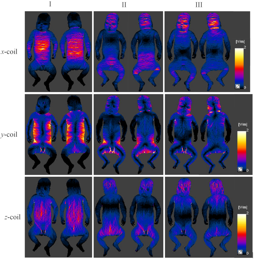
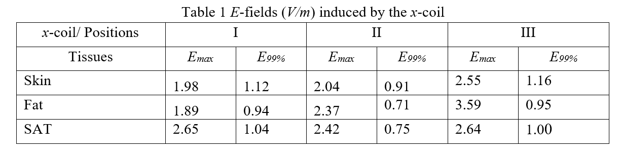
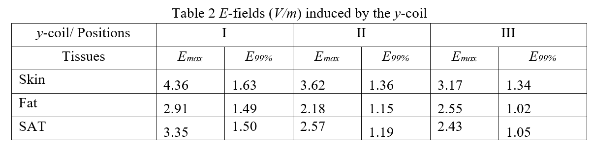
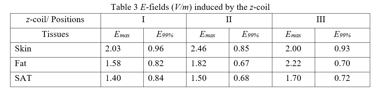
Figure 2 presents the E-fields (front and back views) induced by x,y and z coils at different positions. For x-coil, the E-field profiles are nearly identical in the DSV for different positions but more visible outside the DSV (close the gradient coil ends). The peak E-fields mostly occur in tissues like skin, fat and SAT (subcutaneous fat) and are mainly located in the abdomen and back in position I and in the head for positions II and III. Table 1 collects the Emax and E99% values for x-coil. The maximal Emax was found in fat (3.59V/m), and the maximal E99% was found in the skin (1.16 V/m) in position III. For y-coil, the peak E-fields are mainly observed in the trunk sides and bottom in position I, bottom in position II, and sides of the neck and head in position III, respectively. Similar to x-coil, the peak E-fields mostly occur in skin, fat, SAT and brain. The highest Emax (4.36 V/m) and E99%(1.63 V/m) occur in the skin in position I, which are 3.62 V/m and 1.36 V/m in the skin in position II shown in Table 2. For the z-coil, the E99% is lower than the transverse coils (x and y coils) in most positions (Table 3). The peak E-fields mainly occur in the back and abdomen in position I and the head in position III.Discussion
Simulations of E-fields within an infant model located at different positions inside baby x, y and z coils were performed using SIM4LIFE. As shown in Figure 2, large E-fields can be found near the body surface. Thus, the peripheral nerves in the skin and SAT are exposed to the strongest electric fields. In each case, outside the imaging center (DSV), the maximum E-fields highly correlate with coils and body positions, the shape and heterogeneous tissue conductivity and the local magnetic fields. As reported in some early works [5,7], the y-coil induces a larger E99% than the other two coils. It is noted that the E-fields induced in the infant model exceed the reference level- 0.4 V/m in ICNIRP2010 guidelines for 1 kHz frequency. Since infants' brains are more conductive than adults [11], it raises a concern that it may cause different or extra exposure. Thus, the MRI scan protection of infants' developing nervous system needs to be further investigated.Conclusion
In this paper, we have numerically evaluated the exposure of an infant model inside the dedicated baby x, y, z gradient coils during an MRI scan. It was found that the exposures could exceed the reference level in ICNIRP2010 guidelines during a routine scan. It is urgent to develop an infant-dedicated regulation standard for pediatric MRI, which requires characterizing more realistic field-infant tissue interactions with detailed nerve models.Acknowledgements
No acknowledgement found.References
[1] R. F. Schulte and R. Noeske, "Peripheral nerve stimulation‐optimal gradient waveform design," Magnetic resonance in medicine, vol. 74, no. 2, pp. 518-522, 2015.
[2] J. Lin et al., "ICNIRP Guidelines for limiting exposure to time-varying electric and magnetic fields (1 Hz to 100 kHz)," Health Physics, vol. 99, pp. 818-836, 2010.
[3] J. C. Lin, "A new IEEE standard for safety levels with respect to human exposure to radio-frequency radiation," Antennas and Propagation Magazine, IEEE, vol. 48, no. 1, pp. 157-159, 2006.
[4] I. E. Commission, "Particular requirements for the basic safety and essential performance of magnetic resonance equipment for medical diagnosis," Medical Electrical Equipment, vol. 60601-2-33, pp. Part2-33.[5] F. Liu, H. Zhao, and S. Crozier, "On the induced electric field gradients in the human body for magnetic stimulation by gradient coils in MRI," IEEE Transactions on Biomedical Engineering, vol. 50, no. 7, pp. 804-815, 2003.
[6] P. P. So, M. A. Stuchly, and J. A. Nyenhuis, "Peripheral nerve stimulation by gradient switching fields in magnetic resonance imaging," IEEE Transactions on Biomedical Engineering, vol. 51, no. 11, pp. 1907-1914, 2004.
[7] A. M. Samoudi, G. Vermeeren, E. Tanghe, R. Holen, L. Martens, and W. Josephs, "Numerically simulated exposure of children and adults to pulsed gradient fields in MRI," Journal of Magnetic Resonance Imaging, 2016.
[8] F. Tang et al., "An improved asymmetric gradient coil design for high-resolution MRI head imaging," Physics in Medicine and Biology, vol. 61, no. 24, p. 8875, 2016.
[9] H. Sanchez Lopez, F. Liu, M. Poole, and S. Crozier, "Equivalent magnetization current method applied to the design of gradient coils for magnetic resonance imaging," IEEE Transactions on Magnetics, vol. 45, no. 2, pp. 767-775, 2009.
[10] P. Hasgall et al., "IT’IS Database for thermal and electromagnetic parameters of biological tissues. Version 4.0, May 15, 2018. doi: 10.13099," VIP21000-04-0. Onl: www. itis. ethz. ch/database, 2018.
[11] L. Kheifets, M. Repacholi, R. Saunders, and E. Van Deventer, "The sensitivity of children to electromagnetic fields," Pediatrics, vol. 116, no. 2, pp. e303-e313, 2005.
Figures