4369
Simulation of RF heating for patients with metallic gamma knife stereotaxic headframe in MRI1GE Healthcare, Beijing, China
Synopsis
In this study, electromagnetic simulation was used to calculate RF heating for the patients with metallic gamma knife stereotaxic headframe in MRI. The relationship between headframe structure parameters and patient SAR was investigated. Two effective SAR derating methods of adjusting the headframe structure parameters were proposed, which can significantly decrease 10 g local SAR from 253 W/kg to 68 W/kg and head SAR from 4.2 W/kg to 2.0 W/kg. This study provides the efficient and reliable methods to ensure safety of the patients with metallic gamma knife headframe in MRI.
Introduction
Magnetic resonance imaging (MRI) is a regularly imaging technique in gamma knife (GK) radiosurgery of brain tumor.[1] It can be used to plan patient treatment and assessing treatment response. However, the metallic stereotaxic headframe increases possibility of patient skin injury as they are exposed in the RF field during the MRI scan.[2] Though some GK vendor developed the non-metallic GK headframe, there are still large number of patients with metallic GK headframe which require the safety MRI scan. The values of the patient SAR including 10 g local SAR and head SAR are conservatively estimated to ensure safety. The scan time are therefore very long. Through electromagnetic simulation, this abstract assessed the RF heating for the patients with metallic GK headframe in MRI and proposed two SAR derating methods. Two derating methods are proposed and demonstrated to efficiently decrease both 10 g local SAR and head SAR and have the potential to be implemented into current MRI scan for the patients with metallic GK headframe.Methods
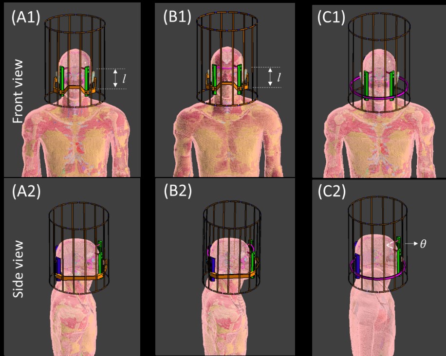
RF heating for the patients with metallic GK stereotaxic headframe in MRI was simulated using a commercial FDTD software (Sim4Life, ZMT, Zurich, Switzerland). Figure 1 shows three models of a standard generic body model (Duke) wearing GK stereotaxic headframe. The birdcage head coil model was adopted to produce the RF field. During simulation, B1rms was normalized with 3.6 µT in the head.The local SAR and head SAR were first calculated for Duke wearing a common Leksell GK stereotaxic headframe as illustrated in Figure 1A. SAR derating effect with decreasing the distance l between the head fixings screw and the rectangular metallic base was simulated.
Besides, the GK stereotaxic headframe with a short circuit cable connecting the fixings screws was proposed (Figure 1B) to decrease both local SAR and head SAR. Two screws in front of head were directly connected by a metal-conductor cable. Two screws behind head were also connected. The SAR derating effect of this structure was simulated. Furthermore, SAR derating effect with decreasing the distance l and using the short circuit cable in the meantime was also simulated.
To provide insight into the RF heating mechanism, Duke wearing a simplified GK stereotaxic headframe in MRI was simulated as well. In the simulation, the standard rectangular metallic base was replaced with a circle metallic base which center is at the coil center (Figure 1C). The central angle θ between two adjacent screws was systematically changed, to investigate the current flowing in Duke’s head and its effect on SAR.
Results and Discussion
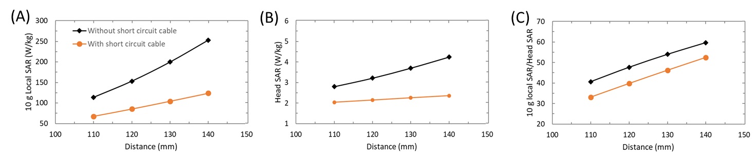
Figure 2A and 2B shows the 10g local SAR and head SAR at four screw-metallic base distances, for Duke wearing Leksell GK stereotaxic headframe with and without short circuit cable. For the standard headframe, 10 g local SAR is in the range of 114–253 W/kg, and head SAR is in the range of 2.8–4.2 W/kg. RF heating increases with the screw-metallic base distance. The ratio between 10 g local SAR and head SAR (Figure 2C) increases with the screw-metallic base distance as well. Further investigation demonstrates the decreased distance makes the current loop forming by head-screws-screw holders-base loop has smaller area in the RF field. Therefore, lower current density and SAR are resulted in.By connecting the screws in front and behind of the head, 10 g local SAR, head SAR, and ratio between 10g local SAR and head SAR are all significantly decreased. 10 g local SAR is even decreased to its half comparing to the headframe without short circuit cable. This is proved due to the current loop propagating along the screw holders and base are shorted, replaced by head-screw-short circuit cable loop. The current loop area reaches minimum in RF field in this condition. For 10 g SAR, the maximum and minimum values in Figure 2A are 253 W/kg and 68 W/kg. For head SAR, the maximum and minimum values in Figure 2B are 4.2 W/kg and 2.0 W/kg.
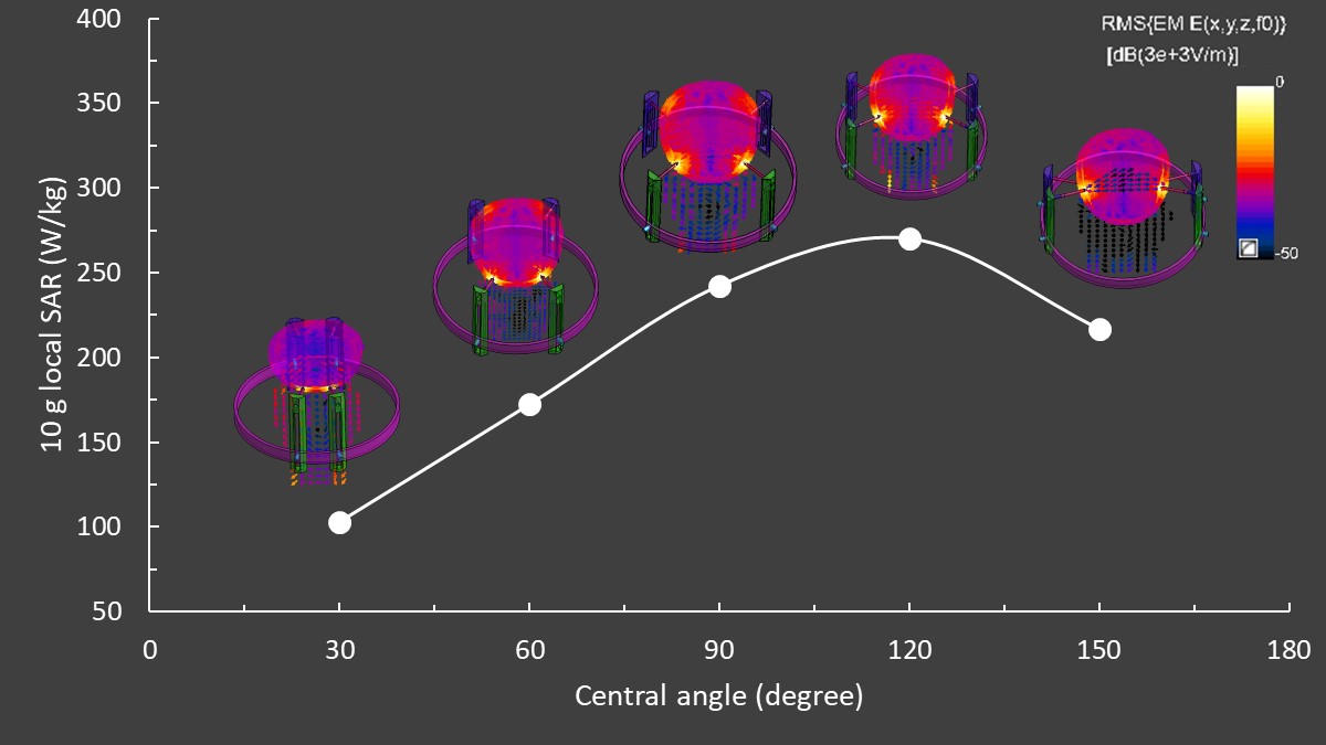
Figure 3 shows 10 g local SAR changing with central angle θ between two adjacent screws in front and behind of the head, relative to the simplified headframe in Figure 1C. The smaller angle has lower SAR due to the smaller current loop. At 120°, there’s a maximum SAR due to the trade-off between the current loop area and the head resistance in the loop. The electric field and its vector direction shown in Figure 3 indicate the induced current related to SAR reaches maximum at 120° and the current loop are mainly in the coronal plane other than the sagittal plane.
Conclusion
This abstract builds a simulation model of RF heating for the patients with metallic GK stereotaxic headframe in MRI. The results demonstrate the RF heating are produced mainly due to the large current loop form between head and headframe. Decreasing the distance between the screw and metallic base results in lower SAR. Further adding a short circuit cable between screws is proposed to significantly decrease local SAR (from 253 W/kg to 68 W/kg) and head SAR (from 4.2 W/kg to 2.0 W/kg).Acknowledgements
No acknowledgement found.References
[1] Gao X, Zhang XN, Zhang YT, Yu CS and Xu DS. Magnetic resonance imaging in assessment of treatment response of gamma knife for brain tumors. Chin Med J 2011;124(12):1906-1910.
[2] Bennett MC, Wiant DB, Gersh JA, Dolesh W, Ding X, Best RC, Bourland JD. Mechanisms and prevention of thermal injury from gamma radiosurgery headframes during 3T MR imaging. J Appl Clin Med Phys. 2012;13(4):54-70.
Figures