4368
RF Coil design for improving human liver fat quantification in a portable single-side MR system1School of Biomedical Engineering, Shanghai Jiao Tong University, Shanghai, China, Shanghai, China, 2Wuxi Marvel Stone Healthcare Co. Ltd., Wuxi, Jiangsu, China, Wuxi, China
Synopsis
Earlier diagnosis of non-alcoholic fatty liver disease (NAFLD) becomes important to prevent the disease progression. Recently, a low-cost portable MR system was developed as a point-of-care screening tool for in vivo liver fat quantification. However, the subcutaneous fat may confound the live fat quantification, particularly in the NAFLD susceptible population. In this work, we propose a novel RF coil design composed of a main target coil sandwiching a set of “saturation” coil to improve human liver fat quantification. We demonstrate the capability and effectiveness of the novel RF design in phantom experiments as well as in-vivo liver scans.
Introduction
Earlier diagnosis of non-alcoholic fatty liver disease (NAFLD) becomes important to prevent the disease progression [1]. Recently, a low-cost portable single-side MR system was developed as a point-of-care screening tool for in vivo liver fat quantification [2, 3]. The system has been demonstrated a good positive correlation between the proton density fat fraction (PDFF) and the fitted fat phantoms. However, there are more-proximal tissues in in-vivo liver evolution compared to the phantom study, such as skin and subcutaneous fat. Those tissues may confound the liver fat quantification. In the work, we propose a novel RF coil design composed of a main target coil sandwiching a set of saturation coil to improve human liver fat quantification. Experiments using the inversion recovery CPMG sequence on phantom and subject show that the saturation coil has a good suppression on the subcutaneous fat signal and improves the fat quantifications.Theory and Methods
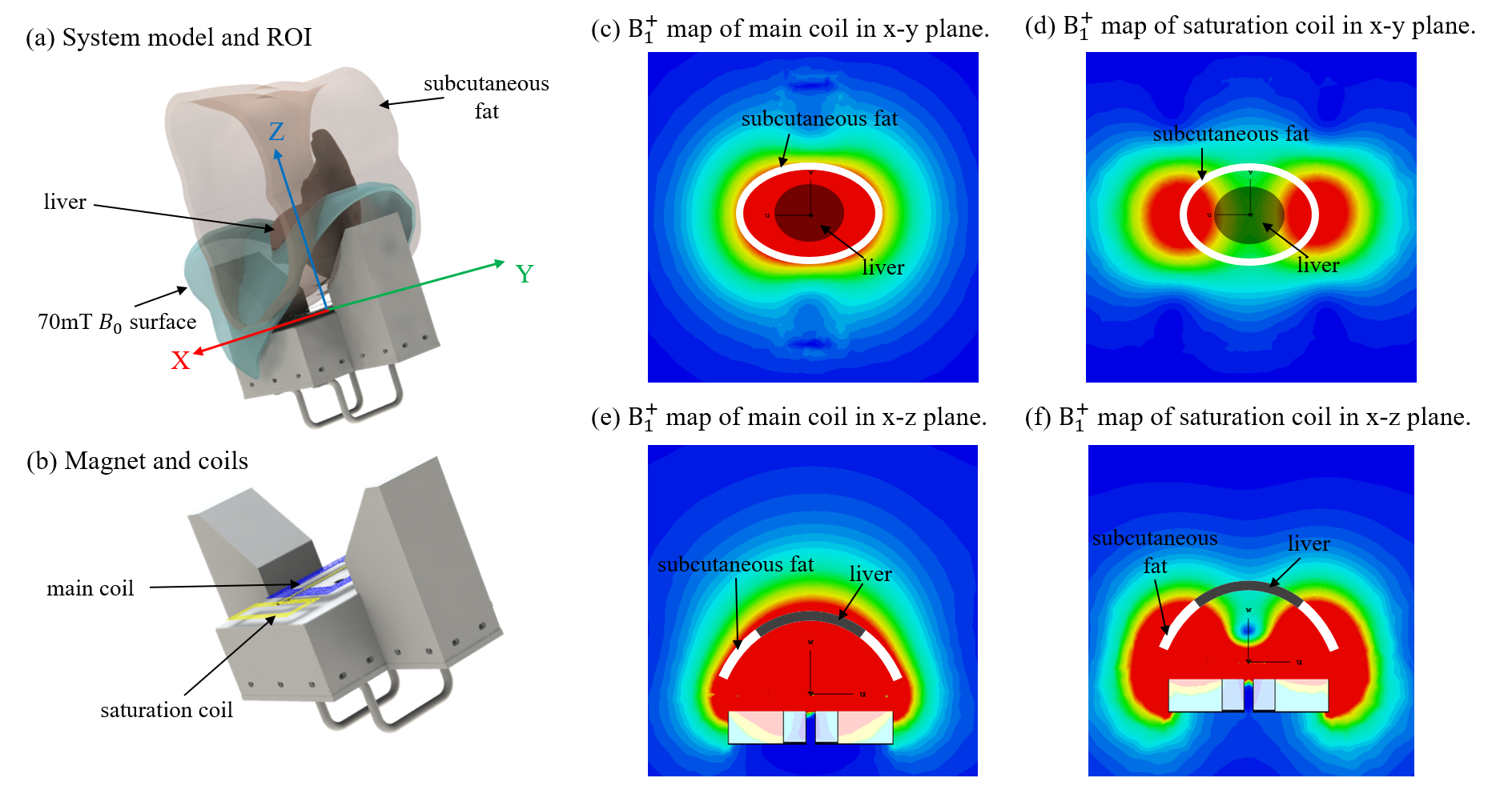
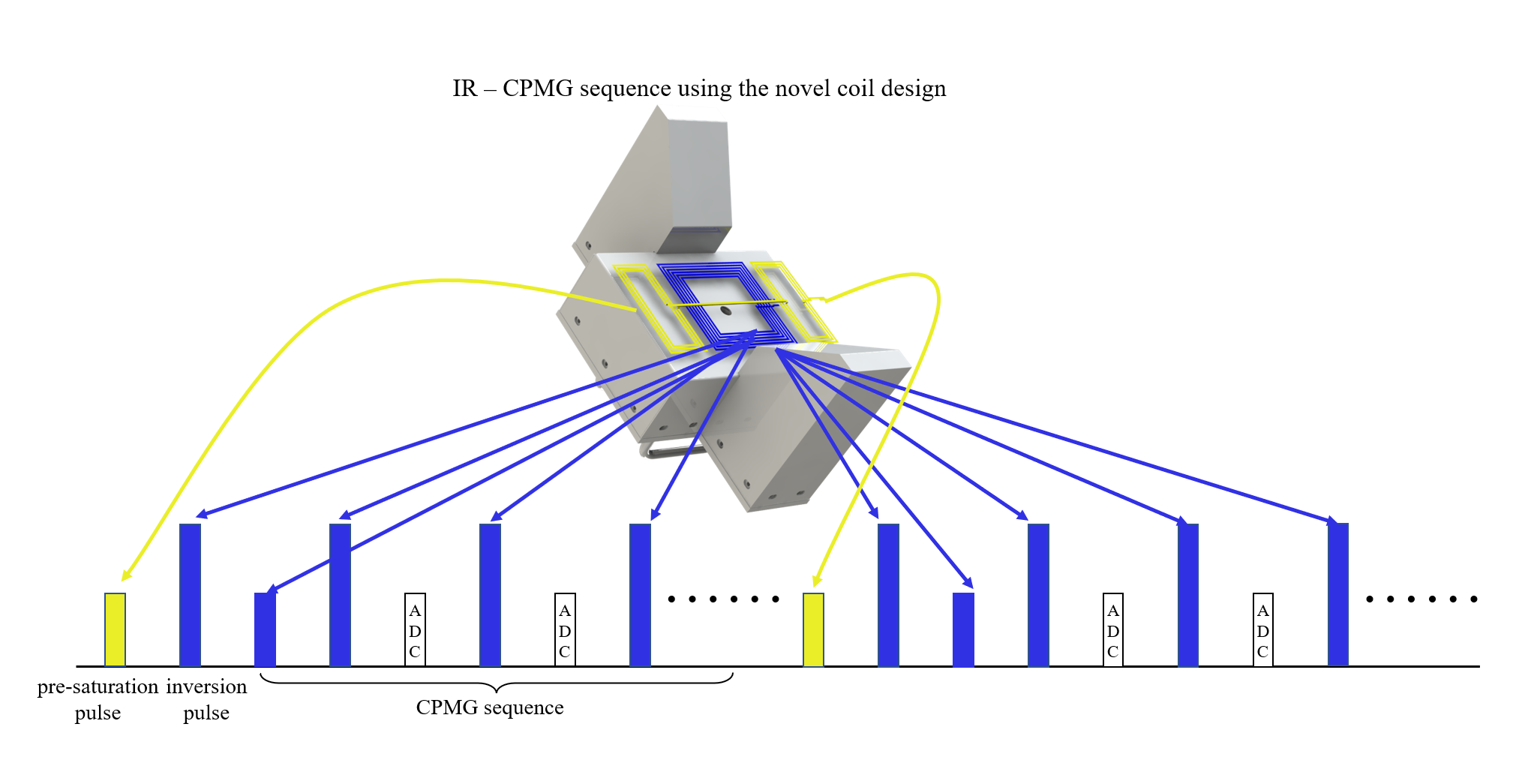
In the single-side MR system with a concave U-shaped magnet, as shown in Fig.1, the targeted imaging region of interest (ROI) is constrained by the B0 field and B1 field. The static magnetic field strength B0 is highly uniform in the direction parallel to the surface of the main coil while decreasing gradually when moving away from its surface. The cyan surface indicates a part of ~70mT iso-surface with a depth of ~8 cm from the main coil center. The B1+ field maps of the main coil projected along the x-y plane and x-z plane are shown in Fig. 1c and e., the subcutaneous fat tissues as indicated by the white line segments are targeted by the main coil. However, the adding saturation coils can be used to suppress those subcutaneous fat signals from the B1+ field projection maps of the butterfly saturation coil as shown in Fig. 1d and f.The sequence used in this study is shown in Fig. 2. A pre-sat pulse transmitted by the butterfly saturation coil is inserted in an IR-CPMG pulse and acquisition train where the pulses are transmitted and the signals are received by the main coil. The signals are acquired with various TEs and TRs (6 × 6) in the IR-CPMG and then fitting into a multiexponential model of 2 substances (fat and liver tissue) [4, 5]. Parameters of relaxation times, diffusion coefficients, and fat fraction are calculated from the fitting solution. The phantom and in-vivo human liver experiments are carried out to demonstrate the capability and effectiveness of the novel RF design for improving fat quantification.
Results and Discussion
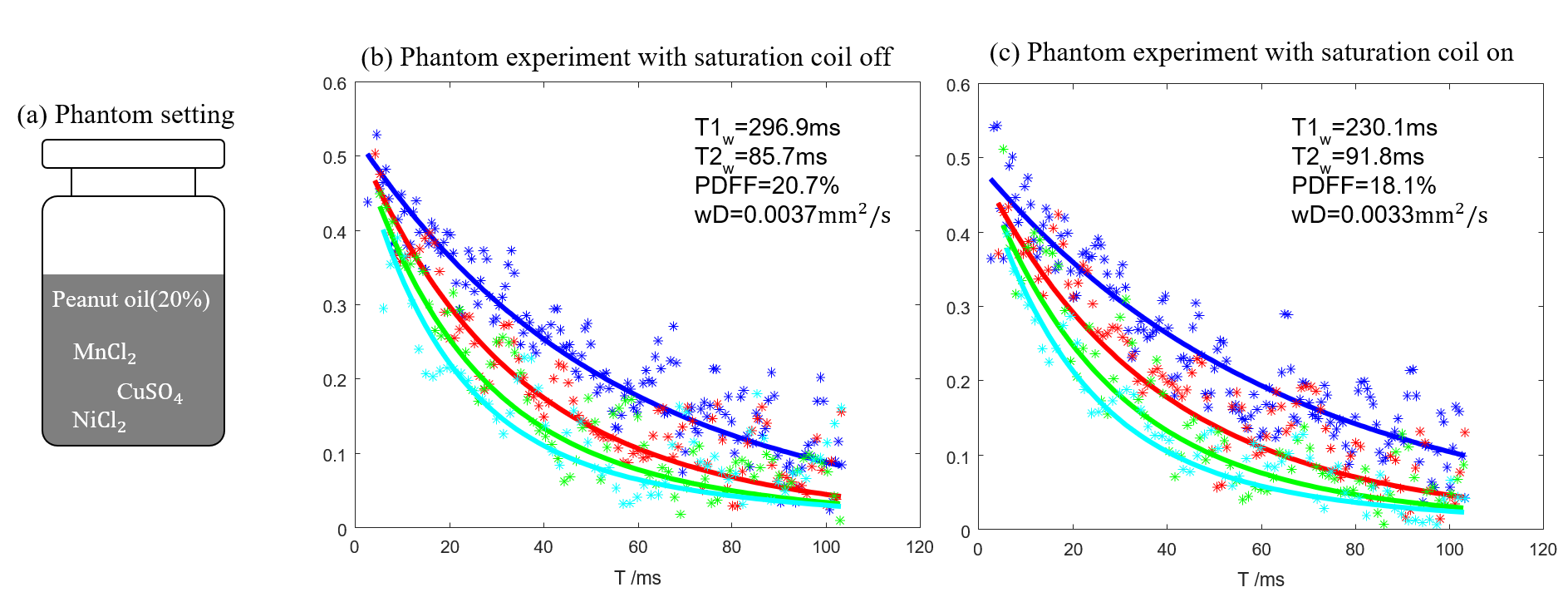
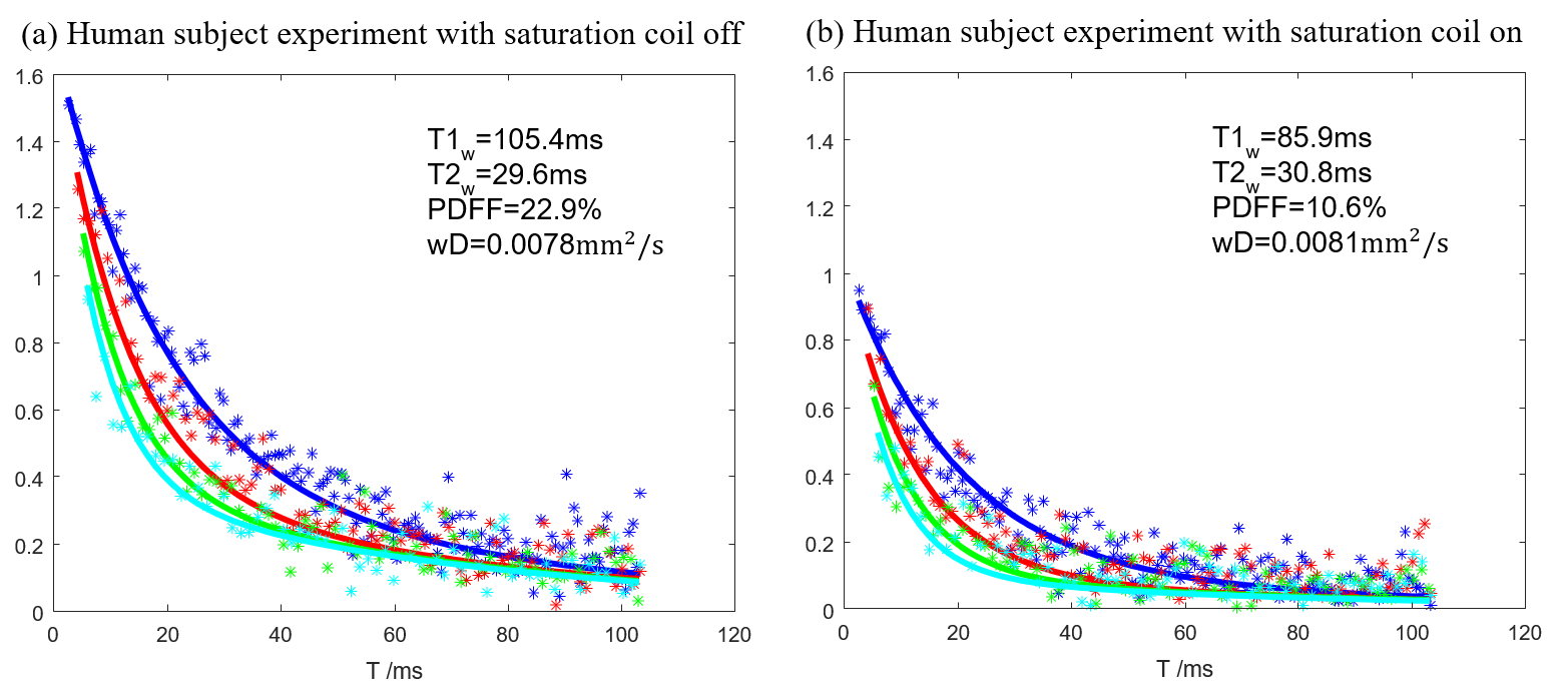
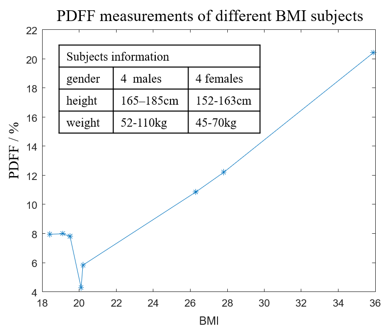
Figure 3 shows the phantom experiment results with and without using saturation coils. The phantom is composed of aqueous MnCl2 solution with 20% peanut oil stabilized with surfactants. Compared to the representative fitted curves and the calculated parameters, those results indicate a consistency due to the fact that phantom size is small and there are no more-proximal components. This demonstrates the adding saturation coils keep the fat assessment of interested target.Figure 4 shows the results of human subject experiments with and without using saturation coils. The signal intensities obtained with saturation coil on are lower than those obtained without using saturation coil since the signal contribution from the subcutaneous fat is significantly suppressed. The PDFF is reduced from 22.9% to 10.6% after the subcutaneous fat suppression. Figure 5 shows the PDFF comparisons of 8 subjects with different Body mass index (BMI), The PDFF has an increasing trend with the increasing BMI but this trend disappears with low BMIs. More work is currently ongoing to further assess the liver fat quantification with golden-standard reference.
Conclusion
A novel RF coil design composed of a main target coil sandwiching a set of saturation coil is developed in a portable single-side MR system to improve human liver fat quantification. The phantom experiments, as well as in-vivo liver scans, demonstrate the capability and effectiveness of the novel RF design.Acknowledgements
This work is supported by the National Science Foundation of China (No. 62001290) and sponsored by Shanghai Sailing Program (20YF1420900).References
[1] Younossi Z, Anstee Q M, Marietti M, et al. Global burden of NAFLD and NASH: trends, predictions, risk factors and prevention[J]. Nature reviews Gastroenterology & hepatology, 2018, 15(1): 11-20.
[2] Bashyam A, Frangieh C J, Raigani S, et al. A portable single-sided magnetic-resonance sensor for the grading of liver steatosis and fibrosis[J]. Nature Biomedical Engineering, 2021, 5(3): 240-251.
[3] Wang Y, Xu Y, Zhang M, et al. A single-sided magnet for deep-depth fat quantification[J]. Journal of Magnetic Resonance, 2021, 331: 107053.
[4] Holmström K, Petersson J. A review of the parameter estimation problem of fitting positive exponential sums to empirical data[J]. Applied Mathematics and Computation, 2002, 126(1): 31-61.
[5] Chevallier O, Zhou N, Cercueil J P, et al. Comparison of tri‐exponential decay versus bi‐exponential decay and full fitting versus segmented fitting for modeling liver intravoxel incoherent motion diffusion MRI[J]. NMR in Biomedicine, 2019, 32(11): e4155.
Figures