4351
Deep learning-accelerated T2-weighted imaging of the female pelvis: reduced acquisition times and improved image quality1Peking Union Medical College Hospital, Peking Union Medical College and Chinese Academy of Medical Sciences, Beijing, China, 2MR collaboration, Siemens Healthineers Ltd., Beijing, China, 3MR Application Predevelopment, Siemens Healthcare GmbH, Erlangen, Germany
Synopsis
Novel deep learning (DL) reconstruction methods may accelerate female pelvis MRI protocols keeping high image quality. The value of a novel DL reconstruction of T2-weighted (T2DLR) turbo spin-echo (TSE) sequences for female pelvis MRI in three orthogonal planes was evaluated. We evaluated examination times, image quality, and lesion conspicuity of benign uterine disease. The T2DLR quantitative parameters remained similar or were significantly improved compared with that of standard T2 TSE (T2S), allowing for a 62.7% reduction in acquisition times. Applying this novel T2DLR sequence achieved better image quality and shorter acquisition time than T2S.
Introduction
Magnetic resonance imaging (MRI) of the female pelvis has been widely used to evaluate malignant and benign diseases of the female pelvis due to its excellent soft-tissue resolution and anatomic detail1-3. T2-weighted imaging (T2WI) is the mainstay for the detection and accurate assessment of the extent of local disease for benign and malignant uterine neoplasms. A basic MRI protocol with at least two T2WI orthogonal planes of the uterus, including a sagittal sequence of the uterine corpus, was recommended for patients with uterine diseases4,5. A disadvantage of this comprehensive protocol were the rather long examination times, ranging from approximately 20 to 30 min. These extended examination times are problematic for elderly and claustrophobic patients, who have trouble staying motionless, resulting in poor image quality. The introduction of novel deep learning reconstruction methods could accelerate the acquisition time and provide high image quality. These methods have shown encouraging results for the abdomen, knee, and prostate MRI protocols6-8. Therefore, this study aimed to investigate the impact of a novel DL reconstruction accelerated T2 TSE sequence applied to female pelvises in regard to examination times, image quality, and lesion conspicuity of benign uterine disease.Materials and methods
Twenty healthy volunteers aged 23–50 years (mean age 26.8 years), and 31 consecutive female patients aged 24–71 years (mean age 41.0 years) with clinically and ultrasound confirmed benign uterine disease were recruited for pelvic MRI (Table 1). All MRI examinations were acquired on a 3T MR system (MAGNETOM Vida, Siemens Healthcare, Erlangen, Germany) using an 18-channel body coil. Standard abdominopelvic MRI, including T2w TSE imaging in three planes, was performed. After completion of standard T2w TSE imaging (T2S), a prototypical T2w TSE sequence with deep learning image reconstruction (T2DLR) (Siemens Healthcare, Erlangen, Germany) was performed. The imaging parameters are listed in Table 2.All sagittal, axial, and coronal T2W images were randomized, anonymized, and independently evaluated. Image quality was evaluated quantitatively (signal-to-noise ratio [SNR], and contrast-to-noise ratio [CNR]) and qualitatively (5-point Likert scale including overall image quality, artifacts, boundary sharpness of the zonal layers, and lesion conspicuity, with 1 as the worst performance and 5 as the best performance). Image quality was compared between two sequences using a paired t-test and Wilcoxon signed-rank test. Inter-observer agreement was calculated using Cohen´s kappa.
Results
The acquisition time was 1:54 min for T2DLR, with a 62.7% reduction compared with T2S. The SNR of the myometrium and junctional zones were significantly higher on T2DLR imaging than on T2S imaging, while no significant differences were found between the SNR of the leiomyoma on T2DLR and that of T2s. T2DLR exhibited significantly higher CNR between the myometrium and gluteal muscle than T2s (p=0.026). There was no significant difference in CNRs between the junctional zone and gluteal muscle and between the leiomyoma and gluteal muscle on T2DLR and T2s (both p>0.05). (Fig. 1)Cohen’s kappa scores regarding image quality parameters between the two readers were good to almost perfect (0.67-0.85 for T2DLR and 0.62-0.87 for T2S). The overall image quality was rated higher for T2DLR compared with T2S on the sagittal, axial, and coronal images (p<0.001). The extent of artifacts in T2DLR was rated as significantly less in all orientations compared with that in T2S (all p<0.05). The boundary sharpness of the zonal layers was rated as significantly better in T2DLR compared with that in T2S (p<0.001). Lesion conspicuity was superior in T2DLR compared with that in T2S with a median of 5 (4–5) vs. a median of 4 (4–4) in the sagittal and coronal images, and a median of 5 (5–5) vs. a median of 4 (4–4) in the axial images (Table 3). A representative case is shown in Fig. 2.
Discussion
In this study, the novel DL T2w TSE sequence was compared with the standard T2w TSE sequence to accelerate female pelvis MRI times in healthy volunteers and patients with benign uterine diseases. We showed that T2DLR exhibited a markedly improved SNR in the myometrium and junctional zone, and contrast between the myometrium and gluteal muscle compared with T2S, which allowed for a 62.7% reduction in acquisition times while maintaining the SNR in the leiomyoma and the contrast between the leiomyoma and gluteal muscle. Furthermore, qualitative image evaluations of image quality, artifacts, and boundary sharpness of the zonal layers in the uterine corpus were superior in T2DLR compared with the T2s. T2DLR also had significantly better lesion conspicuity of benign uterine diseases than that of T2s. However, this study had a small sample size to demonstrate the feasibility of this new technique. Additional studies, including those with larger sample sizes and different pathologies, are warranted to corroborate our findings.Conclusions
This novel T2w TSE sequence with DL reconstruction is an effective and promising approach to optimally reduce the acquisition times of female pelvis MRI examinations compared with standard T2w TSE imaging, and it significantly improved image quality, boundary sharpness, and lesion conspicuity of benign uterine diseases.Acknowledgements
None.References
1. Balcacer P, Shergill A, Litkouhi B. MRI of cervical cancer with a surgical perspective: staging, prognostic implications and pitfalls. Abdominal Radiology. 2019;44(7):2557-2571.
2. Nougaret S, Horta M, Sala E, et al. Endometrial Cancer MRI staging: Updated Guidelines of the European Society of Urogenital Radiology. European radiology. 2019;29(2):792-805.
3. Bazot M, Bharwani N, Huchon C, et al. European society of urogenital radiology (ESUR) guidelines: MR imaging of pelvic endometriosis. European radiology. 2017;27(7):2765-2775.
4. Balleyguier C, Sala E, Da Cunha T, et al. Staging of uterine cervical cancer with MRI: guidelines of the European Society of Urogenital Radiology. European radiology. 2011;21(5):1102-1110.
5. Kubik-Huch RA, Weston M, Nougaret S, et al. European Society of Urogenital Radiology (ESUR) Guidelines: MR Imaging of Leiomyomas. European radiology. 2018;28(8):3125-3137.
6. Herrmann J, Gassenmaier S, Nickel D, et al. Diagnostic Confidence and Feasibility of a Deep Learning Accelerated HASTE Sequence of the Abdomen in a Single Breath-Hold. Investigative radiology. 2021;56(5):313-319.
7. Recht MP, Zbontar J, Sodickson DK, et al. Using Deep Learning to Accelerate Knee MRI at 3 T: Results of an Interchangeability Study. AJR. American journal of roentgenology. 2020;215(6):1421-1429.
8. Gassenmaier S, Afat S, Nickel D, Mostapha M, Herrmann J, Othman AE. Deep learning-accelerated T2-weighted imaging of the prostate: Reduction of acquisition time and improvement of image quality. European journal of radiology. 2021;137:109600.
Figures
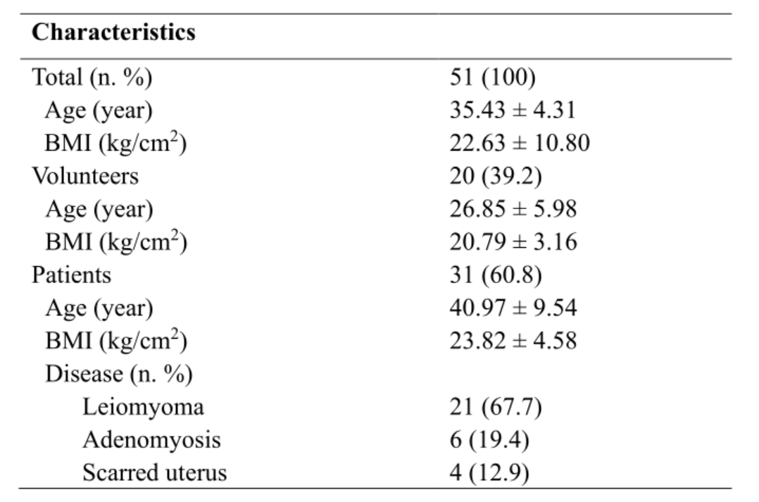
Table 1. Characteristics of the study population. (BMI: Body Mass Index)
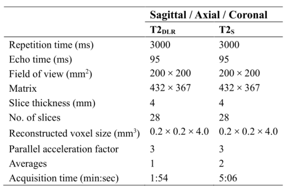
Table 2. MRI acquisition parameters of T2DLR and T2S. (T2DLR: T2-weighted turbo spin-echo with deep learning reconstruction; T2S: standard T2-weighted turbo spin-echo)
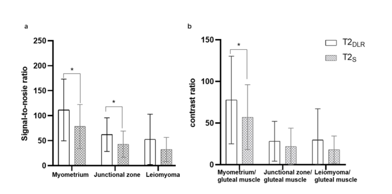
Fig. 1 Box plots of signal-to-noise ratios of the myometrium, junctional zone, and leiomyoma (a), and the contrast-to-noise ratio between the myometrium and gluteal muscle, the junctional zone and gluteal muscle, and the leiomyoma and gluteal muscle (b) on sagittal T2-weighted images obtained with the deep learning reconstruction accelerated T2-weighted turbo spin-echo sequence (T2DLR) and standard T2 turbo spin echo sequence (T2S) (*p<0.05).
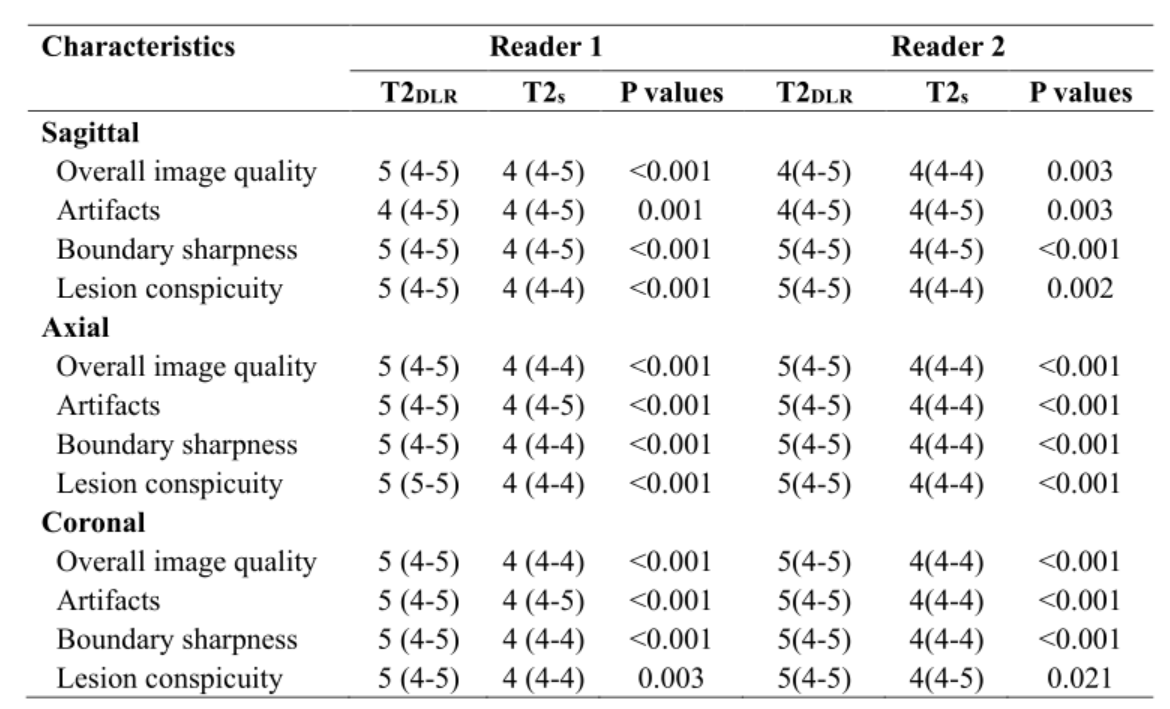
Table 3. Quantitative image evaluations in T2DLR and T2S. (T2DLR: T2-weighted turbo spin-echo with deep learning reconstruction; T2S: standard T2-weighted turbo spin-echo)
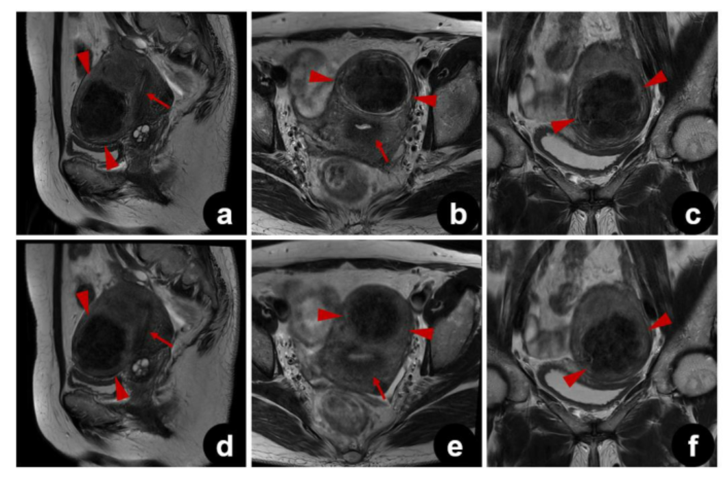
Fig 2. A 47-year-old patient with leiomyoma. Sagittal (a), axial (b), and coronal (c) images of T2DLR and sagittal (d), axial (e), and coronal (f) images of T2s. Less motion artifacts occurred with T2DLR, which shows sharper depiction of the lesion (arrowheads) and the zonal layers of the uterus (arrows). (T2DLR: T2-weighted turbo spin-echo with deep learning reconstruction; T2S: standard T2-weighted turbo spin-echo)