4328
Axial T2-weighted MR imaging of the cervical spine using PROPELLER with deep learning reconstruction to improve image quality
You Seon Song1, In Sook Lee1, Moon jung Hwang2, Kyungeun Jang2, Maggie Fung3, and Xinzeng Wang4
1Pusan National University Hospital, Busan, Korea, Republic of, 2GE Healthcare Korea, Seoul, Korea, Republic of, 3GE Healthcare, New York, NY, United States, 4GE Healthcare, Houston, TX, United States
1Pusan National University Hospital, Busan, Korea, Republic of, 2GE Healthcare Korea, Seoul, Korea, Republic of, 3GE Healthcare, New York, NY, United States, 4GE Healthcare, Houston, TX, United States
Synopsis
We evaluated the utility of PROPELLER T2 FSE with deep-learning (DL) reconstruction in the cervical spine MRI, with the goal of overcoming respiratory and swallowing motion, improving image sharpness, and achieving high-resolution imaging within a comparable scan time to the conventional T2 FSE sequences. With utilization of DL reconstruction, the axial PROPELLER T2 was able to obtain images with high resolution and reduced noise.
Introduction
An axial T2 FSE image in cervical spine MRI is essential to evaluate the spinal cord as well as the disc. In many cases, it is difficult to evaluate the spinal cord because the signal intensity of the spinal cord is inhomogenous due to motion artifact, especially by swallowing or breathing. Motion artifacts may have an important influence on the image quality and the diagnostic value. The periodically rotated overlapping parallel lines with enhanced reconstruction (PROPELLER) technique reduces motion artifacts in MRI of the abdomen, chest, spine and brain. Although PROPELLER image can reduce noise, it is often associated with image blurring by motion artifact. DL have recently been used in medical research and have been reported for improved image quality and noise reduction. The DL network was embedded into the reconstruction pipeline such that both conventional and DL image series could be generated from a single set of raw MRI data. The purpose of this study was to demonstrate that with utilization of a DL reconstruction algorithm, a T2 PROPELLER technique will provide better image quality and decreased image noise or motion artifacts compared to the conventional T2 FSE or non-DL PROPELLER images at cervical spine MRI.Materials and methods
From July to December 2020, 95 patients examined non-enhanced cervical spine MRI on the same 3T scanner (SIGNA Architect, GE healthcare, Waukesha, USA). In addition to routine protocols including axial and sagittal T1- and T2-weighted images(WIs), axial T2WIs were obtained using the PROPELLER fast spin-echo sequence. After excluding cases with metal fixation and severe compression of spinal cord by variable pathologic conditions, finally total 35 patients (age range, 19-76 years; mean 53.4 years; 12 female, 23 male) were included in this study with IRB approval. The imaging parameters for PROPELLER axial T2WI was as follows; auto TR 3650~4050 ms/ effective TE 95~104 ms/echo train length 28/ excitations,2.5; field of view, 140 mm; section thickness 3.0 with 0.3 mm spacing ; matrix, 300x300, bandwidth 50KHz . the imaging time was about 2min and 15s. The images were then reconstructed with conventional reconstruction and a DL. The DL comprised a deep convolutional residual encoder network trained using a prior data. In this study, two different noise reduction factors (DL 50% and 75%) were selected. Total four types of images including FSE, PROPELLER, PROPELLER with DL 50%, and DL 75% were quantitatively and qualitatively reviewed by two musculoskeletal radiologists. For quantitative measurements, we located round ROI at spinal cord, sternocleidomastoid (SCM) muscle, proximal and distal background of each five disc level from C2-3 to C6-7. The size of the ROI was adjusted as much as possible to the spinal cord to be measured, and then copied and placed in the region to be measured. We calculated SNR and CNR of the spinal cord and SCM by using obtained ROI values. Also, the resulting images were scored qualitatively as follows; image noise (none, mild, moderate and severe) and overall image quality (excellent, diagnostic, and imparing diagnostic) about spinal cord, SCM and back muscles at each level.Statistical analysis was done by using ANOVA and Post-hoc t-test (IBM SPSS statistics 25).Results
In the qualitative analysis, all four types of images (FSE, PROPELLER, DL 50, DL 57) showed significant differences at all disc levels for the spinal cord, SCM and back muscles (p < 0.05). In particular, DL50 and DL75 images showed a significant difference in image quality compared to FSE and PROPELLER images (p < 0.0083). For each sequence, the SNR of the spinal cord and SCM muscles showed significant differences at all discs levels (p < 0.0083). The CNR of the spinal cord was significantly higher in PROPELLER, DL50, and DL75 images compared to FSE at C3-4, 4-5, and 6-7 levels. Regardless of patient or disc level, when SNR and CNR were measured for the entire PROPELLER image and DL 75 image, all values showed a significant difference between these two images.Conclusion
Axial T2-weighted MR imaging of the cervical spine using PROPELLER with deep learning reconstruction demonstrated improved image quality with higher CNR, and SNR and reduced noise.Acknowledgements
No acknowledgement found.References
1. Alexander SL, Arvid L. An overview of deep learning in medical imaging focusing on MRI. Z Med Phys 2019;29:102–127
2. Dietrich TJ, Ulbrich, EJ et al. PROPELLER technique to improve image quality of MRI of the shoulder. AJR 2011; 197:1093–1100
Figures
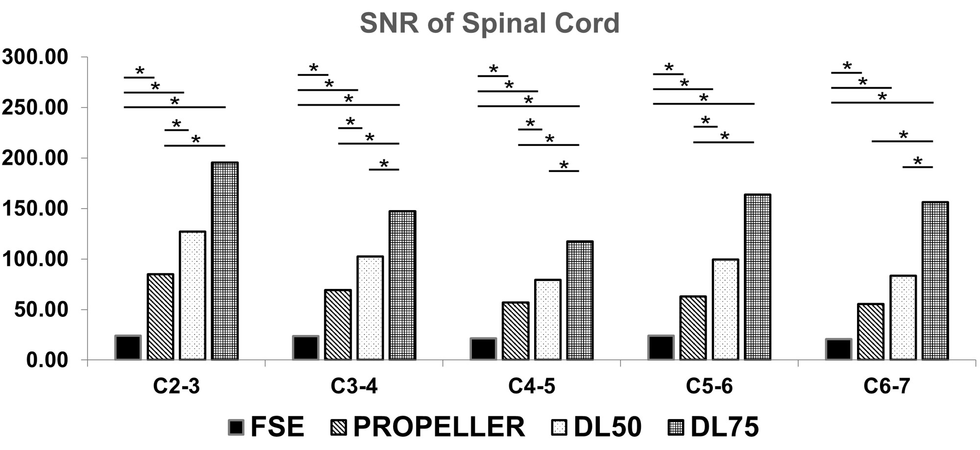
For each sequence, the SNR of the spinal cord showed significant differences at all discs levels (p < 0.0083).
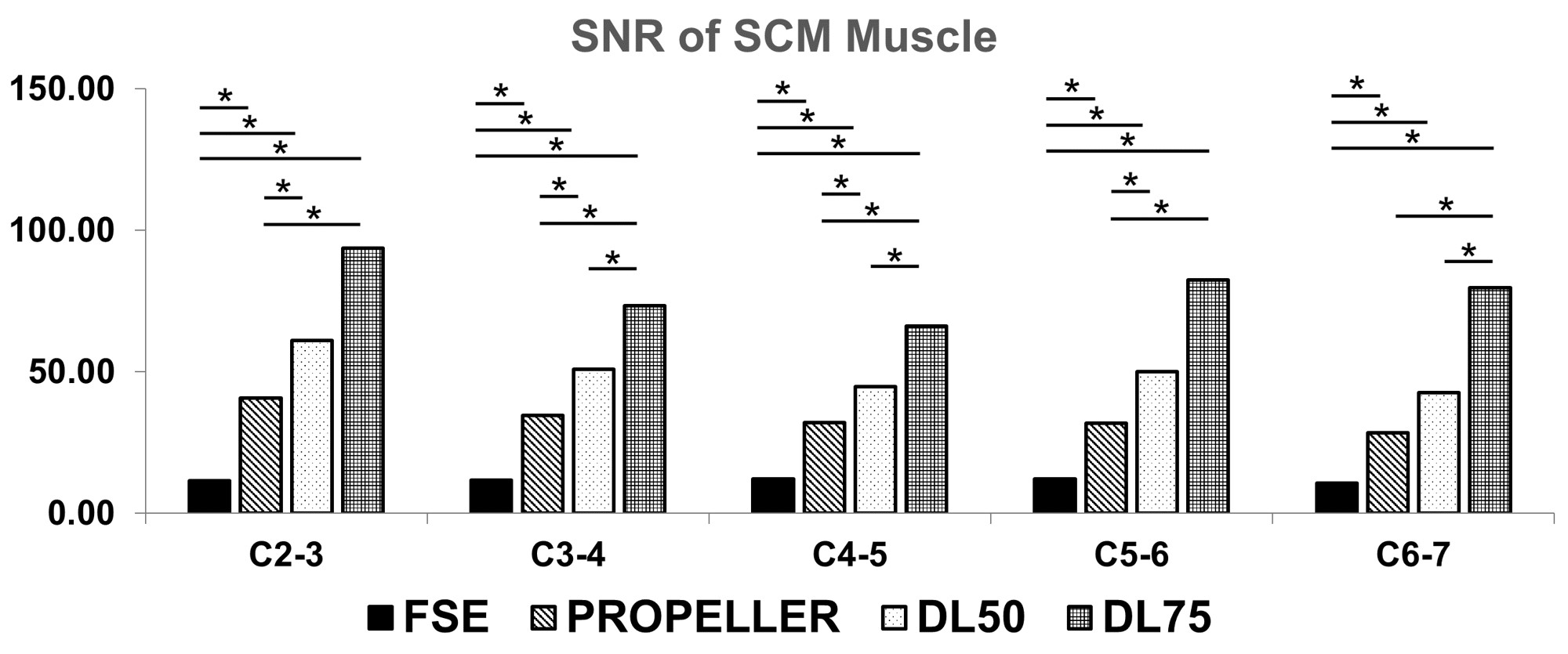
For each sequence, the SNR of the SCM muscles showed significant differences at all discs levels (p < 0.0083).
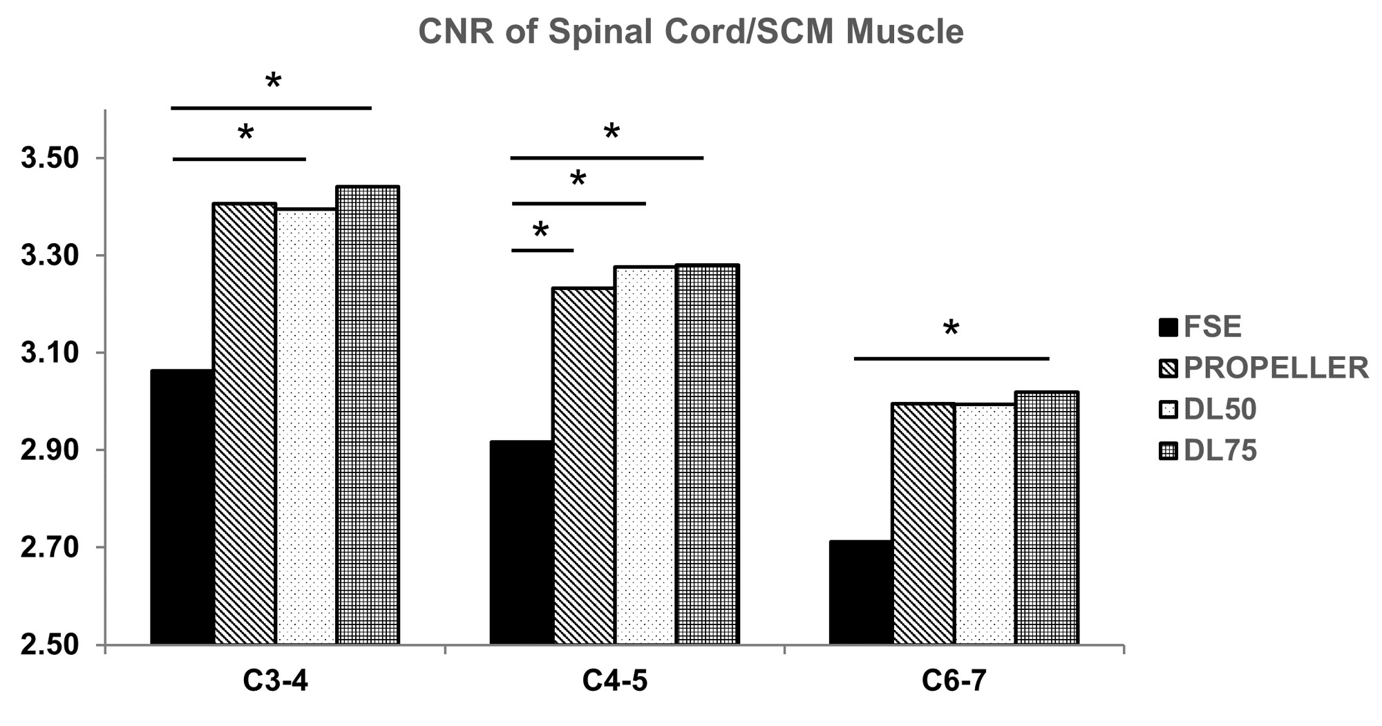
The CNR of the spinal cord was significantly higher
in PROPELLER, DL50, and DL75 images compared to FSE at C3-4, 4-5, and 6-7
levels.
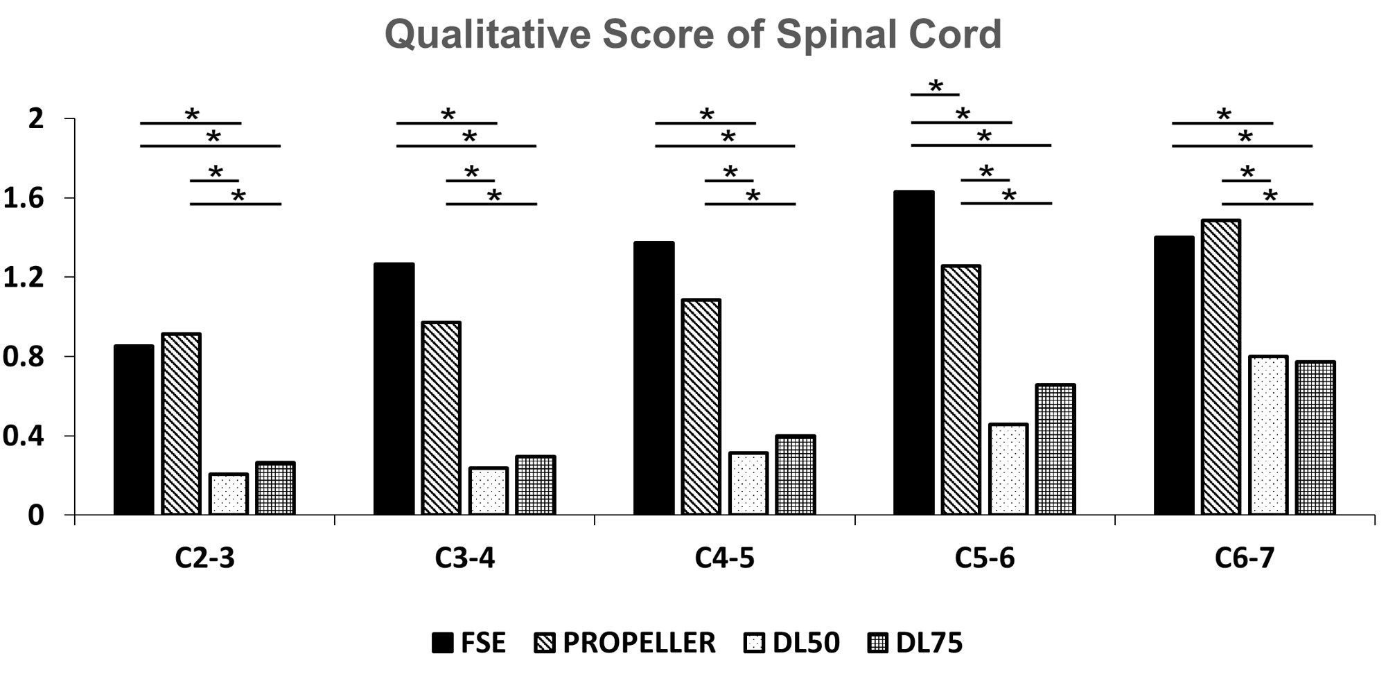
In the qualitative analysis, all four types of
images (FSE, PROPELLER, DL 50, DL 57) showed significant differences at all
disc levels for the spinal cord, SCM and back muscles (p < 0.05). In
particular, DL50 and DL75 images showed a significant difference in image
quality compared to FSE and PROPELLER images (p < 0.0083).
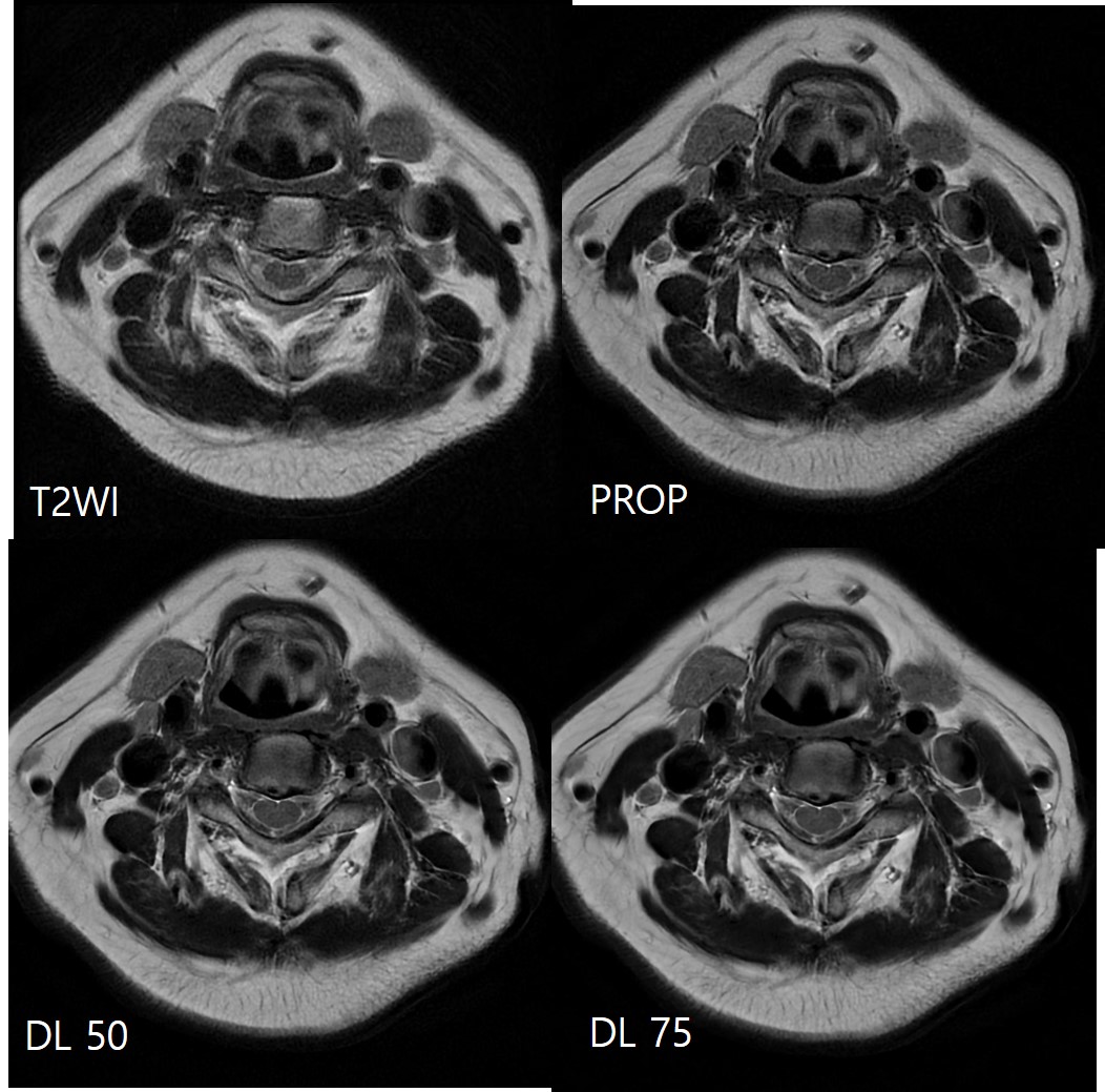
Conventional T2WI has severe motion artifacts. DL images demonstrate reduced blurring and heterogeneity
(coarse) on the spinal cord and surrounding soft tissue structures .
75% DL image is more homogenous overall than 50% DL image, and blurring is
reduced. The sharpness of Margin of 75% DL images seems to be a little better than
50% DL image.
DOI: https://doi.org/10.58530/2022/4328