4322
Improved fat suppression in diffusion MRI using eddy current compensation gradient1Siemens Shenzhen Magnetic Resonance Ltd, Shenzhzen, China, 2Siemens Healthcare GmbH, Erlangen, Germany
Synopsis
In this work, we propose to compensate the residual eddy current field caused by diffusion gradients at the time when chemically selective fat suppression gets applied with an additional gradient applied after the EPI readout. Using an analytic solution, the amplitude of the eddy currents can be cancelled under certain assumptions. The experimental results based on a volunteer scan demonstrate improved fat suppression in diffusion MRI with the proposed method.
INTRODUCTION
Diffusion weighted imaging (DWI) is a valuable complement to traditional techniques and improves the sensitivity for both detection and characterization of diseases. However, it can be affected by eddy current artifacts due to the rapid switching of diffusion gradients, which introduces eddy current in the nearby conductors, inducing local magnetic fields that interfere with the spatial encoding gradients1. The typical eddy current artifacts in DWI include shearing, stretching and rotation, which will lead to misalignment among different volumes acquired by different diffusion gradients. Such effect varies across different diffusion gradient directions and will increase with higher b-values, which usually require diffusion gradients with strong amplitude and long duration. Different techniques have been proposed to improve such eddy current induced distortion2-5. In addition to the common eddy current induced distortion artifacts, the long-term eddy current components can generate a residual field which persists after the image acquisition. This may impact the succeeding chemically selective RF-pulses and lead to insufficient fat suppression.In this study, we propose to compensate such residual eddy current field by introducing an additional gradient, in which the amplitude of the eddy currents can be cancelled by considering an analytical solution under certain assumptions. The approach was validated in a volunteer scan. The results show that the proposed method can improve fat suppression in DWI.
METHODS
Eddy current time dependence can be broadly described by exponential decays with long- and short-time constants. The time constant indicates the rate of exponential build up and decay of field perturbations after a gradient change6. To simplify the analytical calculation, we consider the eddy currents to be mono-exponential with only a single long-time constant. The field generated by the eddy currents after an ideal, rectangular shaped gradient pulse G (amplitude G0, duration TG) can be described by a temporal decay function $$$g(t)$$$:$$g_{0}\approx G_{0}(exp(\frac{-T_{G}}{\tau})-1)$$
$$g(t) = g_{0} exp(\frac{-t}{\tau}) \tag1$$
where τ is the long-time constant, and t = 0 occurs immediately after the end of G with the eddy current field $$$g_{0}$$$.
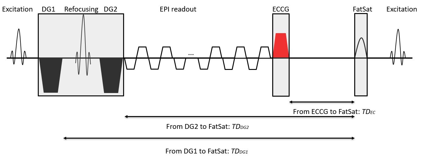
The diffusion pulse sequence with eddy current compensation is shown in Fig. 1. Two monopolar gradients DG1 and DG2 with equal area are used for diffusion encoding, and one chemically selective fat saturation RF-pulse is applied before each excitation. To eliminate the diffusion gradients induced eddy current effect on the succeeding fat saturation pulse, an additional gradient will be applied after the EPI readout. The required compensation parameters can be given by:
$$G_{d}(exp(\frac{-T_{d}}{\tau})-1)exp(\frac{-TD_{DG1}}{\tau}) +G_{d}(exp(\frac{-T_{d}}{\tau})-1)exp(\frac{-TD_{DG2}}{\tau})=G_{ec}(exp(\frac{-T_{ec}}{\tau})-1)exp(\frac{-TD_{EC}}{\tau}) \tag2$$
where Gd is the amplitude of the paired diffusion gradients DG1 and DG2; Td is the duration of DG1 and DG2; Gec is the amplitude of the eddy current compensated gradient (ECCG) applied after the EPI readout; Tec is the duration of ECCG; τ is the eddy current long-time constant; TDDG1, TDDG2 are the durations from the end of DG1 and DG2 to the beginning of the chemically selective fat saturation pulse, respectively; TDEC is the duration from the end of ECCG to the beginning of the chemically selective fat saturation pulse.
To demonstrate improved fat suppression with the proposed method, we calculated the ECCG parameters based on the applied diffusion gradients to generate a prototypic single shot diffusion sequence and assessed the fat suppression performance with a volunteer scan.
EXPERIMENTS AND RESULTS
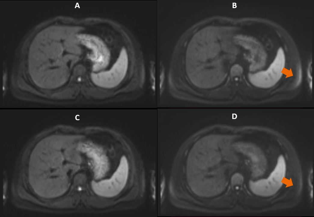
All measurements were performed on a commercial 1.5T scanner equipped with a 6-channel body array and an 18-channel spine array. One volunteer was scanned with informed consent. Conventional and prototypic single shot diffusion sequences were acquired with the same slice thickness and coverage using the following parameters: FOV = 400x275 mm2, matrix size = 134x92, in-plane GRAPPA factor = 2, b = 50 s/mm2 with 1 average, b = 800 s/mm2 with 4 averages, diffusion mode = 4-Scan Trace, 30 slices with 6 mm thickness, 20% slice gap, FatSat mode = SPAIR; TE/TR = 60ms/6300ms with total scan time of 2:23 min. Fig. 2 compares exemplary DW images of the liver acquired using conventional and prototypic diffusion sequences. Inhomogeneous fat suppression was observed in the high-b value image acquired with the conventional sequence. After appropriate eddy current compensation, Fig. 2D shows improved fat suppression in the edge of images, where the eddy current effects are usually more severe.DISCUSSION
We have demonstrated that it is possible to mitigate eddy current related imperfections of fat suppression by adding an extra eddy current compensation gradient into a diffusion sequence. To achieve a more accurate compensation, the eddy current measurement experiment should be done in advance to estimate the long-time constant correctly. Please note that the residual fat signal depends on the diffusion gradient strength, duration between the diffusion gradients and chemically selective fat saturation pulse, and on the eddy current decay. Thus, it exhibits different appearance in low-b and high-b value images, which will also lead to incorrect ADC calculation. The proposed method can also mitigate such incorrection by improving fat suppression.CONCLUSION
The proposed method allows for a practical, accurate compensation of the residual eddy current cancellation after the EPI readout and thus enjoys a more accurate spectral selection of fat signal, which is more attractive in diffusion applications at low field.Acknowledgements
No acknowledgement found.References
1. Jezzard, P., Barnett, A. S., and Pierpaoli, C. Characterization of and correction for eddy current artifacts in echo planar diffusion imaging. Magn. Reson. Med. 1998;39:801-812.
2. Reese TG, Heid O, Weisskoff RM, Wedeen VJ. Reduction of eddy-current-induced distortion in diffusion MRI using a twice-refocused spin echo. Magn. Reson. Med. 2003;49:177-182.
3. Bodammer N, Kaufmann J, Kanowski M, Tempelmann C. Eddy current correction in diffusion-weighted imaging using pairs of images acquired with opposite diffusion gradient polarity. Magn. Reson. Med. 2004;51:188-193.
4. Andersson, J. L. R., and Sotiropoulos, S. N. An integrated approach to correction for off-resonance effects and subject movement in diffusion MR imaging. Neuroimage. 2016;125:1063-1078.
5. Finsterbusch, J. Eddy-Current Compensated Diffusion Weighting with a Single Refocusing RF Pulse. Magn. Reson. Med. 2009;61:748-754.
6. Bernstein, M.A., King, K.F and Zhou, X.J. Handbook of MRI Pulse Sequences. Elsevier, Amsterdam; 2004.
Figures