4318
Molecularly Targeted Magnetic Resonance Imaging and Spectroscopy
Ye-Feng Yao1, Jia-Xiang Xin1, Guang Yang1, Huojun Zhang2, Jiaqi Li1, Caixia Fu3, Jiachen Wang1, Rui Tong1, and Daxiu Wei1
1Shanghai Key Laboratory of Magnetic Resonance, East China Normal University, Shanghai, China, 2Shanghai Changhai Hospital, Shanghai, China, 3Siemens Shenzhen Magnetic Resonance Ltd., Shenzhen, China
1Shanghai Key Laboratory of Magnetic Resonance, East China Normal University, Shanghai, China, 2Shanghai Changhai Hospital, Shanghai, China, 3Siemens Shenzhen Magnetic Resonance Ltd., Shenzhen, China
Synopsis
This study developed a molecularly targeted magnetic resonance imaging/magnetic resonance spectroscopy (MRI/MRS) method to selectively probe a specific metabolite molecule of tissues/organs in vivo. Several biomolecules have been used for molecularly targeted MRI and MRS. We used N-acetyl aspartate (NAA)- and glutamate (GLU)-targeted 1H MRS spectra of the human brain and reported a novel approach to measure the local pH values of tissue in vivo.
Introduction
Magnetic resonance spectroscopy (MRS) signals are often difficult to analyze and quantify due to signal overlap and a corrupted baseline. Various techniques, such as MEGA editing by Mescher and Garwood et al. (1) and double quantum filtered (MQF) MRS (2), can selectively probe signals from specific biomolecules. Although these techniques can probe the specific molecules, they fail to accurately select a specific molecule for truly molecularly targeted imaging/detection. In this work, we reported a series of molecularly targeted MRI/MRS methods in which a spin-singlet order (3-5) is exploited to target specific molecules.Materials and Methods
Sample 1: Four capillary tubes (1 mm diameter), filled with water, and N-acetyl aspartate (NAA), L-alanine-glycine-glycine (AGG), and dopamine (DA) aqueous solutions, were prepared. Then the four capillary tubes were carefully inserted into a 5 mm NMR tube. The experiments for Sample 1 were carried out on a 500 MHz Bruker AVANCE III spectrometer. A Bruker Triple Resonance Broadband Inverse (TBI) probe equipped with a three-dimensional gradient was used.Subjects: Three healthy volunteers were scanned with the molecularly targeted MRS. Experiments were carried out on a 3T MRI scanner (MAGNETOM Prisma, Siemens Healthcare, Erlangen, Germany) with a 64-channel Head/Neck receiver coil.Figure 1a illustrates the scheme of the pulse sequences (YAWX-MRI and YAWX-MRS) developed in this work. The sequences consist of two parts: One for the molecularly targeted signal selection and one for the MRI/MRS acquisition. Sequence parameters used in the spin singlet order (SSO) preparation were calculated using chemical shifts and J-couplings of a spin system in the targeted molecule. Here, the optimal control (OC) strategy is exploited to prepare SSO. Figure 1b shows an OC pulse sequence example that consists of a series of modulated discrete pulses, with each having an optimized amplitude and phase to ensure the highest efficiency of the SSO preparation. MR signals were acquired by the following MRI/MRS block.Results
The routine 1H MRI image of Sample 1 is shown in Figure 2b. Figures 2c, d, and e show the molecularly targeted 1H MRI image of NAA, AGG, and DA, respectively. The capillary containing the targeted molecule can be selectively imaged using YAoWeiXin (YAWX)-MRI. In the conventional 1H MRS spectrum (Figure 2f), the signals from the heavy water (HDO), NAA, AGG, and DA were intense. In the molecularly targeted MRS spectra (Figures 2g, h, and i) acquired using YAWX-MRS, the signals of the targeted molecules are dominant in the spectra. The in-vivo results acquired with the YAWX-MRS sequence are shown in Figures 3 and 4. The routine MRS spectrum (Figure 3a) is featured with severe overlapping signals. The monitored spin system of NAA consists of Ha, Hb, and Hb’ (Figure 3c). The J-coupling between the two 1H spin pairs (Ha, Hb) and (Ha, Hb’), measured via the signal splitting in the spectrum, was Jab = 4.2 Hz and Jab’ = 10.2 Hz, respectively. Figure 4 demonstrates that the CH2 signal of NAA changes with changes in pH. The 1H MRS spectrum of the human brain matched nicely that from the NAA aqueous solution with a pH value of 7.4. Thus, the accurate measurement of the NAA signals provides a way to probe the pH values of the local NAA environment.Discussion and Conclusion
In this work, we developed a series of novel MR methods based on the spin-singlet order to implement molecularly targeted MRI and MRS. Furthermore, we reported NAA- and GLU-targeted 1H MRS spectra of a human brain, demonstrating the feasibility of in vivo probing for various metabolites in people. We believe that the schemes developed in this work will profoundly influence cell/molecular biology, brain science, medicine, pharmacology, medical physics, chemistry, physics, and MRI/NMR methodology.Acknowledgements
This work was supported by Xing-Fu-Zhi-Hua Foundation of ECNU and Microscale Magnetic Resonance Platform of ECNU.References
- Mescher M, Merkle H, Kirsch J, And MG, Gruetter R. Simultaneous in vivo spectral editing and water suppression. NMR in Biomedicine. 1998. doi: 10.1002/(SICI)1099-1492(199810)11:6<266::AID-NBM530>3.0.CO;2-J.
- Choi IY, Lee SP, Merkle H, Shen J. Single-shot two-echo technique for simultaneous measurement of GABA and creatine in the human brain in vivo. Magn Reson Med. 2004;51(6):1115-21. Epub 2004/06/02. doi: 10.1002/mrm.20082.
- Mamone S, Rezaei-Ghaleh N, Opazo F, Griesinger C, Gloggler S. Singlet-filtered NMR spectroscopy. Sci Adv. 2020;6(8):eaaz1955. Epub 2020/03/05. doi: 10.1126/sciadv.aaz1955.
- Devience SJ, Walsworth RL, Rosen MS. Nuclear spin singlet states as a contrast mechanism for NMR spectroscopy. NMR Biomed. 2013;26(10):1204-12. Epub 2013/04/23. doi: 10.1002/nbm.2936.
- Kiryutin AS, Pravdivtsev AN, Yurkovskaya AV, Vieth HM, Ivanov KL. Nuclear Spin Singlet Order Selection by Adiabatically Ramped RF Fields. J Phys Chem B. 2016;120(46):11978-86. Epub 2016/10/28. doi: 10.1021/acs.jpcb.6b08879.
Figures
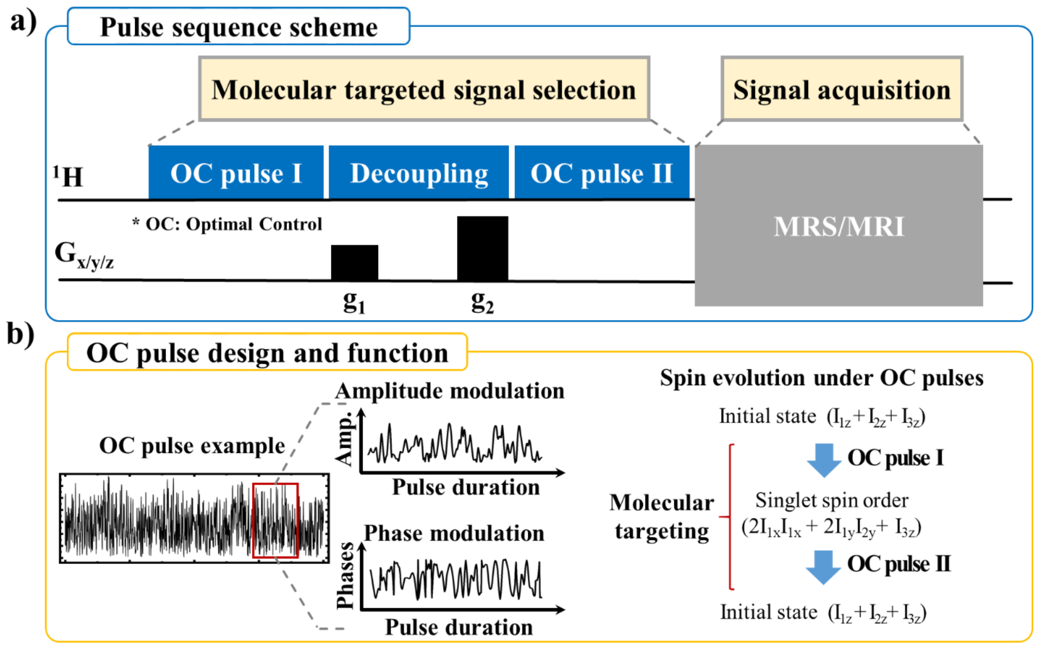
Figure 1: a) Pulse sequence scheme for molecularly targeted MRI/MRS: YAWX-MRI and YAWX-MRS. b) The OC pulse sequence is a series of discrete pulses, amplitudes, and phases calculated to ensure the state transfer efficiency for specific spin systems. Figure 1b shows key spin evolution steps in a three-spin system under OC pulses I and II in Figure 1a.
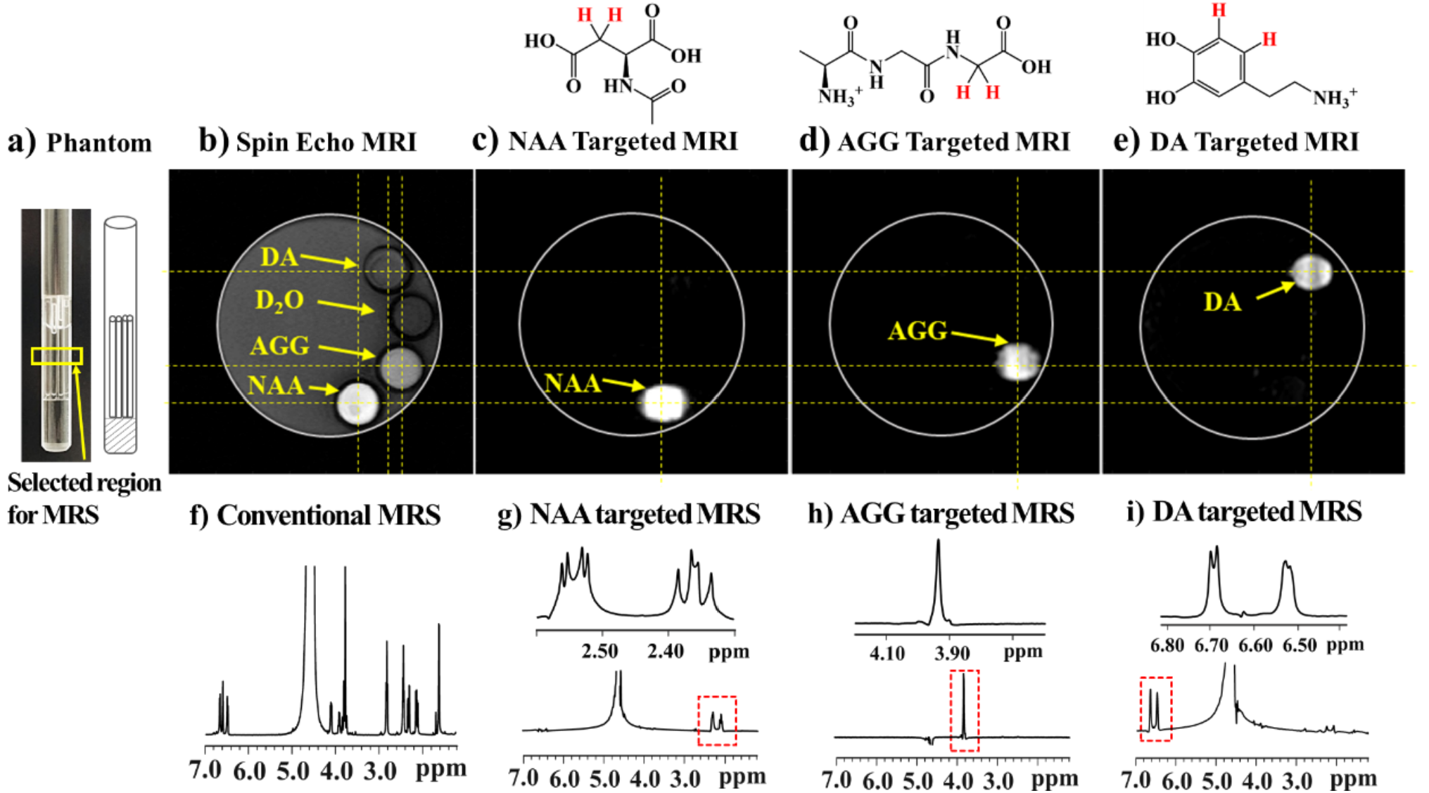
Figure 2: a) A photo of the phantom. The b) spin echo I, c) NAA-targeted, d) AGG-targeted, and e) DA-targeted 1H MRI images of the sample. The f) conventional, g) NAA-targeted, h) AGG-targeted, and i) DA-targeted 1H MRS spectra of the same sample. An 1H spin echo imaging sequence was used to acquire the image in Figure 2b.
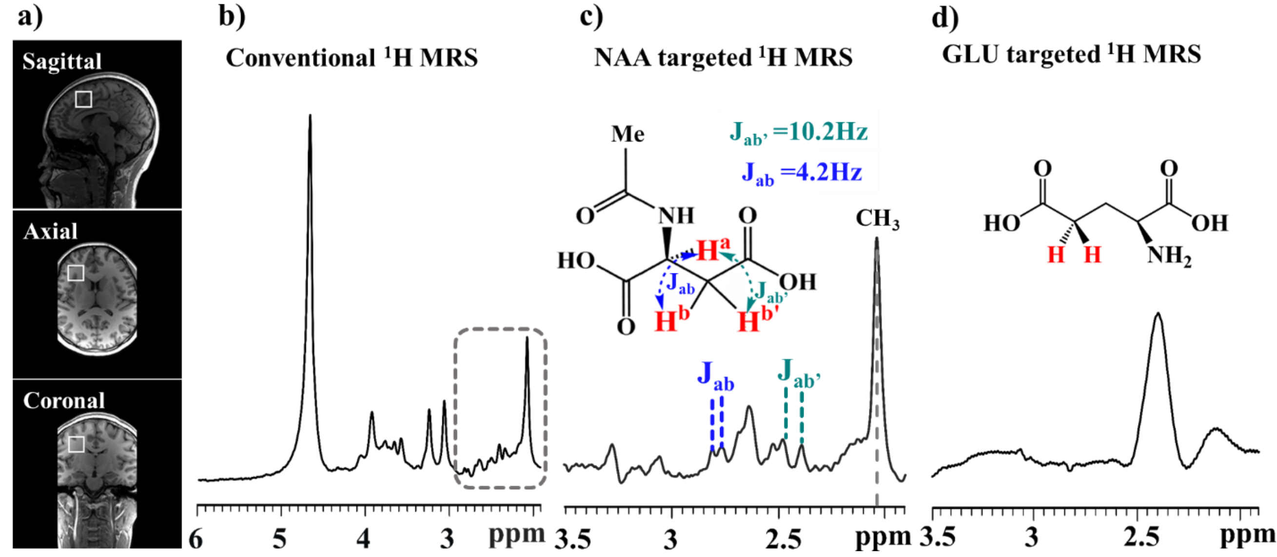
Figure 3: a) Routine magnetic resonance imaging (MRI) images showing the voxel position in a human brain. The following 1H MRS spectra were acquired in this voxel with a size of 20 × 20 × 20 mm3; b) The conventional 1H MRS spectrum of the voxel; The c) NAA and d) GLU targeted 1H MRS spectra of the voxel acquired using YAWX-MRS.
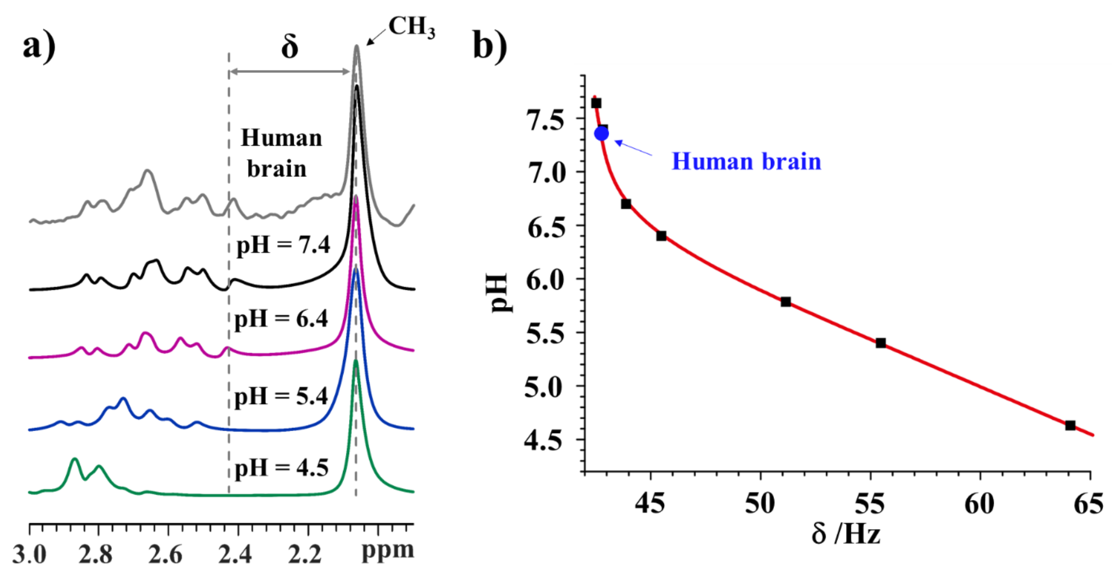
Figure 4: a) The 1H MRS spectra of an NAA aqueous solution containing buffers at the various pH values (black, purple, blue, and green lines) and the NAA-targeted 1H MRS spectrum of a human brain (the grey line); b) a plot of the frequency difference between the CH3 signal and the CH2 signal (see Figure 4a), d, as a function of the pH values.
DOI: https://doi.org/10.58530/2022/4318