4293
Bosniak II-III Cystic Renal Masses: Subtraction MR Images for Improvement of Interobserver Agreement with Bosniak Classification, Version 20191Radiology, the First Medical Center of Chinese PLA General Hospital, Beijing, China, 2Radiology, the Fifth Medical Center of Chinese PLA General Hospital, Beijing, China
Synopsis
The purpose of this study was to investigate whether subtraction MR images can improve the interobserver agreement for Bosniak II, IIF and III cystic renal masses (CRMs) with Bosniak classification, version 2019. The results showed that the interobserver agreement was higher with subtraction MR images than without subtraction MR images (weighted k = 0.73 vs 0.60, respectively; P=.011), which may contribute to the popularization and application of Bosniak classification, version 2019.
Purpose
To investigate whether subtraction MR images can improve the interobserver agreement for Bosniak II-III cystic renal masses (CRMs) with Bosniak classification, version 2019.Methods
The CRMs of Bosniak II, IIF and III were selected from all pathologically confirmed CRMs which had been classified in consensus by two experienced genitourinary radiologists with Bosniak classification version 2019. Blinded to clinical and pathologic information, the other two radiologists independently classified Bosniak II, IIF and III CRMs with and without subtraction MR images, respectively, with an interval of 1 month. Ultimately, by using multirater k statistics with Stata/MP 14.0 software, interobserver agreement was evaluated with comparisons between classifications. P<.05 was considered to indicate a statistically significant difference.Results
A total of 187 patients (mean age ± standard deviation, 50.4 years ± 12.1; 128 male and 59 female patients) with CRMs were included. The weighted k for interobserver agreement among two radiologists with subtraction MR images and without subtraction MR images was 0.73 (95% CI: 0.66, 0.80) and 0.60 (95% CI: 0.53, 0.68), respectively. The interobserver agreement was higher with subtraction MR images than without subtraction MR images (weighted k = 0.73 vs 0.60, respectively; P=.011).Discussion
By eliminating the inherent high signal on the precontrast images, the MR subtraction images may contribute to evaluate whether the lesion has enhancement on the enhanced images, which can improve the interobserver agreement for Bosniak classification.Conclusion
The interobserver agreement for Bosniak II, IIF and III CRMs can be improved using subtraction MR images, which may contribute to the popularization and application of Bosniak classification, version 2019.Acknowledgements
No.References
1. BOSNIAK MA. The Current Radiological Approach to Renal Cysts[J]. 1986, 158(1):1–10.
2. SILVERMAN S G, PEDROSA I, ELLIS J H, et al. Bosniak Classification of Cystic Renal Masses, Version 2019: An Update Proposal and Needs Assessment[J]. Radiology, 2019, 292(2):475–488.
3. ISRAEL G M, BOSNIAK M A. An update of the Bosniak renal cyst classification system[J]. Urology, 2005, 66(3):484–488.
4. BAI X, SUN S-M, XU W, et al. MRI-based Bosniak Classification of Cystic Renal Masses, Version 2019: Interobserver Agreement, Impact of Readers' Experience, and Diagnostic Performance[J]. Radiology, 2020, 297(3):597–605.
5. 康欢欢, 白旭, 王海屹. 2019 版肾脏囊性病变 Bosniak 分级标准解读[J]. 中华放射学杂志, 2020, 54(8):729-736.
6. SEPPALA N, KIELAR A, DABREO D, et al. Inter-rater agreement in the characterization of cystic renal lesions on contrast-enhanced MRI[J]. Abdominal imaging, 2014, 39(6):1267–1273.
7. SCHOOTS I G, ZACCAI K, HUNINK M G, et al. Bosniak Classification for Complex Renal Cysts Reevaluated: A Systematic Review[J]. The Journal of urology, 2017, 198(1):12–21.
8. GRAUMANN O, OSTHER S S, KARSTOFT J, et al. Bosniak classification system: inter-observer and intra-observer agreement among experienced uroradiologists[J]. Acta radiologica (Stockholm, Sweden : 1987), 2015, 56(3):374–383.
9. QUAIA E, BERTOLOTTO M, CIOFFI V, et al. Comparison of contrast-enhanced sonography with unenhanced sonography and contrast-enhanced CT in the diagnosis of malignancy in complex cystic renal masses[J]. AJR. American journal of roentgenology, 2008, 191(4):1239–1249.
10. TAY S-Y, TIU C-M, HU B, et al. Characterization and management of various renal cystic lesions by sonographic features[J]. Journal of the Chinese Medical Association : JCMA, 2018, 81(12):1017–1026.
11. BYUNG KWAN PARK, CHAN KYO KIM AND EUN YOUNG KIM. Differentiation of Bosniak Categories IIF and III Cystic Masses: What Radiologists Should Know[J]. (J Comput Assist Tomogr, 2010, 34(6):847-854.
12. EDNEY E, DAVENPORT M S, CURCI N, et al. Bosniak classification of cystic renal masses, version 2019: interpretation pitfalls and recommendations to avoid misclassification[J]. Abdominal radiology (New York), 2021, 46(6):2699–2711.
13. JACOB COHEN. Weighted kappa nominal scale agreement with provision for scaled disagreement or partial credit[J]. Psychological Bulletin, 1968, 70(4):213–220.
14. GWET K L. Testing the Difference of Correlated Agreement Coefficients for Statistical Significance[J]. Educational and psychological measurement, 2016, 76(4):609–637.
15. LANDIS JR, KOCH GG. The Measurement of Observer Agreement for Categorical Data[J]. Biometrics, 1977, 33(1):159–174.
16. BRENNAN P, SILMAN A. Statistical methods for assessing observer variability in clinical measures[J]. BMJ, 1992, 304:1491–1494.
17. MOCH H, CUBILLA A L, HUMPHREY P A, et al. The 2016 WHO Classification of Tumours of the Urinary System and Male Genital Organs-Part A: Renal, Penile, and Testicular Tumours[J]. European urology, 2016, 70(1):93–105.
18. SEÇIL M, OBUZ F, ALTAY C, GENCEL O, IĞCI E, SAĞOL O, DICLE O. The role of dynamic subtraction MRI in detection of hepatocellular carcinoma[J]. Diagn Interv Radiol, 2008, 14(4):200–204.
19. YU JS, KIM YH, ROFSKY NM. Dynamic subtraction magnetic resonance imaging of cirrhotic liver: assessment of high signal intensity lesions on nonenhanced T1-weighted images[J]. J Comput Assist Tomogr, 2005, 29(1):51–58.
20. ISRAEL G M, HINDMAN N, BOSNIAK M A. Evaluation of cystic renal masses: comparison of CT and MR imaging by using the Bosniak classification system[J]. Radiology, 2004, 231(2):365–371.
21. KRISHNA S, SCHIEDA N, PEDROSA I, et al. Update on MRI of Cystic Renal Masses Including Bosniak Version 2019[J]. Journal of magnetic resonance imaging : JMRI, 2021, 54(2):341–356.
22. HECHT E M, ISRAEL G M, KRINSKY G A, et al. Renal masses: quantitative analysis of enhancement with signal intensity measurements versus qualitative analysis of enhancement with image subtraction for diagnosing malignancy at MR imaging[J]. Radiology, 2004, 232(2):373–378.
23. HINDMAN N M. Approach to Very Small (< 1.5 cm) Cystic Renal Lesions: Ignore, Observe, or Treat?[J]. AJR. American journal of roentgenology, 2015, 204(6):1182-189.
24. LUCOCQ J, PILLAI S, OPARKA R, et al. Complex renal cysts (Bosniak ≥IIF): interobserver agreement, progression and malignancy rates[J]. European radiology, 2021, 31(2):901-908.
25. LAM C J, KAPOOR A. The true malignancy risk of Bosniak III cystic renal lesions: Active surveillance or surgical resection?[J]. Canadian Urological Association journal = Journal de l'Association des urologues du Canada, 2018, 12(6):E276-E280. DOI: 10.5489/cuaj.4960.
26. KIM D Y, KIM J K, MIN G-E, et al. Malignant renal cysts: diagnostic performance and strong predictors at MDCT[J]. Acta radiologica (Stockholm, Sweden : 1987), 2010, 51(5):590–598.
27. ISRAEL G M, BOSNIAK M A. How I do it: evaluating renal masses[J]. Radiology, 2005, 236(2):441–450.
28. TAN I L, VAN SCHIJNDEL R A, FAZEKAS F, et al. Image registration and subtraction to detect active T(2) lesions in MS: an interobserver study[J]. Journal of neurology, 2002, 249(6):767–773.
29. BOSNIAK MA R N M. Problems in the Detection and Characterization of Small Renal Masses[J]. Radiology, 1996, 198(3):638–641.
30. MAKI DD, BIRNBAUM BA, CHAKRABORTY DP, JACOBS JE, CARVALHO BM, HERMAN GT. Renal cyst pseudoenhancement: beam-hardening effects on CT numbers[J]. Radiology, 1999, 213(2):468–472.
Figures
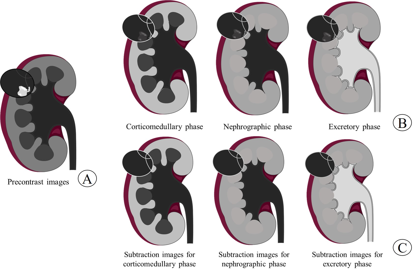
Figure 1. The diagram of subtraction MR images. The mass showed heterogeneous hyperintensity on the precontrast images (a) and two thin (≤2mm) enhancing septa on enhanced MR images (b) (corticomedullary phase, nephrographic phase and excretory phase). However, the subtraction MR images (c) (obtained by subtracting a from b) only showed two thin enhancing septa within the lesion.

Figure 2. 57/F, a cyst with hemorrhage located in the right kidney. On Axial view of T2 weighted imaging (a), the mass showed heterogeneous hyperintensity. On the precontrast images (b), the mass exhibited focal slight hyperintensity (pathologically confirmed hemorrhage). On the excretory phase (c), the lesion showed enhancing wall and one septum. On subtraction images from the excretory phase (d), we can clearly see that the mass showed thin (≤2mm) enhancing wall and one thin septum.