4283
Can whole-tumor histogram based on zoomed EPI diffusion-weighted imaging help predict perineural invasion in rectal cancer?1Department of Radiology, National Cancer Center/National Clinical Research Center for Cancer/Cancer Hospital, Chinese Academy of Medical Sciences and Peking Union Medical College, Beijing, China, 2National Cancer Center/National Clinical Research Center for Cancer/Cancer Hospital, Chinese Academy of Medical Sciences and Peking Union Medical College, Beijing, China, 3MR Scientific Marketing, Siemens Healthineers Ltd., Beijing, China
Synopsis
Perineural invasion (PNI) is a strong prognostic indicator in rectal cancer. Timely and accurate assessment of PNI status can assist in making individualized treatment plans. Histogram features can reflect the heterogeneity of tumors with better reproducibility than higher-order texture features. The zoomed DWI can provide a higher-spatial-resolution with fewer artifacts and distortions. We aim to evaluate the ability of histogram parameters derived from zoomed DWI to identify patients with positive PNI. The results demonstrated that histogram parameters could be used for assessing PNI status in RC, and skewness achieved the highest AUC.
Introduction
Perineural invasion (PNI), which refers to the primary tumor invades the nerves or perineural spaces, has been proven to be significantly associated with increased recurrence and decreased survival. The aim of this study was to explore the value of histogram parameters that derived from high-spatial-resolution zoomed DWI in the assessment of PNI status in rectal cancer.Methods
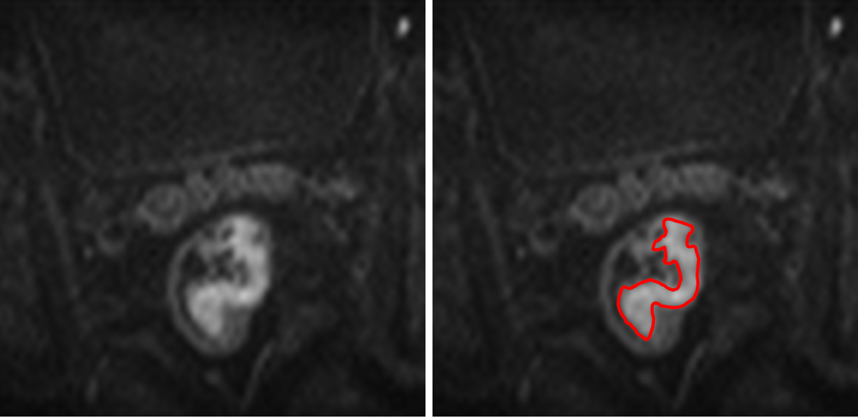
This prospective study included 94 patients with histopathological confirmed rectal adenocarcinoma between July 2020 and July 2021. All patients underwent preoperative rectal MRI examination including the high-spatial-resolution zoomed DWI sequence. A total of ten whole-tumor histogram features (including the 10th percentiles, 90th percentiles, minimum, mean, median, maximum, energy, entropy, kurtosis and skewness) were extracted from zoomed DWI. The significances of clinical-radiological and histogram variables were assessed with univariable analysis of positive PNI and negative PNI groups. Multivariable logistic regression analysis using backward stepwise selection was applied to identify independent predictors for PNI. Receiver operating characteristic (ROC) curve analysis was used to evaluate the diagnostic performance of multivariable model as well as that of the histogram parameters.Results
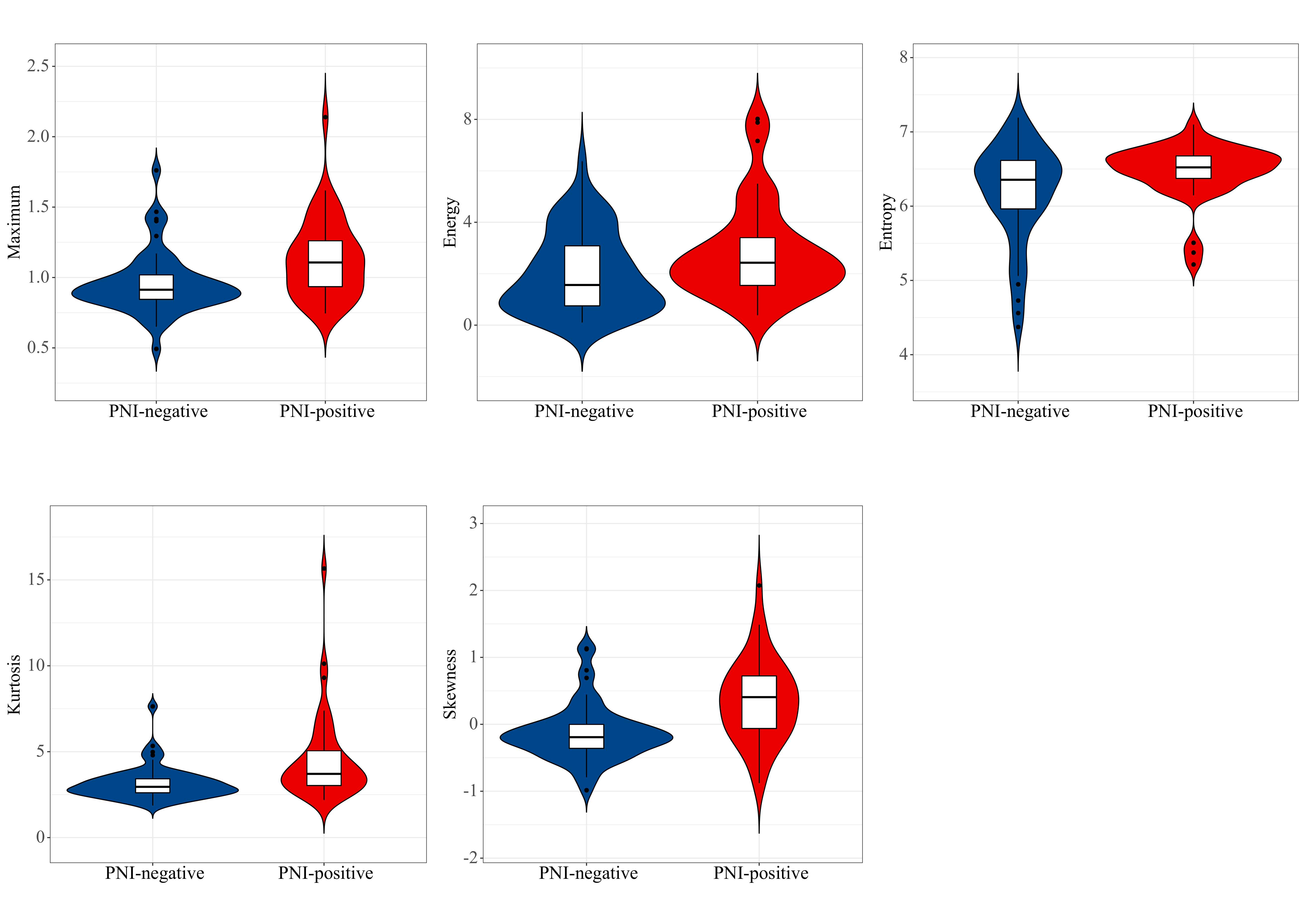
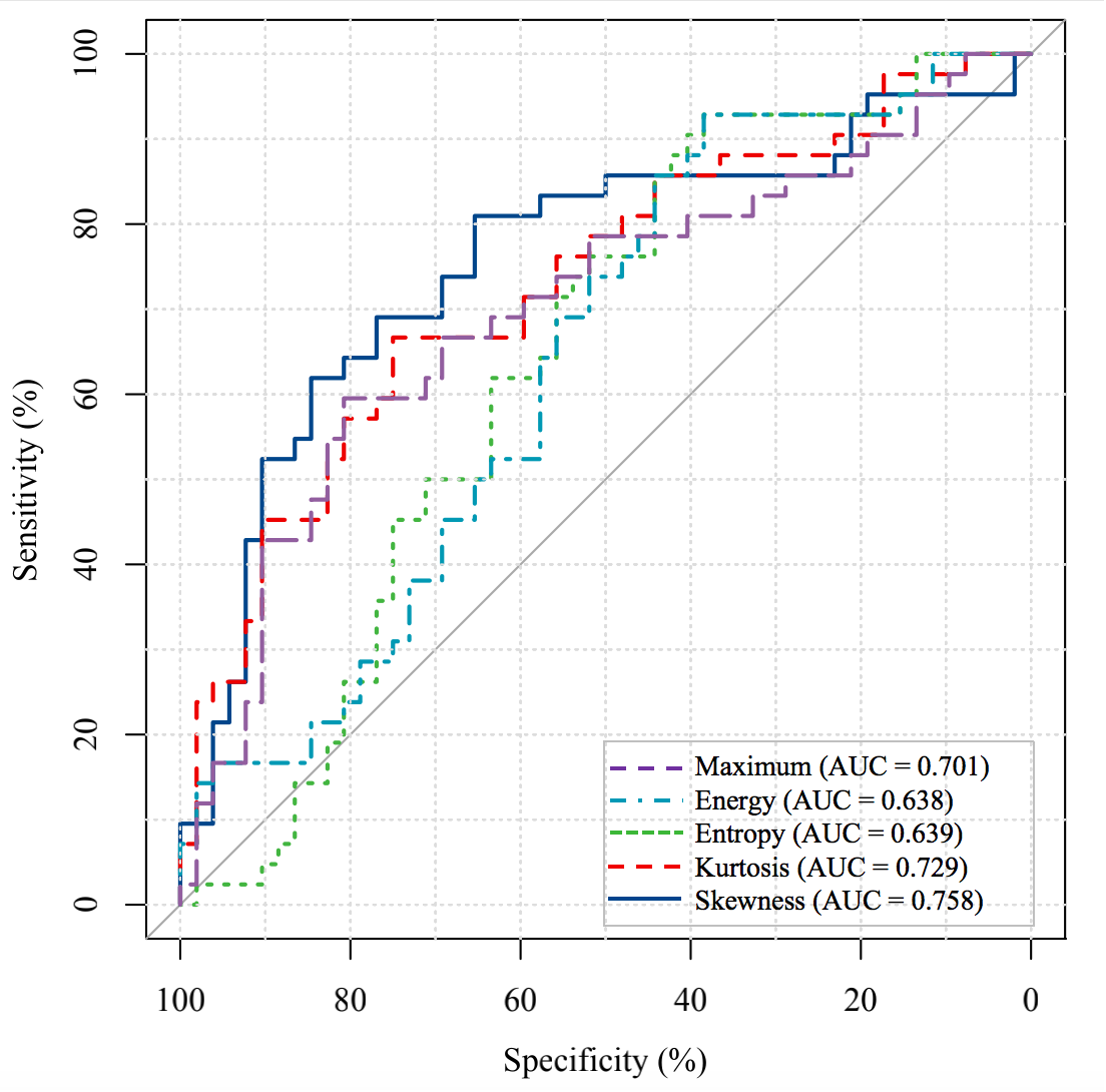
A total of 42 (44.7%) patients presented positive PNI, and 52 (55.3%) patients showed negative PNI. Maximum (p = 0.001), energy (p = 0.021), entropy (p = 0.021), kurtosis (p <0.001), and skewness (p <0.001) were significantly higher in positive PNI group compared to negative PNI group, with respective areas under the curve (AUCs) of 0.638-0.758. Multivariable analysis demonstrated that MRI T stage (p = 0.002) and skewness (p = 0.006) were independent predictors associated with PNI. Patients with positive PNI were more likely to have a higher MRI T stage (β = 2.154; 95%CI: 0.932-3.688) and skewness (β = 0.779; 95%CI: 0.255-1.382). The diagnostic performance of multivariable model for predicting PNI status showed an AUC of 0.811 (95%CI: 0.724-0.899) and achieved a sensitivity of 0.762 (95%CI: 0.605-0.879), specificity of 0.769 (95%CI: 0.632-0.875), and accuracy of 0.766 (95%CI: 0.667-0.847).Discussion
The timely and accurate evaluation of the PNI status of rectal cancer plays an important role in individualized treatment planning and prediction of prognosis. However, both biopsy and morphological assessment based on imaging examinations fail in determining PNI status. In this study, whole-tumor histogram analysis, which contained information of the entire tumor parenchyma and stroma, was performed for predicting PNI. Although in recent years several studies have reported the correlations between different prognostic factors of RC and histogram parameters, our present study is one of few to evaluate the diagnostic performance of histogram in predicting the PNI status. In addition, to obtain more details of the lesions, high-spatial-resolution DWI was used for histogram parameters extraction. High-spatial-resolution can be realized by using a larger matrix size in the conventional ss-EPI DWI, however, that can only be realized by extending echo times and reducing image quality. Zoomed DWI based on the two-dimensional spatially selective radiofrequency excitation pulses technique, can improve the spatial resolution and image quality and decrease artifacts and distortions. Therefore, zoomed DWI was firstly performed for PNI prediction in this study. The results demonstrated that histogram parameters could be used for assessing PNI status in rectal cancer.Conclusion
The histogram parameters that derived from high-spatial-resolution zoomed DWI could be helpful in the prediction of PNI status and allow more individualized treatment decisions.Acknowledgements
This research is supported by the National Natural Science Foundation of China [grant number 81971589].References
1. Liebig C, Ayala G, Wilks J, et al. Perineural invasion is an independent predictor of outcome in colorectal cancer. J Clin Oncol. 2009;27(31):5131-5137.
2. Liu L, Liu Y, Xu L, et al. Application of texture analysis based on apparent diffusion coefficient maps in discriminating different stages of rectal cancer. J Magn Reson Imaging. 2017; 45(6):1798-1808.
3. Riffel P, Michaely HJ, Morelli JN, et al. Zoomed EPI-DWI of the head and neck with two-dimensional, spatially selective radiofrequency excitation pulses. Eur Radiol. 2014; 24(10);24:2507-2512.
Figures