4267
Classification of Benign and Malignant Breast Lesions Using high b-value Diffusion MRI with a Continuous-time Random-walk (CTRW) Model1Peking university first hospital, Beijing, China, 2Central Research Institute, United Imaging Healthcare, Shanghai, China, 3Shanghai Key Laboratory of Magnetic Resonance, School of Physics and Electronics Science, East China Normal University, Shanghai, China
Synopsis
The high b-value DWI with a CTRW Model is useful in classification of benign and malignant proliferative breast lesions. The identification model using the combinations of CTRW parameters, Dm and β, has a moderate efficiency.
Introduction
The high b-value DWI with a CTRW model has been thought as useful for probing intravoxel tissue heterogeneity in cancer imaging1,2. So the purpose of our study was to test whether high b-value DWI with a continuous-time random-walk (CTRW) diffusion model could be used for classification of benign and malignant proliferative breast lesions.Methods
From May 2021 to October 2021, 64 patients (all female) ranging in age from 33 to 77 years (mean age: 53 years) were included in this study. Their breast MRI examination were all performed using 3T MR (uMR790, United Imaging Healthcare, Shanghai, China) with high b-value diffusion. They all had suspicious lesions in breast MRI described as BI-RADS3 4 at least and pathologically confirmed with benign or malignant status by biopsy. Echo-planar diffusion-weighted imaging (DWI) was conducted using 11 b-values (b = 0, 50, 100, 200, 400, 800, 1200, 1500, 2000, 2500, 3000 s/mm2) with total scan time 6 min 13 s covering the whole breast. The ROI containing the entire tumor, excluding cystic or necrotic areas, was drawn on the slice with the maximum diameter of tumor. Three CTRW model4 parameters, including an anomalous diffusion coefficient Dm, and two parameters related to temporal and spatial diffusion heterogeneity α and β, respectively, were obtained. Dm, α and β were compared between benign or malignant group by Mann–Whitney U test and receiver operating characteristic (ROC) analysis was performed.Results
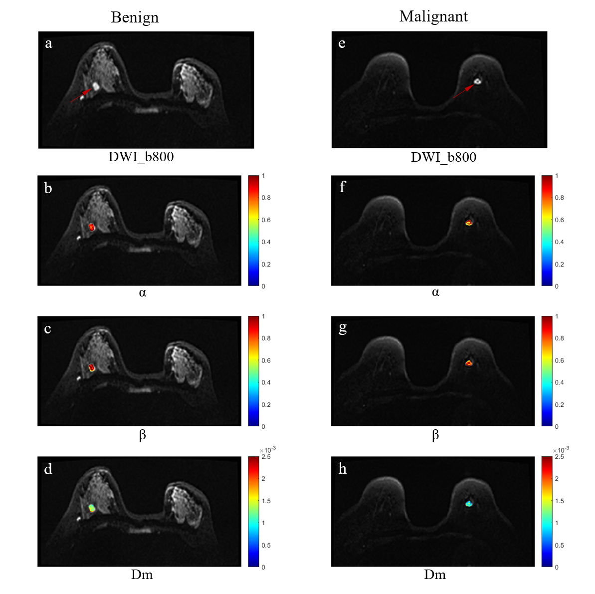
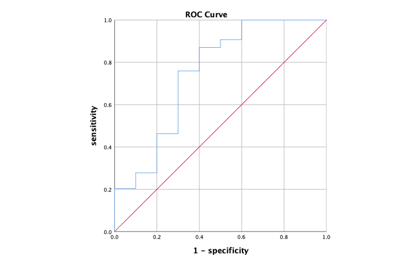
This study included 54 breast cancer and 10 benign breast lesions (5 adenopathy, 3 intraductal papilloma, 1 fibroadenoma and 1 granulomatous mastitis). Representative parametric maps are presented in FIG 1. The Dm and β were significantly lower for malignant breast lesions (Dm: 1.07 ± 0.18 μm2/ms; β:0.829 ± 0.059) than benign breast lesions (Dm: 1.40 ± 0.41 μm2/ms; β:0.882 ± 0.082) (p =0.016, p =0.035). The α between benign (α: 0.891 ± 0.040) and malignant (α: 0.860 ± 0.058) breast lesions were not significantly different (p =0.059). The highest sensitivity, specificity, accuracy, and area-under-the-curve (AUC) of using the combination of the Dm and β were 75.9%, 70.0%, 88.9% and 0.742 for benign and malignant breast lesion classification. ROC analysis result is shown in FIG 2.Discussion
Our results showed that, the Dm and β based on CTRW model had the capability in classification of benign and malignant breast lesion. Dm is an anomalous diffusion coefficient, similar with ADC, may have association with cellularity1. The β is related to spatial diffusion heterogeneity, which represents the mean free length of water molecules diffuse1. As such, the lower β value of malignant breast lesion indicated the more heterogeneous micro-environment in breast cancer. However, the α , another parameter reflecting temporal diffusion heterogeneity, between benign and malignant breast lesion were not significantly different, which might be explained by the reasons that the intrinsic high microstructural complexity with the existence of fibrocystic changes might increase the time of water molecules to make a move and the small number of benign cases might also limit the statistical results. For the ROC analysis, the model using the combination of the Dm and β for benign and malignant breast lesion classification has a moderate performance.Conclusion
It is feasible to use high b-value DWI with a CTRW Model in classification of benign and malignant proliferative breast lesions.Acknowledgements
We sincerely thank the participants in this study.References
[1] Karaman MM, Sui Y, Wang H, et al. Differentiating low- and high-grade pediatric brain tumors using a continuous-time randomwalk diffusion model at high b-values. Magn Reson Med. 2016;76(4):1149-1157.
[2] Karaman MM, Zhang J, Xie KL, et al. Quartile histogram assessment of glioma malignancy using high b-value diffusion MRI with a continuous-time random-walk model. NMR Biomed. 2021;34(4):e4485.
[3]Morris EA, Comstock CE, Lee CH, et al. ACR BI-RADS® Magnetic Resonance Imaging. In: ACR BI-RADS® Atlas, Breast Imaging Reporting and Data System. Reston, VA, American College of Radiology; 2013.
[4] Ingo C, Magin RL, Colon‐Perez L, et al. On random walks and entropy in diffusion‐weighted magnetic resonance imaging studies of neural tissue. Magnetic resonance in medicine. 2014; 71(2): 617-627.
Figures