4255
Robust fat saturation by combination of SPIR with gradient reversal for TSE at large FOV and coverage1Philips Healthcare, Beijing, China, 2MR Clinical Science, Philips Healthcare (Suzhou), Suzhou, China, 3MR R&D, Philips Healthcare (Suzhou), Suzhou, China
Synopsis
Robust fat suppression remains essential in clinical MRI to improve tissue signal contrast, minimize fat-related artifacts, and enhance image quality. It’s still a challenge to suppress the fat signal when the FOV and coverage is large, especially for abdomen imaging, where uneven fat suppression become common owing to both B0 and B1 field inhomogeneity. We propose a new solution that combines the optimized gradient reversal technique and Spectral Presaturation with Inversion Recovery (SPIR) simultaneously to overcome these challenges. This framework allows to suppress the fat signal robustly in large FOV with whole liver and kidney coverage.
Purpose
The goal of this work is to provide a new solution, which combines the optimized gradient reversal technique and SPIR simultaneously, for robust fat suppression at large coverage for TSE.Introduction
Fat suppression is widely used in diagnostic MRI to enhance image contrast for the detection of pathological lesions and changes1. Several techniques have been widely used for fat saturation: 1) Spectral Presaturation with Inversion Recovery (SPIR)2; 2) short inversion time (TI) inversion recovery (STIR)3; 3) chemical shift based water–fat separation (Dixon) methods4; 4) the slice-selection gradient-reversal (SSGR) method5,6. Despite the wide success of these techniques in clinical practice, fat suppression remains a challenge in abdominal imaging, especially when whole liver and kidney coverage is required, due to both B0 and B1 inhomogeneities. Particularly, SPIR is sensitive to both B0 and B1 inhomogeneity, SPAIR is sensitive to the B0 inhomogeneity, STIR has low SNR although it is not sensitive to both B0 and B1 inhomogeneity, Dixon needs considerably longer scan time, and SSGR is currently only applicable to echo-planar-based imaging6. SPIR is widely used for TSE7, it could efficiently suppress the fat signal, it uses the chemical shift but SPIR is very sensitive to B0 and B1 inhomogeneity, especially for the abdomen imaging, the fat suppression often failed. The SSGR method suppresses any off-resonance signal, not only those due to chemical shift, it is a good supplementary for other fat suppression techniques. Due to the long RF pulse duration, it will increase the echo spacing of TSE, SSGR have been used for diffusion imaging based on echo planar imaging6. We hereby propose a new scheme that combines the optimized SSGR technique and SPIR to produce robust fat suppression, and we demonstrate its application with turbo spin echo (TSE) acquisition.Methods
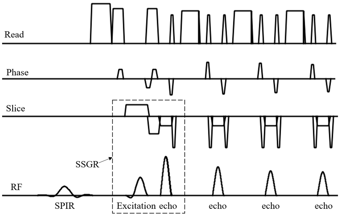
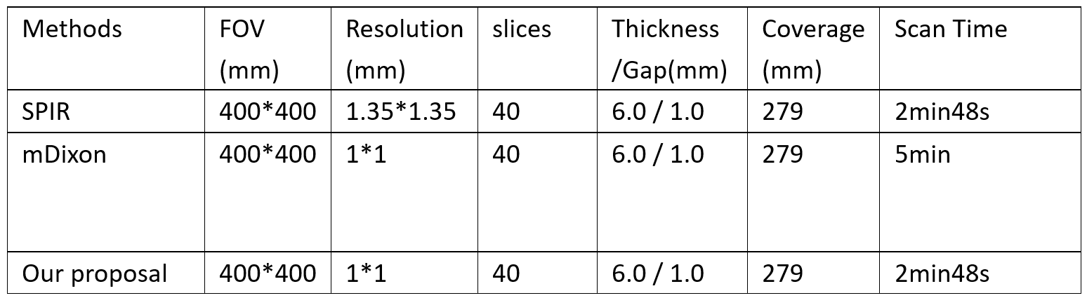
To improve the fat saturation performance for TSE on large FOV and coverage, we combined SPIR and SSGR simultaneously for TSE, the sequence diagram as Fig 1. In this scheme the excitation uses minimum phase pulse to improve the slice profile and shorten the echo spacing. The slice selection gradient of echo pulse was reversed to implement SSGR, with the optimized pulse, it could be combined with TSE.To evaluate the performance of our proposal, Multivane XD TSE with SPIR only, mDixon, and the combination of SPIR with SSGR were acquired on a Philips 3.0T Elition system (Philips Healthcare, Suzhou, China) with a 32-ch torso and spine coil. Detailed scan parameters were summarized in Table 1. Since mDixon8 is more robust on B0 and B1 inhomogeneity than SPIR or SSGR, despite its prolonged imaging time, it was used as the reference fat suppression scheme in this study. We used an FOV of 400x400 mm2 and a coverage of 279 mm in FH direction to mimic the common abdominal imaging in clinical scenarios for full liver and kidney coverage.
Results
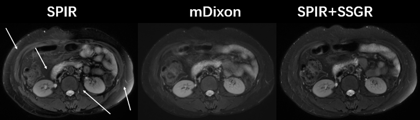
Fig. 2 shows the comparison between different fat suppression techniques, including (A) SPIR only, (B) mDixon and (C) the combination of SPIR and SSGR, where the combination of SPIR and SSGR had similar performance with mDixon and had more uniform fat suppression than SPIR. The proposed method had better contrast than SPIR only or mDixon. These observations were consistent in all slices, while 4 representative slices were shown here to demonstrate the result in upper abdomen (slice 1), middle abdomen (slice 10 and 25), and lower abdomen (slice 40). Fig.3 shows that the contrast and sharpness of kidney tissues was better in the proposed technique than SPIR only or mDixon. While the proposed method did not increase the scan time (2min48s) when compared to SPIR only, it’s more time efficient than mDixon scheme which cost ~5min.Discussion and conclusions
The proposed method showed robust fat suppression in FOV and coverage as large as 400x400 mm2 and 279 mm, with uniform fat suppression in all slices. The fat suppression was comparable to mDixon and was superior to SPIR only. Meanwhile, it showed improved tissue sharpness in kidney when compared to mDixon, due to the reduced imaging time, reduced number of acquisitions, and hence less sensitiveness to physiological motions. The proposed method retained a similar imaging time as in the SPIR only scan, which was ~50% shorter than that of the mDixon scan. Considering the image quality improvement, the relatively short scan time, and the straightforward implementation, this technique holds the potential for wide clinical applications, especially when large FOV is required.Acknowledgements
NoReferences
1. Thorsten AB, Oliver W, Christopher JG, Jean HB, Scott BR, Fat and Water Magnetic Resonance Imaging, J Magn Reson Imaging 2010; 31:4-18.
2. Haase A, Frahm J, Hanicke W, Matthaei D. 1H NMR chemical shift selective (CHESS) imaging. Phys Med Biol 1985;30:341–344.
3. Bydder GM, Steiner RE, Blumgart LH, Khenia S, Young IR. MR imaging of the liver using short TI inversion recovery sequences. J Comput Assist Tomogr 1985;9:1084–1089.
4. Dixon WT. Simple proton spectroscopic imaging. Radiology 1984; 153:189–194.
5. Gomori JM, Holland GA, Grossman RI, Gefter WB, Lenkinski RE. Fat suppression by section-select gradient reversal on spin-echo MR imaging. Work in progress. Radiology 1988;168:493–495.
6. Zoltan N and Nikolaus W, Efficient Fat Suppression by Slice-Selection Gradient Reversal in Twice-Refocused Diffusion Encoding, Magn Reson Med 60:1256–1260 (2008).
7. Hennig J, Nauerth A, Friedburg H. RARE imaging - a fast imaging method for clinical MR. Magn Reson Med 1986; 3: 823-833.
8. Eggers, H and Börnert P, Chemical shift encoding-based water–fat separation methods. J Magn Reson Imaging, 2014; 40(2): p. 251-268
Figures