4254
The feasibility of using magnetic resonance elastography for differentiation of benign and malignant solid pancreatic masses1Zhongshan Hospital affiliated to Fudan University, Shanghai, China, 2Central Research Institute, United Imaging Healthcare, Shanghai, China
Synopsis
Magnetic resonance elastography (MRE) is an emerging functional MR technique. Pancreatic ductal adenocarcinoma (PDAC), as one of the most lethal diseases, accounts for 85–90% of all malignant pancreatic tumors. Histologically, PDAC is a hard mass characterized by a marked desmoplastic reaction and build-up of fibrotic tissues. In this study, we aim to evaluate the usefulness of 2D spin-echo echo-planar-imaging (SE-EPI) MRE for differentiation of solid pancreatic masses. Our results showed that compared with benign pancreatic masses, PDAC demonstrated higher mass stiffness and stiffness.
Introduction:
PDAC is a highly fatal malignancy, the 5-year survival of PDAC is only about 10% and approximately 80–85% of patients present with either unresectable or metastatic disease (1). The early detection and diagnosis of PDAC is essential to get complete R0 surgical resection, thus reaching better prognosis. However, the significant overlap in radiological findings lead to the confusion of differentiation of PDAC and other benign solid pancreatic masses. MRE is an MR-based technique that could noninvasively assesses mechanical tissue properties and provides quantitative information about soft tissue stiffness in vivo (2). Consider of PDAC is a hard mass which have rich fibrotic tissues, we hypothesis MRE might be helpful to provide additional information about mechanical tissue properties and differentiate PDAC from other solid pancreatic masses. Hence, this study aims to explore the diagnostic value of using MRE in preoperatively differentiation of PDAC and other benign pancreatic masses.Methods:
10 patients with pathological results after surgery of endoscopic ultrasound (EUS)-guided biopsy were included in this prospective study. All the examinations were acquired using a 3.0 T MR scanner (uMR 790, United Imaging Healthcare Co Ltd, Shanghai, China). An ergonomic soft pancreatic MRE passive driver with an active acoustic actuator to generated external mechanical shear waves (60Hz) was placed against the upper abdomen, centred on the xiphisternum, and secured with a 20-cm-wide elastic band that wrapped around the body to ensure good coupling between the driver and the body (3). A 32 slice, flow-compensated, SE-EPI pulse sequence was modified to include additional MRE motion-encoding gradients. The detailed parameters of SE-EPI MRE sequence are as followings: TR/TE/FA: 3800 ms/86.2 ms/90°; Matrix/FOV: 300×380/300×380 mm2. The Mann-Whitney U test were used to compare the stiffness of PDAC and other benign pancreatic masses.Results:
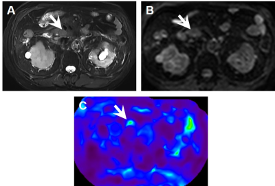
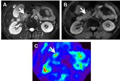
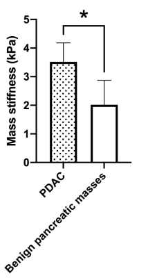
Representative MR images of a patient with benign pancreatic masses and PDAC were displayed in Fig1 and Fig2, respectively. Between 10 selected patients (4 PDAC, 1 serous cystadenoma, 1 solid-pseudopapillary tumor and 4 mass-forming pancreatitis), compared with benign pancreatic masses, PDAC demonstrated significantly higher stiffness (P=0.033).Discussion:
Our results showed that PDAC demonstrated significantly higher stiffness compared with benign pancreatic masses, indicating that MRE is feasible for assessing the mechanical properties of solid pancreatic masses. The preoperative diagnosis of PDAC remains difficult when the absence of definite features of malignancy, such as signs of vascular invasion or liver metastases. However, PDAC has obvious fibrous tissue infiltrating and enveloping the neoplasm (4). This can result in significant changes in the mechanical tissue properties and can be detected by MRE, thus making MRE a useful tool for the diagnosis and discrimination of PDAC from other benign pancreatic masses.Conclusion:
The stiffness value detected by MRE may serve as noninvasive and quantitative imaging markers for non-invasive differentiation between PDAC and other benign pancreatic masses.Acknowledgements
NoneReferences
1. Mizrahi JD, Surana R, Valle JW, Shroff RT. Pancreatic cancer. Lancet 2020;395:2008-2020.
2. Glaser KJ, Manduca A, Ehman RL. Review of MR elastography applications and recent developments. J Magn Reson Imaging 2012;36:757-774.
3. Shi Y, Gao F, Li Y, Tao S, Yu B, Liu Z, Liu Y, et al. Differentiation of benign and malignant solid pancreatic masses using magnetic resonance elastography with spin-echo echo planar imaging and three-dimensional inversion reconstruction: a prospective study. Eur Radiol 2018;28:936-945.
4. Armstrong T, Packham G, Murphy LB, Bateman AC, Conti JA, Fine DR, Johnson CD, et al. Type I collagen promotes the malignant phenotype of pancreatic ductal adenocarcinoma. Clin Cancer Res 2004;10:7427-7437.
Figures


