4245
Application of synthetic MRI to diagnose benign and malignant breast masses: APTw imaging combined with T1 Mapping and DWI imaging1Medical Imaging Center, People's Hospital of Ningxia Hui Autonomous Region, Yinchuan, China, 2Philips Healthcare, Beijing, China
Synopsis
Breast cancer has surpassed lung cancer to become the world's largest cancer for the first time, and the cancer mortality rate ranks second, showing a younger trend in recent years. APTw imaging, as a noninvasive molecular imaging technique, can measure the concentration of free proteins and polypeptides in tissues without the use of exogenous contrast agents, based on a chemical exchange between amide protons and water protons. The aim of study was to explore the diagnostic efficacy for benign and malignant breast tumor by using synthetic MRI.
Introduction
2020 WHO's International Agency for Research on Cancer (IARC) will release the latest global cancer data. Breast cancer has surpassed lung cancer to become the world's largest cancer for the first time, and the cancer mortality rate ranks second, showing a younger trend in recent years. Conventional MRI imaging cannot reflect metabolism and blood flow of lesions. Multi-parameters functional MRI technology, such as Amide Proton Transfer Weighted imaging (APTw), T1mapping and MR diffusion weighted imaging (DWI) may provide new indicators for the diagnosis of benign and malignant breast tumor. APTw imaging is a noninvasive molecular imaging technique that can measure the concentration of free proteins and polypeptides in tissues without the use of exogenous contrast agents, based on a chemical exchange between amide protons and water protons. The aim of study was to explore the diagnostic efficacy for benign and malignant breast tumor by using synthetic imaging, including APTw imaging, DWI and T1 mapping.Methods
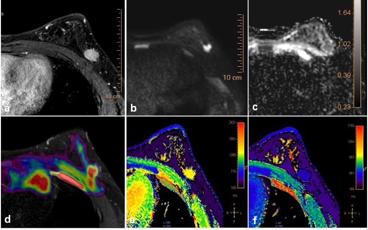
APT imaging, T1 mapping and DWI were performed in 53 patients with breast mass lesions, and all lesions were confirmed by pathology. All MRI were scanned on a 3.0T MR scanner (Ingenia CX, Philips Healthcare) with a 16-channel breast coil. APTw imaging was performed using a saturation pulse with duration of 2s and strength of 2µT. The APT signal intensity (SI) (APT SImean), T1 mapping change rate and average ADC value of benign and malignant breast lesions were measured, calculated and compared by two experienced radiologists, blinded to clinical data. The ROC curve was used to evaluate the diagnostic efficacy of various imaging techniques in differentiating breast lesions. Logistic regression was used to calculate the diagnostic probability of the combination of two or three methods, and the probability was used to draw the ROC curve and calculate the area under the curve.Results
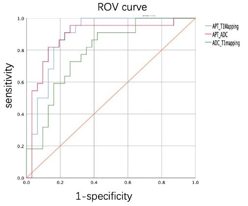
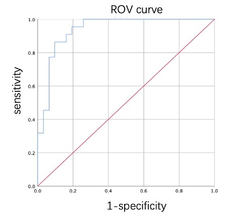
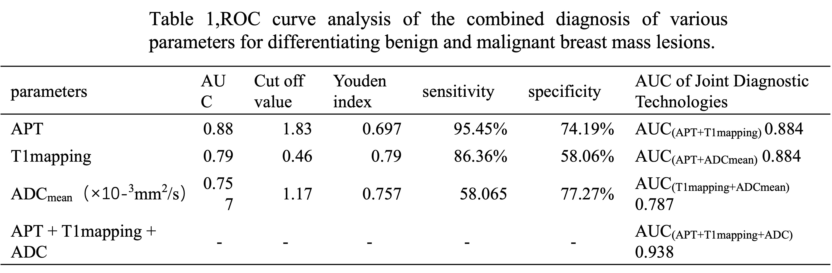
A total of 53 lesions were included, including 22 benign lesions and 31 malignant lesions. Benign group: APT SImean value: 1.71±1.66, T1 mapping rate: 0.44±0.23ms, average ADC value: 1.42±0.37mm2/s×10-3. Malignancy group: APT SImean value: 3.63±1.20, T1 mapping rate: 0.68±0.18ms, ADC value: 0.99±0.43mm2/s×10-3. The APT SImean value and T1 Mapping change rate in malignant group were higher than those in benign group, and the average ADC value was lower than that in benign group. The areas under the ROC curve of APT SImean value, T1 mapping change rate and ADC value were (0.880, 0.790 and 0.757), respectively. The areas under the curve of APTw combined with T1 mapping, APTw combined with ADC, and ADC combined with T1 mapping were (0.884, 0.884, 0.787), respectively, P>0.05. The AUC of APTw combined with T1 mapping and ADC was 0.94.Discussion
APT imaging technology is not limited by the blood-brain barrier without contrast agent, and it can non-invasively detect the process of the exchange between protein amino protons and water molecules. Besides, it can detect the content of endogenous free proteins and polypeptides in tissues, which can reflect the metabolism and physiological information of cells more accurately from the cellular and molecular level. Through the joint diagnostic performance of different imaging technologies, it is found that three joint imaging diagnosis than two joint diagnosis technique can significantly increase the efficiency of the diagnosis. (Table1, Figure 1 and Figure 2).Conclusion
APTw imaging was feasible for breast cancer. The results shown that APT had a better diagnostic performance than ADC in distinguish the benign or malignant of breast mass lesions. APTw combined T1 mapping and ADC imaging techniques could be used to diagnose and significantly improve the diagnostic efficiency compared with the combined of pairwise.Acknowledgements
No acknowledgement found.References
[1] Debnath A, Gupta RK, Singh A. Evaluating the role of amide proton transfer (APT)-weighted contrast, optimized for normalization and region of interest selection, in differentiation of neoplastic and infective mass lesions on 3 T MRI. Mol Imaging Biol, 2020, 22(2): 384-396.
[2] Lisa C, Adams, Bernhard, Ralla, Philipp, Jurmeister, Keno K, Bressem, Ute L, Fahlenkamp, Bernd, Hamm, Jonas, Busch, Marcus R, Makowski. Native T1 Mapping as an In Vivo Biomarker for the Identification of Higher-Grade Renal Cell Carcinoma: Correlation With Histopathological Findings.[J].Investigative radiology,2019,54(2):118-128.
[3] Kim, Ji Eun, Kim, Hyun Ok, Bae, Kyungsoo, Choi, Dae Seob,Nickel, Dominik.T1 mapping for liver function evaluation in geodetic acid-enhanced MR imaging: comparison of look-locker inversion recovery and B-1 inhomogeneity-corrected variable flip angle method[J].European radiology,2019,29(7):3584-3594.
[4] Shuyi,Yang,Fei,Shan,Qinqin,Yan,Jie,Shen,Peiyan,Ye,Zhiyong,Zhang,Yuxin,Shi,Rengyin,Zhang.A pilot study of native T1-mapping for focal pulmonary lesions in 3.0 T magnetic resonance imaging: size estimation and differential diagnosis.[J].Journal of thoracic disease,2020,12(5):2517-2528.
[5] Adrienne N, Dula, Blake E, Dewey, Lori R, Arlinghaus, Jason M, Williams, Dennis, Klomp, Thomas E, Yankeelov, Seth, Smith. Optimization of 7-T chemical exchange saturation transfer parameters for validation of glycosaminoglycan and amide proton transfer of fibro glandular breast tissue.[J].Radiology,2015,275(1):255-61.
Figures