4229
Age-related changes in elasticity, fat degeneration, and morphology of the pancreas: evaluation using multiparametric MR imaging
Akihiko Kanki1, Kazuya Yasokawa1, Hidemitsu Sotozono1, Kiyoka Maeba1, Atsushi Higaki1, Akira Yamamoto1, and Tsutomu Tamada1
1Radiology, Kawasaki Medical School, Kurashiki, Japan
1Radiology, Kawasaki Medical School, Kurashiki, Japan
Synopsis
We evaluated relationships between age and shifted apparent diffusion coefficient, proton density fat fraction (PDFF), and pancreas size using 3-T MRI. PDFF increased with age, and anterior-posterior diameters decreased with age. Interestingly, elasticity of the pancreas decreased with age. Based on these results, decreases in the size of the pancreatic parenchyma with age appear attributable to not only fibrosis, but also fatty degeneration, resulting in decreased elasticity. Fat content should be evaluated at the same time as fibrosis of the pancreatic parenchyma.
INTRODUCTION
Pancreatic fibrosis is a risk factor for eventual progression to chronic pancreatitis and pancreatic ductal adenocarcinoma (PDAC)1. Various techniques for the noninvasive evaluation of pancreatic fibrosis have been described in recent years, including magnetic resonance elastography and ultrasound elastography2,3. However, these techniques need to be performed in addition to regular clinical laboratory tests, and require both more time to perform and dedicated hardware and software. In recent years, diffusion-weighted imaging (DWI)-based virtual elastography has been suggested for noninvasive staging of liver fibrosis without any need for special hardware or software4. In addition, recent reports have suggested pancreatic fat infiltration as a risk factor for primary PDAC5. Fukui et al. showed that proton density fat fraction (PDFF) correlated with histological pancreatic fat fraction, demonstrating that PDFF was higher in the PDAC group than in the control group5. It is important to clarify the age-related changes in findings that represent development risks for PDAC, such as fibrosis and fatty degeneration. The aim of this study was to evaluate the relationship between age and shifted apparent diffusion coefficient (sADC), PDFF, and pancreas size using 3-T MRI.METHODS
This retrospective study received institutional review board approval. Participants comprised 62 patients (mean age, 60.9±14.5 years) with no history of pancreatic disease. Values of sADC were calculated from DWI performed using b-values of 200 and 1500 s/mm2. Values of sADC, PDFF on mDIXON Quant and anterior-posterior (AP) diameters on opposed-phase T1-weighted imaging at the head, body, and tail of the pancreas were measured and mean values of those indices were used for analyses. Comparisons of sADC, PDFF and AP diameters were made among three age ranges: Group 1, <50 years old (n=12); Group 2, 50–69 years old (n=32); and Group 3, ³70 years old (n=18).RESULTS
Significant correlations were identified between age and each of sADC, PDFF and AP diameters (r=0.36, P=0.004; r=0.46, P<0.001; and r=-0.36, P=0.004, respectively). Significant differences in each of sADC, PDFF, and AP diameters were seen among the 3 groups (P=0.016, P=0.004, and P=0.012, respectively). In pairwise comparisons, sADC was significantly lower in Group 1 (0.89±0.06 x 10⁻³mm2/s) than in Group 2 (0.94±0.08 x 10⁻³mm2/s, P=0.043) or Group 3 (0.98±0.10 x 10⁻³mm2/s, P=0.008). PDFF was also significantly lower Group 1 (4.61±4.75 %) than in Group 2 (8.04±4.96 %, P=0.003) or Group 3 (8.49±4.99 %, P=0.001). AP diameters were significantly higher in Group 1 (21.2±3.9 mm) than in Group 2 (17.9±3.5 mm, P=0.02 or Group 3 (16.3±3.9 mm, P=0.007). No indices showed significant differences between Groups 2 and 3 (P=0.108–0.628).DISCUSSION
In our study, PDFF increased with age, and AP diameters decreased with age. Interestingly, sADC increased with age, reflecting decreasing elasticity of the pancreas with age. These results suggest that decreases in the size of the pancreatic parenchyma with age are due to not only fibrosis, but also fatty degeneration resulting in decreased elasticity.CONCLUSION
PDFF increased with age, and AP diameters decreased with age. Interestingly, our study suggested that elasticity of the pancreas using sADC decreases with age. These results suggest that decreases in the size of the pancreatic parenchyma with age are due to not only fibrosis, but also fatty degeneration resulting in decreased elasticity. Fat content should be evaluated at the same time as fibrosis of the pancreatic parenchyma.Acknowledgements
No acknowledgement found.References
- Ceyhan GO, Friess H. Pancreatic disease in 2014: pancreatic fibrosis and standard diagnostics. Nat Rev Gastroenterol Hepatol 2015;12(2):68-70.
- Tang A, Cloutier G, Szeverenyi NM, Sirlin CB. Ultrasound elastography and MR elastography for assessing liver fibrosis: part 2, diagnostic performance, confounders, and future directions. Am J Roentgenol 2015;205(1):33-40.
- Yoshimitsu K, Mitsufuji T, Shinagawa Y, et al. MR elastography of the liver at 3.0 T in diagnosing liver fibrosis grades; preliminary clinical experience. Eur Radiol 2016;26(3):656-63.
- Le Bihan D, Ichikawa S, Motosugi U. Diffusion and intravoxel incoherent motion MR imaging-based virtual elastography: a hypothesis-generating study in the liver. Radiology 2017;285(2):609-19.
- Fukui H, Hori M, Fukuda Y, et al. Evaluation of fatty pancreas by proton density fat fraction using 3-T magnetic resonance imaging and its association with pancreatic cancer. Eur J Radiol 2019;118:25-31.
Figures
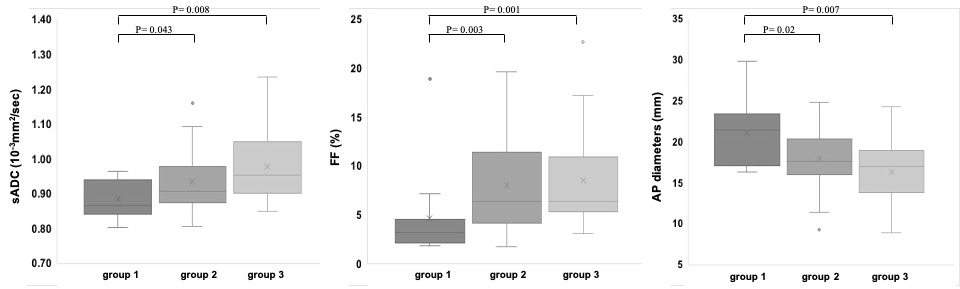
Fig. 1 Box plot comparing shifted apparent diffusion coefficient (sADC), proton density fat fraction (PDFF), and anterior-posterior (AP) diameters among the three groups (Group 1, <50 years old; Group 2, 50–69 years old; Group 3, ≥70 years old).
DOI: https://doi.org/10.58530/2022/4229