4228
The value of DCE-MRI quantitatively predicting vascular invasion of rectal cancer1Radiology, the First Affiliated Hospital of Dalian Medical University, Dalian, China, 2the First Affiliated Hospital of Dalian Medical University, Dalian, China, 3GE Healthcare, MR Research, Beijing, China
Synopsis
Vascular invasion has been proved to be closely related to the poor prognosis of rectal cancer. The current gold standard for the diagnosis of rectal cancer vascular tumor thrombus is postoperative pathology. MRI is a common technology in evaluating malignant tumor. In this work, we explored the feasibility of DCE-MRI in quantitatively predicting vascular invasion of rectal cancer. Results showed that the Ktrans and Kep can differentiate the vascular invasion from normal status accurately. Therefore, DCE-MRI may serve as a feasible and non-invasive way in predicting vascular invasion of rectal cancer preoperatively, that is of great significance for clinical diagnosis.
Introduction
According to the 2018 world cancer statistics [1], the incidence and mortality of colorectal cancer ranked fourth and second among all malignant tumors, respectively, and kept rising. Colorectal cancer is mostly found in the rectum. Invasion of tumor cells into lymph nodes or blood vessels is a key step in the process of metastasis [2]. DCE-MRI is a functional sequence, which can not only provide the morphological characteristics of the tumor, but also obtain the hemodynamic quantitative parameters of the lesion tissue perfusion or microcirculation permeability through post-processing of the image, so as to objectively evaluate the enhancement of the lesion. DCE-MRI includes Ktrans, Kep and Ve quantitative parameters. At present, it is widely used in the research of prostate cancer and cervical cancer [3]. Here, we explored whether DCE-MRI can identify vascular invasion of rectal cancer.Materials and Methods
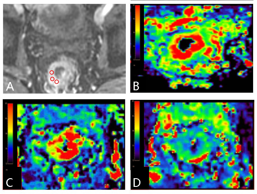
Data of 45 patients with rectal cancer ( age range from 44 to 82 years, mean age: 64 ± 7.83; 12 women) were retrospectively collected. The cases were divided into two groups (group A: 10 cases with vascular invasion, and group B: 35 cases without vascular invasion). All patients underwent pelvic MR scans (including DCE sequence and some routine scans) on a 3.0T MR scanner (GE Signa HDxt, America). Scanning parameters for the DCE-MRI were as follows: using LAVA fast volume scan sequence, FOV=40mm×32mm, TR/TE=3.5/1.5ms, slice thick/gap=3.6/0mm, NEX=0.69, matrix=256×192, scanning suration=4min, 40 periods are scanned totally. Quantitative parameter value of DCE-MRI (Ktrans, Kep and Ve) were measured by two radiologists. Three ROIs were placed at the largest level of the lesion and thier mean value were calculated as the value of the whole lesion (Figure 1). The intra-class correlation coefficient (ICC) was used to test the consistency between measurements by the two radiologists. The Mann Whitney U test was used to analyze the difference of each parameter value between the two groups. Logistic regression was used to combine significantly different parameter valus, and got predicted probabilities. The receiver operating characteristic (ROC) curve was used to test potency of the predictive value in distinguishing the two groups.Results
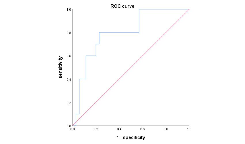
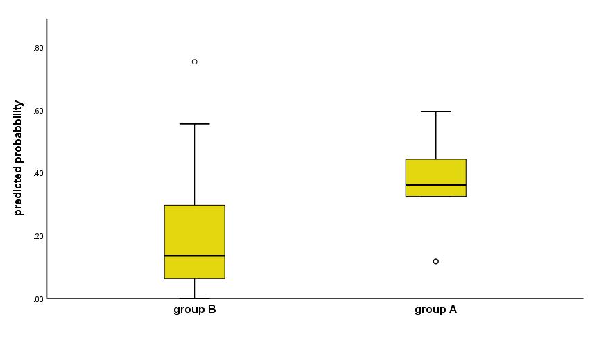
Measurements by the two radiologists were in good agreement (ICC=0.893), and the average of them was taken for subsequent analysis. The value of Ktrans and Kep were significantly lower in group A than group B (P<0.05, respectively) (Table 1). Ktrans combined with Kep has a high efficacy, with area under the curve (AUC) of 0.8, sensitivity of 80%, specificity of 77.1%, threshold value of 0.31 (Figure 2), and the Ktrans-Kep combined value was significantly higher in group A than group B (Figure 3).Discussion and Conclusion
Ktrans represents the capillary permeability-surface area product per unit volume, which depends on blood flow, vascular endothelial cell permeability and endothelial cell surface area. Kep is the flow rate of contrast medium from extracellular space to intravascular flow, which is closely related to vascular wall permeability. In our work, the Ktrans-Kep combined value were significantly higher in the group of vascular invasion than the other. And the ROC curve indicated that the combined value can differentiate the them with a high accuracy. In summary, DCE-MRI may serve as a feasible and non-invasive way in predicting vascular invasion of rectal cancer preoperatively, that is of great significance for clinical diagnosis.Acknowledgements
No acknowledgementReferences
[1] Bray F, Ferlay J, Soerjomataram I et al. Global cancer statistics 2018: GLOBOCAN estimates of incidence and mortality worldwide for 36 cancers in 185 countries[J]. CA Cancer J Clin, 2018, 68(6): 394-424.
[2] Bayar S, Saxena R, Emir B et al. Venous invasion may predict lymph node metastasis in early rectal cancer[J]. Eur J Surg Oncol, 2002, 28(4): 413-417.
[3] Liu Bing, Sun Zhen, Ma Wan-Ling et al. DCE-MRI Quantitative Parameters as Predictors of Treatment Response in Patients With Locally Advanced Cervical Squamous Cell Carcinoma Underwent CCRT[J]. Front Oncol, 2020, 10: 585738.
Figures