4218
Evaluation of the advantage of free-breathing liver DCE-MRI with golden-angle radial sparse parallel using different contrast agents1Department of Radiology, Sun Yat-Sen Memorial Hospital, Sun Yat-Sen University, Guangzhou, China, 2MR Scientific Marketing, Siemens Healthineers, Guangzhou, China
Synopsis
To evaluate the image quality between two sequences (GRASP vs. CDT-VIBE), as well as two kinds of contrast agents (Gd-EOB-DTPA vs. gadobutrol) in liver DCE-MRI. The qualitative and quantitative evaluations regarding of the image quality, and the detection rates of lesions among four groups were analyzed and compared. The results showed that MR images with GRASP sequence had significantly better image quality and less artifacts than that with CDT-VIBE sequence especially in arterial phase, and MR images with injection of gadobutrol could improve the hepatic vessel clarity and reduce motion artifacts compared with that of Gd-EOB-DTPA in CDT-VIBE sequence.
Introduction
Dynamic contrast-enhanced magnetic resonance imaging (DCE-MRI) is a valuable imaging technique for the detection and differential diagnosis of liver lesions by observing the patterns of the contrast agent uptake in liver lesions after using contrast agent 1, 2. At present, the CAIPIRINHA-Dixon-TWIST-volumetric interpolated breath-hold examination (CDT-VIBE) was the most common used sequence in liver DCE-MRI because of its less acquisition time. Nonetheless, it is sensitive to respiratory motion and can reduce image quality in patients who can’t adequately hold breath 3. In addition, transient dyspnea associated with injection of hepatobiliary specific contrast agent ethoxybenzyl diethylenetriaminepentaacetic acid (Gd-EOB-DTPA) might aggravate the respiratory motion and obviously reduce the image quality of DCE-MRI images, especially in arterial phase 4. Recently, an advanced sequence named the golden-angle radial sparse parallel (GRASP) was preliminarily applied for liver DCE-MRI. It overcame respiratory motion compared to conventional CDT-VIBE sequence due to the motion averaging effect from the repeated sampling of k-space center 4. Nevertheless, the image quality and lesion detection rate on liver DCE-MRI images between GRASP and CDT-VIBE, and the influence of different contrast agents are still not well comprehensively understanded. This study was designed to verify the advantage of GRASP in free-breathing liver DCE-MRI in comparison with CDT-VIBE. The influence of different contrast agents, including Gd-EOB-DTPA and a common gadolinium contrast agent, on the image quality was also been determined.Materials and Methods
A total of 80 patients with liver disease were enrolled prospectively, and they were divided into four groups randomly with 20 cases in each group. All patients underwent epigastric MRI examination using a 3T MR scanner (MAGNETOM Vida, Siemens Healthcare, Erlangen, Germany). For dynamic enhanced scanning, first, all patients were randomly assigned to use a free-breathing GRASP sequence or a breath-holding CDT-VIBE sequence to form a free-breathing (FB) group and a breath-holding (BH) group respectively. For different contrast agents, the free-breathing group and the breath-holding group were then randomly divided into Gd-EOB-DTPA or gadobutrol injections, and finally four groups including free-breathing-Gd-EOB-DTPA (FB-Gd) group and gadobutrol (FB-Ga) group, breath-holding-Gd-EOB-DTPA (BH-Gd) group and gadobutrol (BH-Ga) group were formed. Images of non-contrast, arterial, portal venous and delay phases in four groups of patients were acquired, and all images were evaluated by two radiologists qualitatively using a five-point system respectively, in the terms of the edge sharpness and the contrast of liver, lesion, hepatic vessel, and overall image quality, diagnostic confidence, as well as motion artifacts and parallel acquisition technique (PAT) artifacts. Higher score indicated better image quality. Besides, signal-to-noise ratio (SNR), contrast-to-noise ratio (CNR), and the detection rates of lesions relative to T2-weighted image (T2WI) for different sizes (d ≤ 5 mm, 5 mm < d ≤ 10 mm, d > 10 mm) were calculated as quantitative evaluation indicators for image quality. Moreover, the detection rates of small lesions (d ≤ 5 mm) in arterial and portal venous phases in the same group were compared.Results
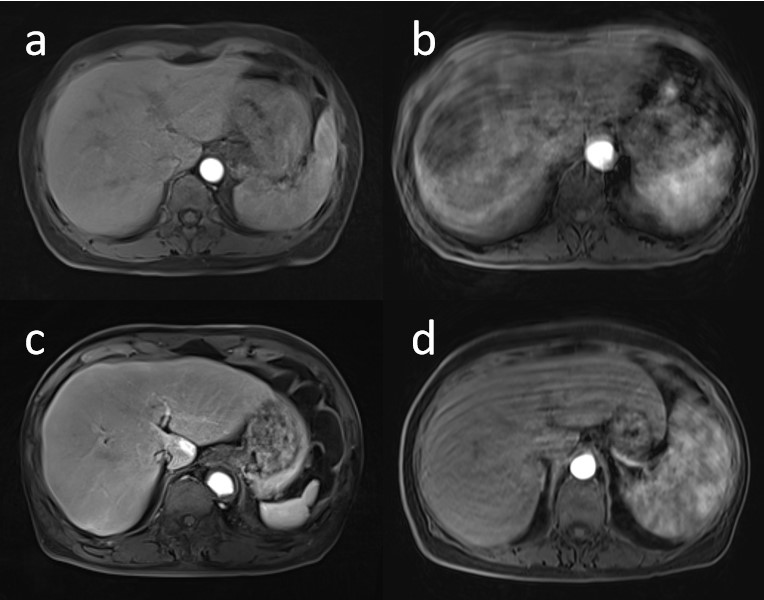
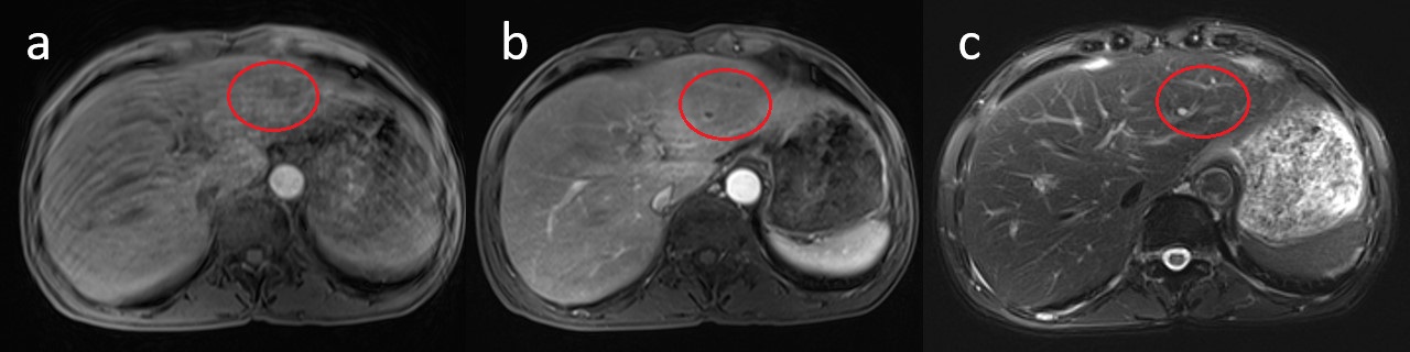
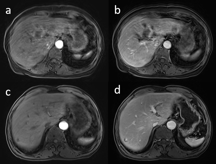
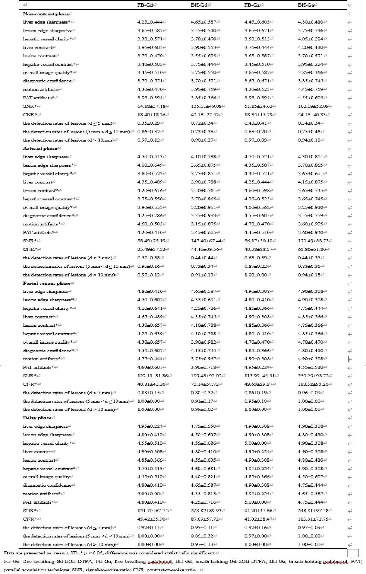
Table 1 showed the qualitative and quantitative evaluation of four groups. The scores of the edge sharpness and the contrast of liver, lesion and hepatic vessel in non-contrast phase images of CDT-VIBE were higher than those of GRASP. For the arterial phase images, GRASP had significantly higher overall image quality and diagnostic confidence compared with CDT-VIBE (P < 0.05) regardless of the different contrast agents, while there were no significant differences of those between images of GRASP and CDT-VIBE in the portal venous and delay phases (P > 0.05). In addition, the motion artifacts and PAT artifacts in all post-contrast images of both breath-holding groups were obviously more serious than those of free-breathing groups (P < 0.05), especially in arterial phase (P < 0.001), as shown in the representative cases in Figure 1. The SNR and CNR for all phases of CDT-VIBE were significantly higher than those of GRASP (P < 0.05). The detection rates of lesions regardless of sizes had no significant differences in all post-contrast images (P > 0.05). Compared with portal venous phases, the detection rates of small lesions (d ≤ 5 mm) in arterial phases of FB-Gd group (P < 0.05) and BH-Ga group (P < 0.001) were significantly lower, as shown in Figure 2. With regard to different contrast agents, the hepatic vessel clarity and contrast in gadobutrol-enhanced images were significantly improved in comparison to Gd-EOB-DTPA-enhanced images (P < 0.05). Besides, as shown in Figure 3, the administration of Gd-EOB-DTPA significantly aggravated motion artifacts in arterial and portal venous phases images of BH-CDT-VIBE compared with gadobutrol (P < 0.05).Discussion and Conclusion
Respiratory motion in DCE-MRI data results in blurring of the reconstructed images and lower value of clinical diagnosis due to misalignment among the acquired frames 2. Radial acquisition has overcome the motion effects, and hence is a promising approach for free-breathing abdominal MRI 4, especially for patients who can’t cooperate with breath-holding. The results of this study showed that GRASP sequence in free-breathing liver DCE-MRI had advantages for higher image quality as well as less motion artifacts and PAT artifacts in comparison with CDT-VIBE, especially in arterial phase. And the administration of gadobutrol could display hepatic vessel more clearly compared with Gd-EOB-DTPA and decrease motion artifacts in CDT-VIBE.Acknowledgements
We sincerely thank the participants in this study.References
1. Mansour, R., et al., Impact of temporal resolution and motion correction for dynamic contrast-enhanced MRI of the liver using an accelerated golden-angle radial sequence. Phys Med Biol, 2020. 65(8): p. 085004.
2. Shahzadi, I., et al., Respiratory motion compensation using data binning in dynamic contrast enhanced golden-angle radial MRI. Magn Reson Imaging, 2020. 70: p. 115-125.
3. Chandarana, H., et al., Free-breathing contrast-enhanced multiphase MRI of the liver using a combination of compressed sensing, parallel imaging, and golden-angle radial sampling. Invest Radiol, 2013. 48(1): p. 10-6.
4. Chandarana, H., et al., Respiratory Motion-Resolved Compressed Sensing Reconstruction of Free-Breathing Radial Acquisition for Dynamic Liver Magnetic Resonance Imaging. Invest Radiol, 2015. 50(11): p. 749-56.
Figures