4200
Evaluation of interstitial lung diseases using free-breathing 3D isotropic ZTE-MRI in comparison with High-resolution computed tomography
Qiuxi Lin1, Yu Deng1, Xinchun Li1, Weiyin Vivian Liu2, Lei Zhang2, Ziyi Zhang3, Qun Luo3, Qi Wan1, and Chongpeng Sun1
1Department of Radiology, The first affiliated hospital of Guangzhou Medical University, Guangzhou, China, 2GE Healthcare, Beijing, China, 3Department of Respiratory and Critical Care Medicine, The first affiliated hospital of Guangzhou Medical University, Guangzhou, China
1Department of Radiology, The first affiliated hospital of Guangzhou Medical University, Guangzhou, China, 2GE Healthcare, Beijing, China, 3Department of Respiratory and Critical Care Medicine, The first affiliated hospital of Guangzhou Medical University, Guangzhou, China
Synopsis
Pulmonary MRI provides qualitative structural and quantitative functional images of the lungs without ionizing radiation. In this study, free-breathing 3D isotropic high-resolution pulmonary zero echo time (ZTE) imaging was applied to detect pulmonary abnormalities and make diagnosis of interstitial lung diseases (ILDs). The pulmonary interstitial abnormalities including ground-glass opacity, reticular opacity, honeycombing, traction bronchiectasis and consolidation can be well depicted. The diagnosis performance of ZTE in ILDs is comparable with HRCT. ZTE might assist long-term follow-ups in ILDs evaluation without radiation exposure.
Introduction and purpose
Interstitial lung diseases (ILDs) are a set of heterogeneous respiratory diseases with limited therapeutic option. Frequent high-resolution computed tomography (HRCT) has been used to long-term follow-ups in evaluation of disease progression or treatment response, leading to a large amount of cumulative radiation exposure to the patients. Pulmonary MRI provides qualitative structural and quantitative functional images of the lungs without ionizing radiation1. Three-dimensional isotropic high-resolution using a free-breathing zero-echo time (ZTE) technique has gained increasing interest in lung imaging recently and with good image quality such as visibility of pulmonary arteries (up to the 7th generation) and the bronchi (up to the 5th generation) 2,3. The aim of this study was to compare ZTE with HRCT in terms of the demonstration of the pulmonary interstitial abnormalities and inter-methods agreement of diagnosis in ILD.Materials and Methods
The study was approved by the ethics committee of our hospital. 47 patients (20 males, 27 females; age from 29 to 70 years; mean: 54 years) consisted of 23 connective tissue disease-associated interstitial lung disease (CTD-ILD), 9 idiopathic pulmonary fibrosis (IPF), 7 interstitial pneumonia with autoimmune features (IPAF) , 7 idiopathic nonspecific interstitial pneumonia (iNSIP), 1 (hypersensitive pneumonia, HP) , were included in the study and given written informed consent. All patients underwent both HRCT and ZTE scan on a 3.0T MRI scanner (Discovery MR750W, GE Healthcare, Waukesha, WI) within an interval of a week. The imaging features including the ground-glass opacity, reticular opacity, honeycombing, traction bronchiectasis and consolidation for each pulmonary segments were evaluated independently by two radiologists on both HRCT and ZTE (Fig1-3). The radiology patterns of usual interstitial pneumonia (UIP), nonspecific interstitial pneumonia (NSIP), organizing pneumonia (OP), mixed NSIP and OP, unclassified were determined for each case on both HRCT and ZTE independently by two radiologists. The receiver operating characteristic analyses was performed to evaluate ability of ZTE in the demonstration of aforementioned imaging features, using HRCT as the reference standard. The inter-method agreement of the ILD radiology patterns was determined by means of kappa statistics. Statistical analyses were performed using SPSS 25.0. P value < 0.05 was regarded as statistically significant.Results
Representative images of a patient respectively with anti-synthetase syndrome and IPF were shown in Figure 1- 3. Using HRCT as the reference standard, areas under the curve (AUC) for ground-glass opacity, reticular opacity, honeycombing, traction bronchiectasis and consolidation ranged from 0.71 to 0.80 (p < 0.001) (Table 1). The performance of ZTE in the depiction of consolidation was highest (AUC=0.80), which may due to increased protons in air space. Inter-method agreements of radiology patterns between pulmonary ZTE and HRCT was moderate (κ = 0.76) (Table 2).Discussion and conclusion
The pulmonary interstitial abnormalities including ground-glass opacity, reticular opacity, honeycombing, traction bronchiectasis and consolidation can be well demonstrated on ZTE-MRI. The diagnosis performance of ILD radiology patterns of ZTE is comparable with HRCT. In conclusion, free-breathing 3D isotropic high-resolution pulmonary zero echo time imaging might assist long-term follow-ups in ILDs evaluation without radiation exposure.Acknowledgements
No acknowledgement found.References
1. HATABU H, OHNO Y, GEFTER WB, et al. Expanding Applications of Pulmonary MRI in the Clinical Evaluation of Lung Disorders: Fleischner Society Position Paper . Radiology, 2020, 297(2): 286-301.2. Yu Deng, Qiuxi Lin, Xinchun Li, et al. Feasibility of free-breathing 3D isotropic whole-lung zero echo time imaging. ISMRM & SMRT Annual Meeting 2021
3. GIBIINO F, SACOLICK L, MENINI A, et al. Free-breathing, zero-TE MR lung imaging . Magma (New York, NY), 2015, 28(3): 207-215.
Figures
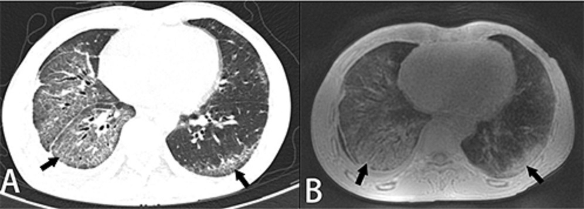
Figure 1 A-B. A 47-year-old male patient with anti-synthetase syndrome. (A) An axial HRCT image showed typical NSIP features of reticular and ground-glass opacities on both lung (arrows). (B) The ZTE image of the same level well depicted the imaging features which were consistent with HRCT (arrows).
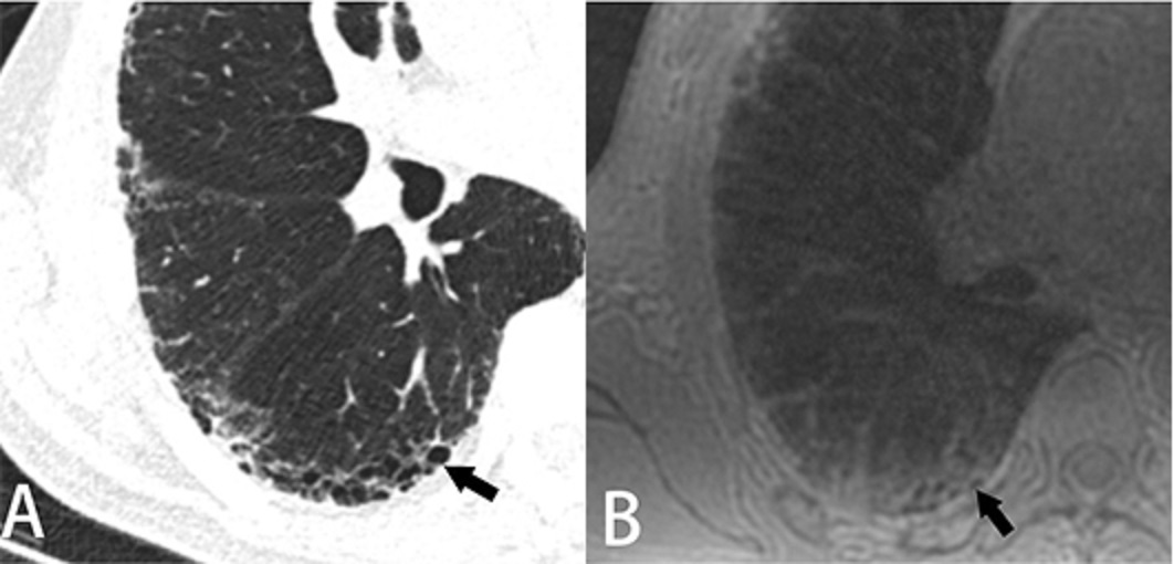
Figure 2 A-B. A 69-year-old male patient with IPF. (A) A zoom-in HRCT showed subpleural multi-layer honeycombing (arrow). (B) A zoom-in ZTE image of the same level showed the honeycombing as a cluster of cysts with no signal (arrow).
(Note: The ZTE image was acquired at the end of expiratory phase, therefore the size of honeycombing decreased along with the whole lung volume in comparison with HRCT).
(Note: The ZTE image was acquired at the end of expiratory phase, therefore the size of honeycombing decreased along with the whole lung volume in comparison with HRCT).
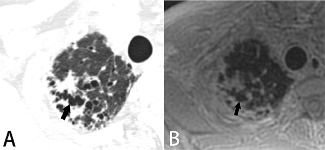
Figure.3 A-B. A 70-year-old male patient with IPF. (A) A zoom-in HRCT showed traction bronchiectasis surrounded by irregular consolidation in the lung apex. (B) A zoom-in ZTE image on the same level demonstrated the traction bronchiectasis and consolidation identical to HRCT.
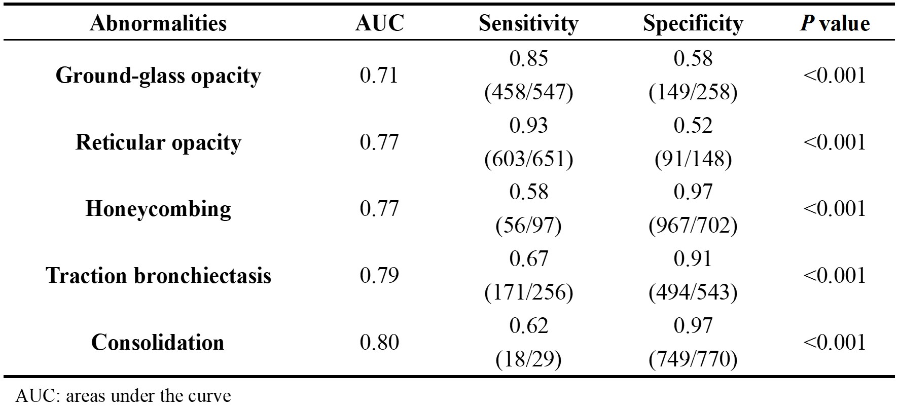
Table 1. Diagnostic performance of ZTE in demonstration of imaging features of ILDs
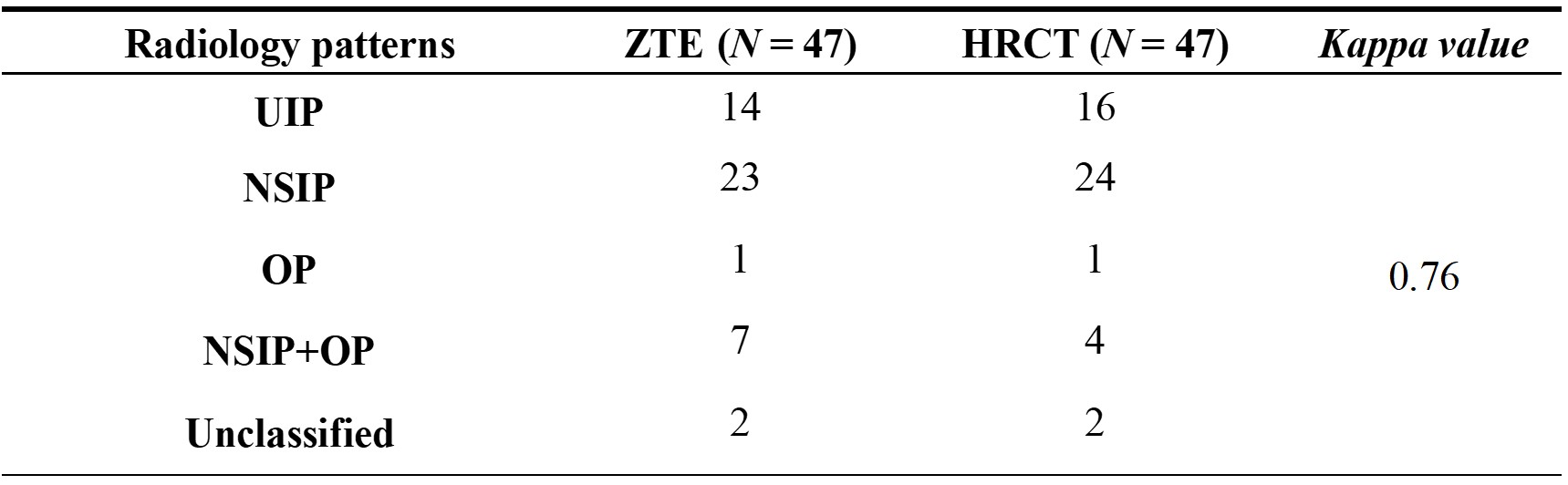
Table 2. Inter-method agreements of radiology patterns between ZTE and HRCT
DOI: https://doi.org/10.58530/2022/4200