4189
Assessment of fibrotic liver regeneration with IVIM imaging: an experimental study in a rat model
Shuangshuang Xie1, Caixin Qiu1, Yajie Sun1, Jinxia Zhu2, Robert Grimm3, and Wen Shen1
1Tianjin First Central Hospital, Tianjin, China, 2MR Collaboration, Siemens Healthcare Ltd., Beijing, China, 3MR Application development, Siemens Healthcare GmbH, Erlangen, Germany
1Tianjin First Central Hospital, Tianjin, China, 2MR Collaboration, Siemens Healthcare Ltd., Beijing, China, 3MR Application development, Siemens Healthcare GmbH, Erlangen, Germany
Synopsis
This study investigated the feasibility of IVIM to evaluate fibrotic liver regeneration and function recovery after partial hepatectomy in a rat model. In fibrotic rats, significant increase in D values and decrease in D* and PF values followed with recovery to pre-PH levels were shown. PF values correlated well with Ki-67 indices, D* and PF values correlated well with ALT, AST, and TBil values. IVIM may be an noninvasive and promising approach to monitor liver regeneration and functional recovery after PH.
Introduction and purpose
Liver regeneration (LR) is critical for the recovery of liver function after partial hepatectomy (PH). In fibrotic liver, total hepatocytes are decreased, which delays and impairs LR resulting in liver dysfunction[1]. An accurate prediction of LR when the liver is compromised would enable clinicians to optimize PH approaches and improve liver surgery success rate. Liver volume measurements are commonly used as a measure of regeneration. But the relevance of this crude measure is not clear in the setting of more subtle interventions[2]. Post-PH, sudden hemodynamic change plays an important role in the initiation of LR[3, 4]. In the process of LR, hepatocyte hypertrophy and cell division may affect the diffusion of water molecules. Intravoxel incoherent motion (IVIM) imaging provides perfusion and diffusion measurements of the liver simutaneously[5, 6], which correspond to possible changes during LR. In this study, we used IVIM to longitudinally observe liver changes in the control and carbon tetrachloride (CCl4)-induced fibrosis rat model of PH. The primary purpose was to investigate LR differences between control and fibrotic livers post-PH. Secondarily, our goal was to determine whether IVIM could be used as a noninvasive method to monitor the progress of LR and liver functional recovery post-PH.Materials and Methods
A total of 62 adult male Sprague-Dawley rats (250-280g) were used for experimentation. All liver MRI examinations were performed with a 3T MR scanner (MEGNETOM Prisma, Siemens Healthcare, Erlangen, Germany) with an eight-channel rat-specific coil (Chenguang, Shanghai, China). IVIM imaging, using a single-shot echo-planar imaging (EPI) sequence, was performed with the following parameters: TR/TE=2300/74 ms; FOV=120 × 98 mm2; slice thickness=3 mm; matrix=120 × 98; reconstructed voxel size=0.5×0.5×3 mm3; acceleration factor=4 (GRAPPA=2; slice acceleration factor=2); 10 b values (0, 10, 20, 30, 50, 75, 100, 300, 500, and 800 s/mm2) were applied in 3 diffusion directions, and acquisition time=7min 12s. MR imaging (n = 10 per group) and histologic and blood biochemical analyses (n = 3/group/time point) were performed pre- and post- left lateral lobe resection (on days 1, 2, 3, 5, 7, 14, and 21) in two groups of rats. The treatment group received CCl4 injection for 8 weeks, and the control group was weight-matched controls (Figure 1). Differences in IVIM parameters were tested with repeated-measures ANOVA and multivariate analysis of variance. Correlation between IVIM parameters and histologic and biochemical indices were analyzed with a Pearson/Spearman analysis.Results
Pre-PH, liver D, and D* values in the fibrosis group were significantly lower than in controls (P ≤ .001). Post PH, an increase in liver D values and a decrease in liver D* and PF values followed with recovery to pre-PH levels were shown in both the fibrosis and control rats. PF values in the fibrosis group were significantly higher than in controls from 3 days to 21 days post-PH (P < .05) (Figure 2). In fibrosis rats, both hepatocyte ki-67 indices and hepatocyte diameters increased post-PH, and a strong correlation was found between liver PF values and ki-67 indices (r = -0.756; P = .03), and D* and PF values and ALT, AST, and TBil values (r = -0.762 to -0.905; P < .05). In control rats, only hepatocyte diameters increased post-PH, and all IVIM parameters correlated well with hepatocyte diameters (r = -0.779 to -0.935; P < .05) and alanine transaminase, aspartate aminotransferase, and total bilirubin values (r = -0.810 to -0.976; P < .05) (Figure 3).Discussion and Conclusion
This study assessed the usefulness of IVIM imaging for the noninvasive assessment of liver regeneration and liver functional recovery after PH in control and fibrotic livers. After left lateral lobe resection, fibrotic livers regenerated by increasing hepatocyte size and cell division, but control liver regenerated only by increasing hepatocyte size. Liver D values increased, and PF values decreased immediately post-PH, then gradually returned to baseline levels. Liver PF values were higher in fibrotic livers compared with control livers post-PH. IVIM-derived MR parameters correlated well with LR-related histology and liver function biochemical results. IVIM imaging is a noninvasive and reliable method for monitoring the process of LR and functional recovery in control and fibrotic livers after PH.Acknowledgements
This study was supported by grants from National Nature Science foundation of China (81901710).References
1. Ozaki M. Cellular and molecular mechanisms of liver regeneration: Proliferation, growth, death and protection of hepatocytes. Semin Cell Dev Biol 2020;100:62-73.2. Forbes SJ, Newsome PN. Liver regeneration - mechanisms and models to clinical application. Nat Rev Gastroenterol Hepatol 2016;13:473-485.3. Yagi S, Hirata M, Miyachi Y, Uemoto S. Liver Regeneration after Hepatectomy and Partial Liver Transplantation. Int J Mol Sci 2020;21:8414.4. Michalopoulos GK, Bhushan B. Liver regeneration: biological and pathological mechanisms and implications. Nat Rev Gastroenterol Hepatol 2021;18:40-55.5. Le Bihan D. What can we see with IVIM MRI. Neuroimage. 2019. 187: 56-67.6. Le Bihan D. Intravoxel incoherent motion perfusion MR imaging: a wake-up call. Radiology 2008;249:748-752.Figures
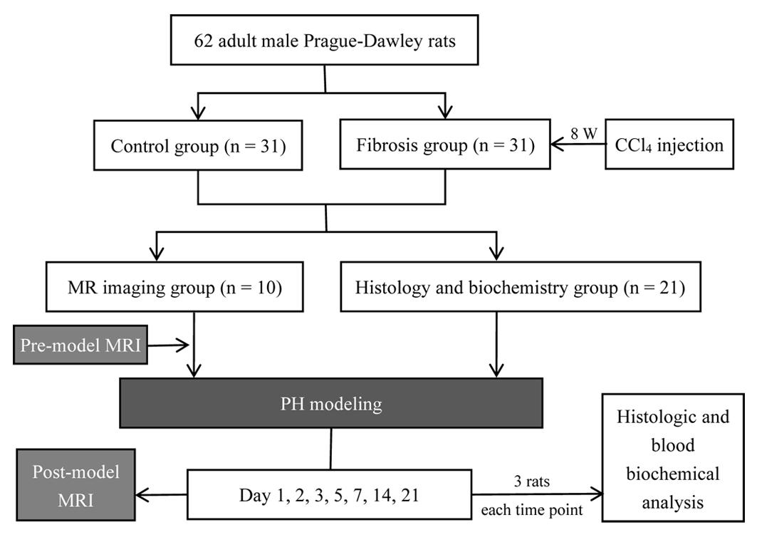
Figure 1. Flowchart of the experimental procedure
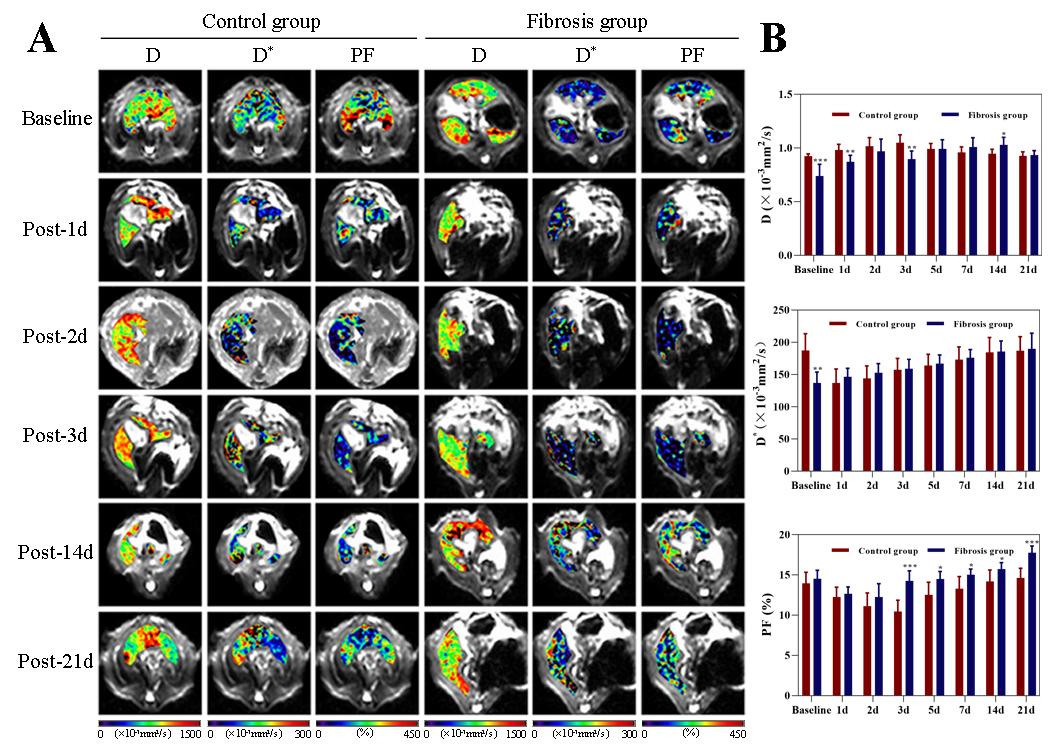
Figure 2. Liver changes showed in D, D*, and PF maps derived from IVIM imaging. A, Changes in control and fibrosis groups on baseline and day 1, 2, 3, 14, and 21 post-PH. B, Changes in liver D, D,* and PF values are shown at all time points before and after surgery in the control and fibrosis groups. IVIM, intravoxel incoherent motion; PF, perfusion fraction; PH, partial hepatectomy. * P < .05; ** P ≤ 0.01
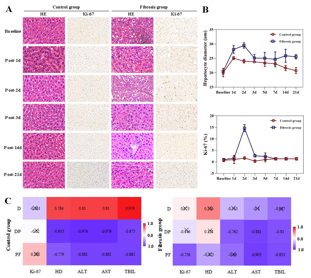
Figure 3. Liver histopathologic changes and correlation analysis. A, Hematoxylin and eosin stain (HE) showed enlarged hepatocytes post-PH in both the control and fibrosis groups. Immunohistochemical staining of Ki-67 (brown) showed increased Ki-67+ cells in the fibrosis group 2 days after surgery. B, Changes in hepatocyte diameters and Ki-67 indices post-PH. C, Correlation hot maps between liver IVIM parameters and LR-related histologic results and liver function biochemical indices
DOI: https://doi.org/10.58530/2022/4189