4152
Microstructural alterations in projection and association fibers in neonatal hypoxia-ischemia1College of Biomedical Engineering & Instrument Science, Zhejiang University, HangZhou, China, 2Department of Neonatal Intensive Care Unit, Children's Hospital affiliated to Zhejiang University School of Medicine, HangZhou, China, 3Department of Radiology, Children's Hospital affiliated to Zhejiang University School of Medicine, HangZhou, China, 4Department of Radiology, New York University School of Medicine, New York, NY, United States
Synopsis
Diffusion MRI (dMRI) is known to be sensitive to hypoxic-ischemic encephalopathy (HIE) , however, the existing dMRI studies only used diffusion tensor metrics in a few selected brain regions and primarily focus on severe-to-moderate HIE. Here we investigated microstructural alterations using multi-shell dMRI across the whole-brain in severe, moderate, and mild HIE babies (n=5/13/13) in comparison with control neonates (n=11) with region-of-interest-based, tract-based, and fixel-based analysis. We found microstructural alternations in projection and association fibers, especially the inferior fronto-occipital fasciculus and inferior longitudinal fasciculus that are important for visual functions.
Introduction
Hypoxic-ischemic encephalopathy (HIE) is one of the most common neonatal diseases that leads to devastating neurological outcomes. Neonatal HIE is clinically classified into mild, moderate, severe HIE according to the Sarnat staging system1. Diffusion MRI (dMRI) is known to be sensitive to Hypoxic-ischemic encephalopathy (HIE) , however, the existing dMRI studies only used diffusion tensor metrics in a few selected brain regions and primarily focus on severe-to-moderate HIE. In this study, we performed a systematic evaluation of the microstructural changes of white matter structures across the whole-brain at region-of-interest (ROI) level, tract level, and fixel levels based on multi-shell dMRI data, in a spectrum of neonatal HIE from severe to mild injuries.Methods
Data acquisition: A total of 31 term-born HIE neonates (13 mild, 13 moderate, 5 severe) and 11 healthy term-born neonates were included in this study. The neonates were scanned at postmenstrual age at scan (PMA) of 0-3 weeks on a 3T Philips Achieva system with an 8-channel head coil. dMRI data was acquired using a diffusion-weighted echo-planar imaging sequence: TR/TR = 7177/73ms, FOV = 179.2×179.2×120 mm3, in-plane resolution = 1.4×1.4 mm2, 60 slices with a slice-thickness of 2mm, two b values of 800/1500 s/mm2, and 15 noncolinear diffusion directions for each b-value.ROI-based analysis: After preprocessing, the dMRI data were registered to the JHU Neonatal Atlas2 and segmented into 40 ROIs bilaterally. Mean fractional anisotropy (FA), mean diffusivity (MD), and fixel-based metrics were obtained from each ROI.
Tract-based analysis (TBA): Except for the severe HIE group as the serious brain damage impeded fiber tracking, six fasciculi were tracked and segmented using Automatic Fiber Quantification3, including the inferior fronto-occipital fasciculus (IFOF), inferior longitudinal fasciculus (ILF), anterior thalamic radiation (ATR), corticospinal tract (CST), genu of corpus callosum (GCC), and splenium of corpus callosum (SCC). Each tract was divided into 100 segments to obtain the tract profiles.
Fixel-based analysis (FBA): For tracts that were found to significantly injured in HIE from the aforementioned TBA, FBA was performed to obtain microstrucal parameters of fiber density (FD), fiber cross-section (FC), and fiber density and cross-section (FDC)4 of individual fixels in MRtrix35.
Statistical analysis: ANCOVA was performed for detecting the difference between the 3 HIE groups (severe, moderate, mild) and control group separately, with gender, birth weight, gestational age (GA) at birth, and PMA at scan as covariates. The p values of ROI-based analysis were adjusted using the FDR method. The significance level was set to 0.05 for all analysis.
Results
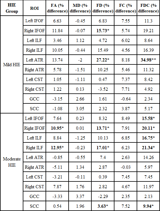
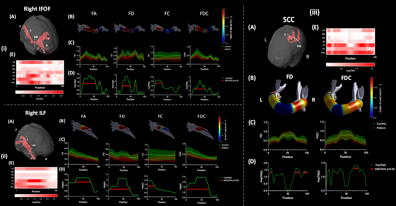
For ROI-based analysis (Figure 1), extensive WM and deep GM regions showed reduced FA bilaterally in the severe HIE babies compared with healthy neonates. In the moderate HIE group, a few selected ROIs closed to the posterior watershed zone demonstrated significantly reduced FC, including the left stria terminalis (ST), right tapetum, right posterior thalamic radiation (PTR), and right sagittal stratum (SS); while the other tensor-based and FBA-based parameters did not show group difference. No significant difference was found between the mild HIE and control group.The moderate HIE neonates showed significantly reduced FA, FD, and FDC over the whole-track in the right IFOF, right ILF, and SCC compared with the control; while in mild HIE, was FD and FDC in the left ATR found to be reduced (Figure 2). Segmentation analysis further revealed the injury significantly altered the posterior-to-middle part of right IFOF, posterior part of right ILF, and central-to-right part of SCC in moderate HIE neonates (Figure 3). While no significant difference were found for any segment between mild HIE groups and controls.
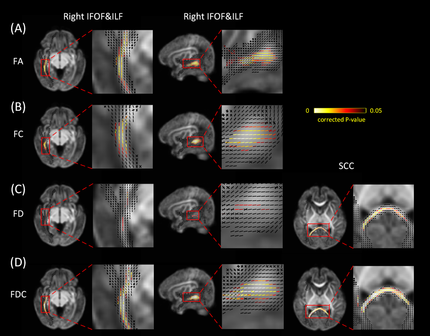
Finally, FBA was performed on the three tracts that were detected by TBA, which revealed consistent but more extensive fiber damage in moderate HIE compared with TBA findings. In the right inferior-posterior IFOF&ILF, a number of fixels along the tract orientation showed statistically reduced FA, FC, FD, and FDC (adjusted p<0.05), as shown in both axial and sagittal views (Figure 4). An extensive segments of the SCC towards the central-to-right part demonstrated significantly altered fixels with reduced FD and FDC (Figure 4).
Discussion
In this study, we performed a systematic analysis of the brain microstructural alternations in severe, moderate, and mild HIE at ROI, tract, and fixel levels. Results revealed that : (1) while severe HIE babies showed extensive injury, the moderate and mild HIE patients exhibited selected regions of WM injury that can be detected by fixel-based metrics; (2) the right IFOF, right ILF, and SCC that are associated with the occipital and temporal lobes were found to be selectively altered in moderate HIE, which agreed with the fact that HIE preferencially impacts visual function; (3) the tensor-based metrics and fixel-based metrics demonstrated varying sensitivities to different WM regions depending on the injury group and may be complementary to each other.Conclusion
ROI analysis revealed extensively reduced FA across the brain in severe HIE and reduced fiber cross-section in selected WM close to the posterior watershed zone in moderate HIE. Tract- and fixel-based analysis pointed to a unique injury pattern associated with reduced FA, FD, and FC in association fibers in moderate HIE, indicating selective injury associated with the temporal and occipital lobes in these neonates.Acknowledgements
Ministry of Science and Technology of the People’s Republic of China (2018YFE0114600), National Natural Science Foundation of China (61801424, 81971606, 82122032), and Science and Technology Department of Zhejiang Province (202006140).References
1.Sarnat HB, Sarnat MSJAon. Neonatal encephalopathy following fetal distress: a clinical and electroencephalographic study. Archives of neurology. 1976;33(10):696-705.
2.Oishi K, Mori S, Donohue PK, Ernst T, Anderson L, Buchthal S, et al. Multi-contrast human neonatal brain atlas: application to normal neonate development analysis. Neuroimage. 2011;56(1):8-20.
3.Yeatman JD, Dougherty RF, Myall NJ, Wandell BA, Feldman HM. Tract profiles of white matter properties: automating fiber-tract quantification. PLoS One. 2012;7(11):e49790.
4.Raffelt DA, Tournier JD, Smith RE, Vaughan DN, Jackson G, Ridgway GR, et al. Investigating white matter fibre density and morphology using fixel-based analysis. Neuroimage. 2017;144(Pt A):58-73.
5.Tournier JD, Smith R, Raffelt D, Tabbara R, Dhollander T, Pietsch M, et al. MRtrix3: A fast, flexible and open software framework for medical image processing and visualisation. Neuroimage. 2019;202:116137.
Figures
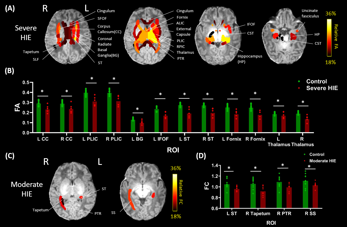
The WM and deep GM regions show significant changes in HIE. (A) Percentage differences in FA in the ROIs with statistically lower FA in the severe HIE group compared to the controls. (B) FA values in representative ROIs of the severe HIE and control groups. (C) Percentage differences in FC in the ROIs with statistically lower FC in the moderate HIE group compared to the controls. (D) FC values in the significantly altered regions of the moderate HIE compared with the controls. *p<0.05, **p<0.01.
Abbreviations: L-left; R-right.