4150
Can HR-VW-MRI effectively evaluate the treatment effect on patients with MCA atherosclerotic stenosis after recanalization?1Department of Radiology, The First Affiliated Hospital of Shandong First Medical University, Jinan, China, 2MR Research, GE Healthcare, Beijing, China
Synopsis
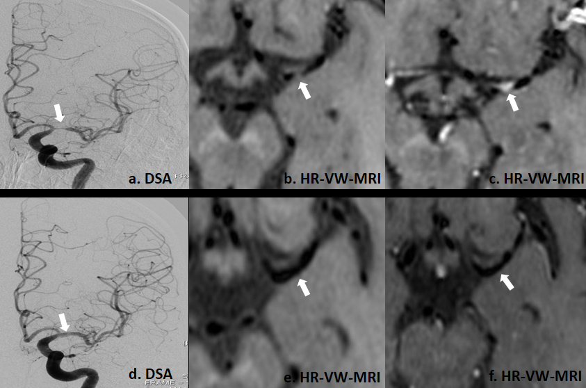
This study mainly explored the feasibility of high-resolution vascular wall magnetic resonance imaging(HR-VW-MRI)in evaluating the treatment effect for patient with middle cerebral artery (MCA) recanalization. We included 21 patients with MCA stenosis diagnosed by DSA. All patients were treated with SeQuent Please drug-coated balloon recanalization and performed with HR-VW-MRI examination. Decreased responsible plaque area, length, enhancement index and lumen stenosis degree were found in MCA postoperatively. Additionally, the lumen stenosis degree evaluated by HR-VW-MRI was highly correlated with DSA. Therefore, HR-VW-MRI could be used as a valuable method to diagnose the effect of vascular recanalization.
Introduction
Middle cerebral artery(MCA)atherosclerotic stenosis is a common independent risk factor for stroke[1]. In clinic,interventional surgery, adopting balloon expansion or stent implantation has been applied to alleviate luminal stenosis with varying degrees [2], and the postoperative prognosis is mainly evaluated relying on the stenosis rate measured with traditional DSA, MRA or CTA. However, these methods are not able to observe the changes of atherosclerotic plaques which are underlying cause of the first or recurring ischemic stroke [3,4].High-resolution vessel wall magnetic resonance imaging (HR-VW-MRI) has been proposed in intracranial vascular wall imaging [4]. This technique can not only evaluate the vascular stenosis degree, but also recognize the basic characteristics of atherosclerotic plaque (e. g. area, length, and enhancement index) [5]. With these properties, we hypothesize that HR-VW-MRI also has a clinical potential to evaluate the responsible plaque characteristics and lumen stenosis degree changes for MCA stenotic patients after recanalization.
Therefore, the main goal of this study was to investigate if HR-VW-MRI was feasibility in evaluating the treatment effect on patients with MCA atherosclerotic stenosis after recanalization.
Materials and Methods
Subjects21 patients (15 males vs 6 females; age: 47.87±15.83 years old) with MCA stenosis were included in this study. At admission and discharge, all patients were scored using the National-Institutes-of-Health-Stroke-Scale (NIHSS) and the modified Rankin-Scale (mRS) to assess the damage to central nervous system. All patients have undergone SeQuent Please drug-coated balloon dilation, and received HR-VW-MRI examinations before and after operation.
MRI experiments
All HR-VW-MRI experiments were performed on a 3.0T MRI scanner (GE Discovery 75, USA) equipped with a 32-channel head coil. HR-VW-MRI included CUBE T1WI at sagittal view with and without injecting contrast agent. The corresponding scan parameters were described as follows: FOV=240m×240mm,slice thickness=1mm,TR/TE=600/13.50ms. Total scan time was 8 mins 32s. The contrast agent was gapenate meglumine injection, and the dosage was 0.2ml/kg.
Image Analysis
All imaging analyses were performed using software of Osirix MD, v.9.02. Two senior radiologists were employed for measuring T1-weighted HR-VW-MRI, including lumen diameter, lumen area and outer wall area at the maximal stenotic site and reference site of MCA. The reference site was defined using the MCA segment of normal appearance proximal to the stenotic segment. MCA plaque area and stenotic degree were calculated by the following formulas[4,6]: 1). Wall area = outer wall area - lumen area;Plaque area= stenotic wall area - reference wall area; 2). Stenotic degree = (1- stenotic lumen diameter/ reference lumen diameter) ×100%. Three ROIs with area of (0.4-1.0) mm2 were delineated on the maximum level of plaque area of pre-contrast and post-contrast images. The signal intensities (SI) of ROIs were measured and the mean values of three ROIs were calculated. Therefore, the calculation formula of enhancement index was as follows[7]: Enhancement index = (SI post-contrast /SI pre-contrast - 1)×100%. We counted the total number of images with plaques shown as the plaque length.
Statistical Analysis
All statistical analyses were performed using GraphPad Prism 9. Paired t-test or Kruskal‑Wallis was separately used to compare NIHSS, mRS, plaque area, length, enhancement index and lumen stenosis degree difference before and after operation. Receiver-operating-characteristic curve (ROC) analysis and area-under-the-curve (AUC) were used to evaluate the diagnostic efficacy of plaque area, length and enhancement index for postoperative efficacy. Bland-Altman images and Pearson correlation analysis were used to test the consistency of HR-VW-MRI and DSA in measuring the lumen stenosis degree before and after recanalization. Significant threshold was set as p <0.05.
Results
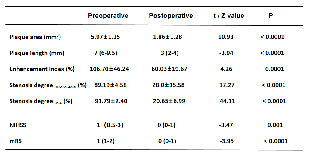
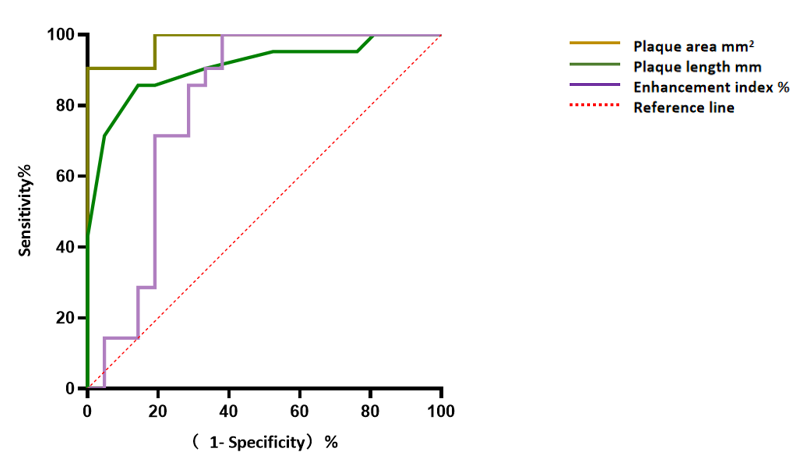
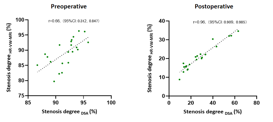
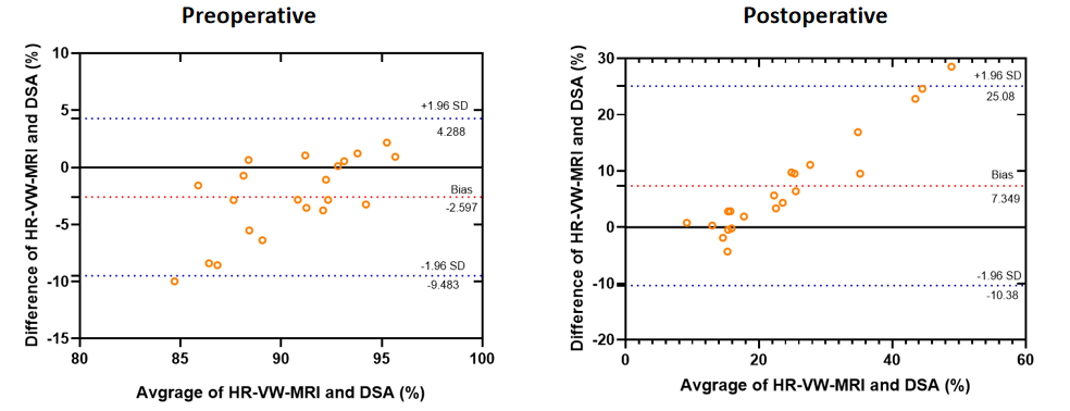
After recanalization, plaque area, length, enhancement index, lumen stenosis degree, NIHSS and mRS scores were significantly lower than the respective ones before operation (all P<0.05; Table1). Using ROC analysis, plaque area, length, enhancement index showed robust prognostic prediction for SeQuent Please drug-coated balloon recanalization (AUC=0.98, 0.91, 0.80 ; P<0.05; Fig1). With Pearson correlation analysis,stenosis degree measured by HR-VW-MRI was highly correlated with DSA (r=0.66; r=0.96; P<0.0001;Fig2). Bland-Altman scatter plot showed that the measurement bias between HR-VW-MRI and DSA was small. The mean difference between HR-VW-MRI and DSA in preoperative and postoperative was -2.597 %and7.349% respectively, and 95% (20/21) of these values were within the 95% confidence interval of the Bland–Altman analysis (Fig3).Discussion and conclusions
In this study, we explored the feasibility of HR-VW-MRI in evaluating changes of basic plaque characteristics and stenosis degree after recanalization for patients with MCA stenosis. After recanalization, the degree of lumen stenosis was significantly improved. Moreover, the area, length and enhancement index of responsible plaque have shown close relationships with the risk and severity of stroke [8,9]. Decreased responsible plaque area, length, enhancement index not only indicates improved stenosis etiology, but also reduces risk of stroke recurrence. Additionally, using ROC curve, these indexes showed robust diagnostic value in the evaluation of postoperative treatment effect. Finally, the lumen stenosis degree evaluated by HR-VW-MRI was proven highly consistent with DSA.In conclusion, HR-VW-MRI can be demonstrated an effective tool for evaluating treatment effect on MCA stenotic patients after recanalization.
Acknowledgements
NoneReferences
[1] Wang W, Jiang B, Sun H, et al. Prevalence, Incidence, and Mortality of Stroke in China: Results from a Nationwide Population-Based Survey of 480687 Adults. Circulation, 2017, 135(8): 759-771.
[2]. Choi JW, Kim JK, Choi BS, et al. Adjuvant revascularization of intracranial artery occlusion with angioplasty and/or stenting. Neuroradiology, 2009,51(1):33-43.
[3] Mo L, Ma G, Dai C, et al. Endovascular recanalization for symptomatic subacute and chronically occluded internal carotid artery: feasibility, safety, a modified radiographic classification system, and clinical outcomes. Neuroradiology, 2020, 62(10):1323-1334.
[4]. Xu WH, Li ML, Gao S, et al. In vivo high-resolution MR imaging of symptomatic and asymptomatic middle cerebral artery atherosclerotic stenosis. Atherosclerosis, 2010,212(2):507-511.
[5]. Natori T, Sasaki M, Miyoshi M, et al. Intracranial Plaque Characterization in Patients with Acute Ischemic Stroke Using Pre- and Post-Contrast Three-Dimensional Magnetic Resonance Vessel Wall Imaging. J Stroke Cerebrovasc Dis, 2016, 25(6):1425-1430.
[6] Samuels OB, Joseph GJ, Lynn MJ, et al. A standardized method for measuring intracranial arterial stenosis. AJNR Am J Neuroradiol, 2000, 21(4):643-646.
[7] Lou X, Ma N, Ma L,et al. Contrast-enhanced 3T high-resolution MR imaging in symptomatic atherosclerotic basilar artery stenosis. Am J Neuroradiol, 2013,34(3):513-517.
[8] Zhang X, Chen L, Li S, et al. Enhancement Characteristics of Middle Cerebral Arterial Atherosclerotic Plaques Over Time and Their Correlation With Stroke Recurrence. J Magn Reson Imaging, 2021, 53(3):953-962.
[9] Ran Y, Wang Y, Zhu M, et al. Higher Plaque Burden of Middle Cerebral Artery Is Associated With Recurrent Ischemic Stroke: A Quantitative Magnetic Resonance Imaging Study. Stroke, 2020, 51(2):659-662.
Figures