4149
Comparison of the Accuracy between High-resolution B-TFE and 3D T1-FFE MR Sequences on Human Spinal Artery Lesions1Department of Radiology, the Third Xiangya Hospital, Central South University, Changsha, China, 2Philips Healthcare, Guangzhou, China
Synopsis
Spinal artery imaging has long been difficult in clinical detection and assessment of spinal vascular lesions, due to its anatomical complexity and very small diameter. Compared with DSA and CT, CE-MRA technology has the advantages of safety, noninvasive and radiation free. Traditional MR sequences, such as TOF and PC, perform poorly with lower SNR. Here, accuracy of B-TFE and 3D T1-FFE sequences were compared to provide radiologists a better choice in the spinal artery scenario. The results indicated that 3D T1-FFE would be preferred than B-TFE. And B-TFE should be used merely when patients are sensitive to contrast media
INTRODUCTION
Since the anatomical complexity of spinal artery with very small diameter, spinal artery imaging has long been difficult in clinical detection and assessment of spinal vascular lesions [1]. Digital Subtraction Angiography (DSA) has long been the GOLD criteria in diagnosing spinal artery lesions, while it is time consuming (about 2~3 h) and causes risk of radiation exposure. Computed tomography angiography (CTA) requires high dose of iodine contrast medium additionally, besides radiation. Magnetic resonance imaging (MRI) should be a safer alternate in such scenario. However, traditional MR sequences, such as Time-of-flight (TOF) sequence [2] and phase contrast (PC) sequence [3], showed poor performance with lower signal-to-noise ratio (SNR), due to a weak inflow-related enhancement effect of spinal artery. Besides, due to the relatively smaller size of spinal arteries, i.e., the mean diameter of the anterior spinal artery is 134 ± 20 μm and the circular epidural artery is 150 ± 26 μm [4], conventional MR sequences could hardly reconstruct such structures clearly. In this study, balanced turbo field-echo sequence (B-TFE) [5] without contrast medium and high-resolution 3D fast field echo sequence (3D T1-FFE) with gadolinium contrast were employed, to provide precise visualization of human spinal arteries. Comparison of accuracy between these two techniques was performed to suggest radiologists a better protocol while choosing to use spinal MR angiography (MRA)METHODS
30 patients with thoracic enhancement examination in our hospital (Mar 1-Aug 31, 2021) were enrolled. Informed consent forms were signed. The average age of those patients was 45.25 ± 6.52 years. MRI acquisition was performed on Philips ingenia 3.0T scanner with phased array spinal and abdominal coils. High resolution B-TFE and CE-MRA3D T1-FFE sequences were employed to scan the thoracic spinal cord. Two deputy chief physicians were devoted to evaluating the capacity of 3D T1-FFE sequence in displaying spinal artery vessels as well as vascular lesions. Results of this evaluation were set as reference to further evaluating the accuracy of B-TFE sequence. B-TFE settings: FOV = 300 * 150 mm, voxel size = 0.5 * 0.5 * 1 mm, layer spacing = -0.5 mm, number of layers = 70, phase direction: RL, sense: 0, NSA = 1, TR/TE/Flip = 6.7 ms/2.7 ms/45°. Total scanning time was 5:50 min. 3D T1-FFE settings: phase direction: FH, sense: 1.5, NSA = 2, dyn = 2, 53s/phase, TR/TE/Flip = 9.1 ms/4.4 ms/20°, Keyhole = 22%, and other settings were consistent with B-TFE. Total scanning time was 5:45 min.
RESULTS
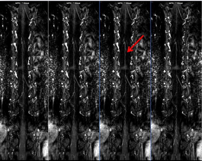
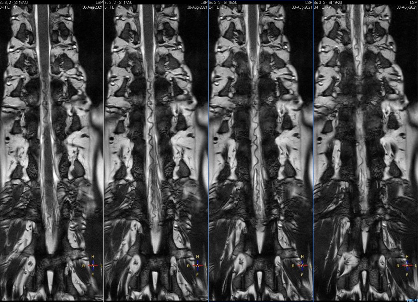
3D T1-FFE and B-TFE images of an example patient are shown in Figure 1 and Figure 2.3D T1-FFE sequence clearly showed the anterior spinal artery lesions in 29 of 30 cases, and the posterior spinal artery lesions in 27 of 30 cases.
B-TFE sequence clearly showed the anterior spinal artery lesions in 23 of 30 cases and the posterior spinal artery lesions in 21 of 30 cases.
3D T1-FFE sequence helped displaying anterior spinal artery more effectively than B-TFE (P < 0.05), and there was no significant difference between genders (P > 0.05), indicating that 3D T1-FFE would be preferred than B-TFE in such scenario.
DISCUSSION & CONCLUSION
In this study, the comparison of MRA performance on detecting spinal artery lesions between 3D T1-FFE and B-TFE was investigated. Results indicated that the involvement of 3D T1-FFE sequence with Keyhole could better delineate the spinal artery and accurately evaluate the lesions, compared to B-TFE. B-TFE sequence can also clearly display the morphology of small arteries. However, B-TFE sequence mainly depends on the background of cerebrospinal fluid in the spinal canal, which can be used as an effective alternate to such examination, and the injection of gadolinium contrast agent is not necessary. It should be only a plan-B for patients allergy to contrast media.Acknowledgements
No acknowledgement found.References
1. Shinji Yamamoto, Hideaki Kanaya, and Phyo Kim. Spinal intraarterial computed tomography angiography as an effective adjunct for spinal angiography. J Neurosurg Spine. 2015 Sep;23(3):360-7
2. Sumi T, Sumi M, Van Cauteren M, Kimura Y, Nakamura T. Parallel imaging technique for the external carotid artery and its branches: comparison of balanced turbo field echo, phase contrast, and time-of-flight sequences. J Magn Reson Imaging. 2007 May;25(5):1028-34
3. David T. Wymer, Kunal P. Patel, William F. Burke III, and Vinay K. Bhatia. Phase-Contrast MRI: Physics, Techniques, and Clinical Applications. RadioGraphics. Vol. 40, No. 1, 2020
4. Christian D Etz, Fabian A Kari, Christoph S Mueller, Daniel Silovitz, Robert M Brenner, Hung-Mo Lin, and Randall B Griepp. The collateral network concept: a reassessment of the anatomy of spinal cord perfusion. J Thorac Cardiovasc Surg. 2011 Apr;141(4):1020-8
5. Kenneth L. Coenegrachts, Romhild M. Hoogeveen, Johan A. Vaninbroukx, Hilde T. Bosmans, Didier J. Bielen, Geert Maleux, Frederik Maes, Pascal Hamaekers, Raymond H. Oyen, and Guy J. Marchal. High-Spatial-Resolution 3D Balanced Turbo Field-Echo Technique for MR Angiography of the Renal Arteries: Initial Experience. Radiology, 2004, Vol. 231, No. 1
Figures