4137
iZoom with 2nd order flow compensated diffusion for Improving cardiac diffusion imaging: a preliminary study1Philips Healthcare, Beijing, China, 2MR Clinical Science, Philips Healthcare (Suzhou), Suzhou, China, 3MR R&D, Philips Healthcare (Suzhou), Suzhou, China, 4Philips Healthcare, Precision Diagnosis, Hillmount, ON, Canada
Synopsis
Diffusion MRI could provide unique information non-invasively. However, it is still a technical challenge due to the intrinsic non-rigid motion during the cardiac cycle, displacement of the myocardium due to respiratory motion, field inhomogeneity, and short T1 and T2 values. Parallel imaging and Zoom imaging based on 2D RF (iZoom) could both reduce the distortion dramatically, second order flow compensated diffusion could be used to decrease the impact of motion. We propose a solution that combines iZoom, SENSE and the 2nd flow compensated diffusion to generate a robust cardiac diffusion MRI, its robustness was validated by a preliminary study.
Purpose
The goal of this work is to provide a new scheme which combines Zoom imaging based on 2D RF (iZoom), SENSE and 2nd order motion compensated diffusion to generate a robust cardiac diffusion imaging sequence with high resolution, less distortion, and decreased motion artifacts, a pilot study was also explored.Introduction
Diffusion weighted imaging (DWI) provides unique information on the structure, organization, and integrity of the myocardium without the need for exogenous contrast agents1-2. DWI of the heart is still a technical challenge because of the intrinsic non-rigid deformation during the cardiac cycle, displacement of the myocardium due to respiratory motion, B0 inhomogeneity1-2. The conventional single-shot EPI (ssEPI) sequence suffers from geometric distortion, signal loss and blurring. Parallel imaging such as SENSE3 and Zoom imaging based on 2D RF (iZoom)4 could both be used to reduce the distortion dramatically. 2nd order compensated diffusion gradient-encoding(FC2nd) was used which could decrease the sensitivity to cardiac motion and improve the robustness of diffusion MRI in the myocardium5-7. It should be very helpful to find a solution which could combine all of these techniques to generate a more robust cardiac diffusion imaging.Methods
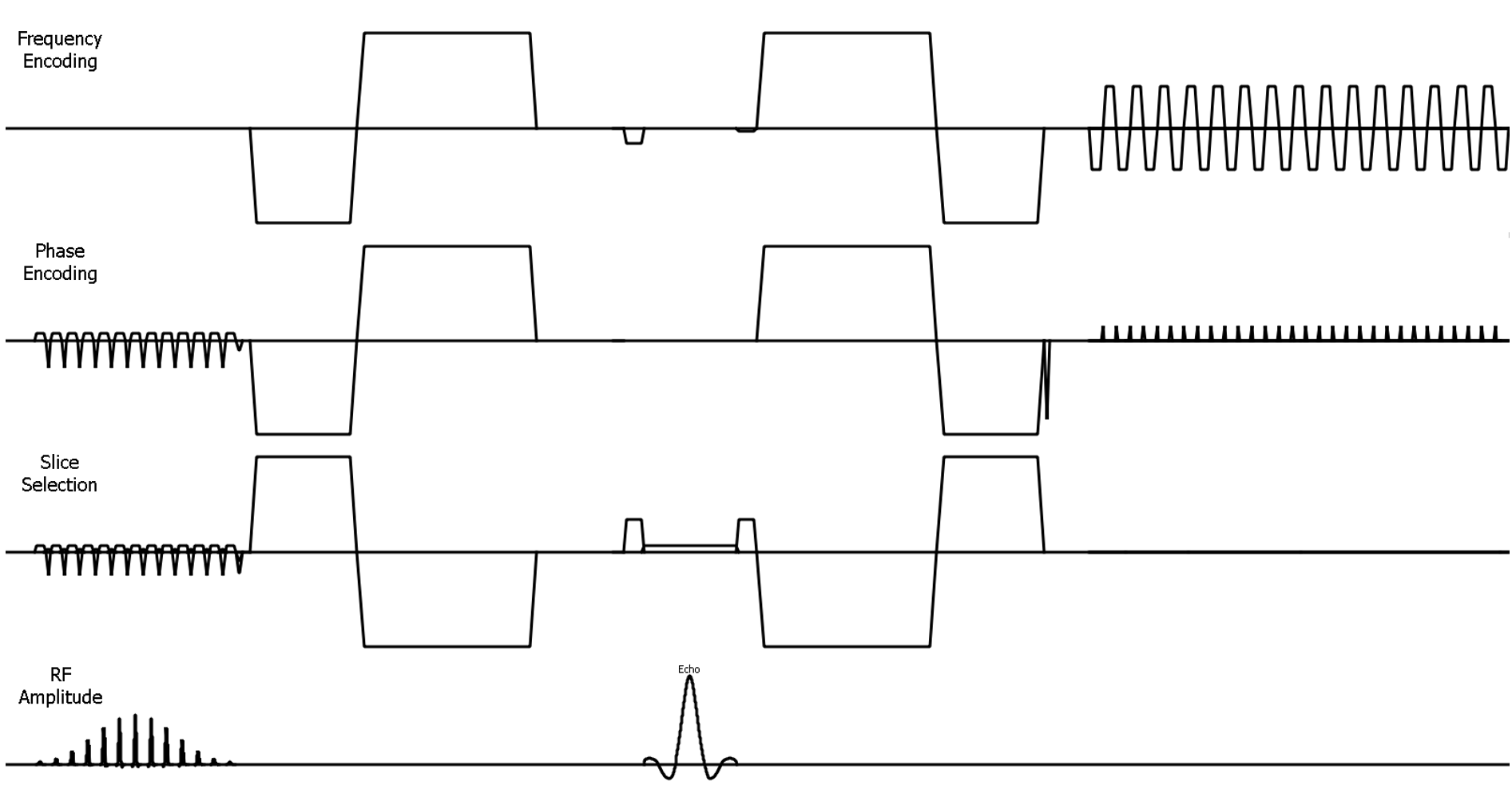
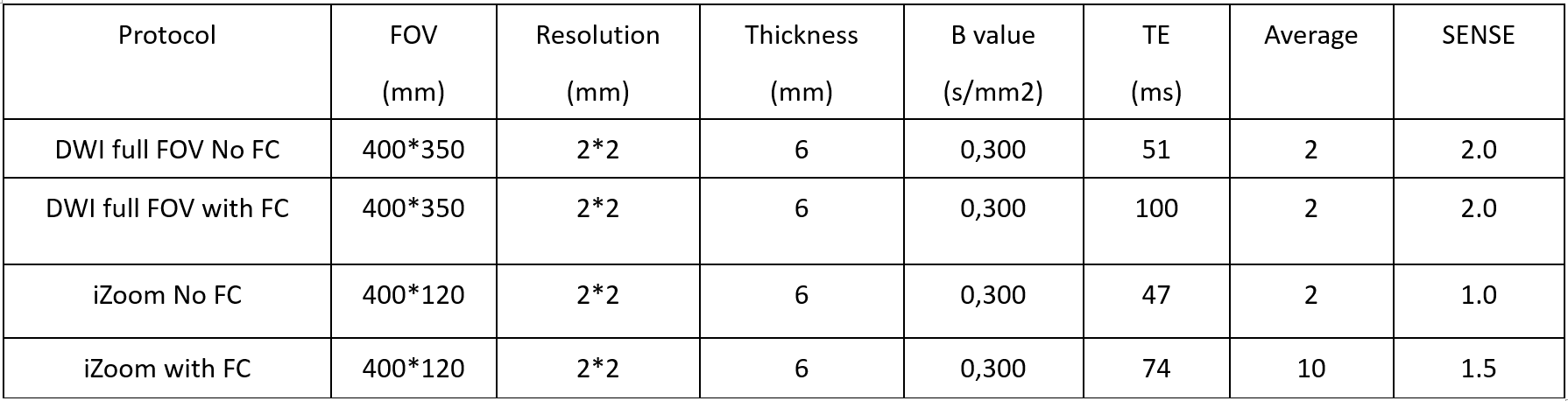
Since the distortion of DWI based on single-shot echo planar imaging is proportional to the FOV size, iZoom could be used to reduce the distortion and signal loss. Fig 1 shows the sequence diagram of our proposed scheme for cardiac diffusion imaging using a 2D RF pulse to excite a region of interest, which could contain only the cardiac region. Given the local excitation, it could reduce the motion from surrounding tissues, especially respiratory related motion artifacts. One remaining challenge is the local B0 field inhomogeneities which is caused by the chest cavity; SENSE could be used to reduce the distortion and signal void further. FC2nd could reduce the motion impact on cardiac diffusion, FC2nd was implemented as reference7. We combine the iZoom, SENSE and 2nd order compensated diffusion for the first time. To evaluate the feasibility of our proposal, we performed a pilot study on volunteers. DWI Full FOV with and without iZoom and with and without FC2nd were compared, both for diffusion weighted images and ADC maps. All scans were performed on a Philips 3.0T Elition system (Philips Healthcare, Best, Netherland) with a 32-ch torso and spine coil. Detailed scan parameters were summarized in Table 1.Results
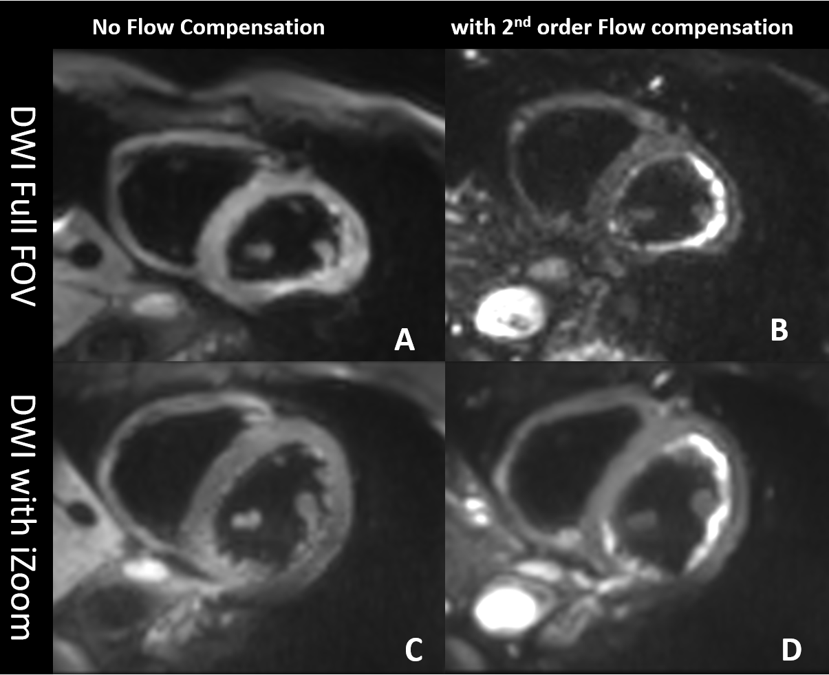
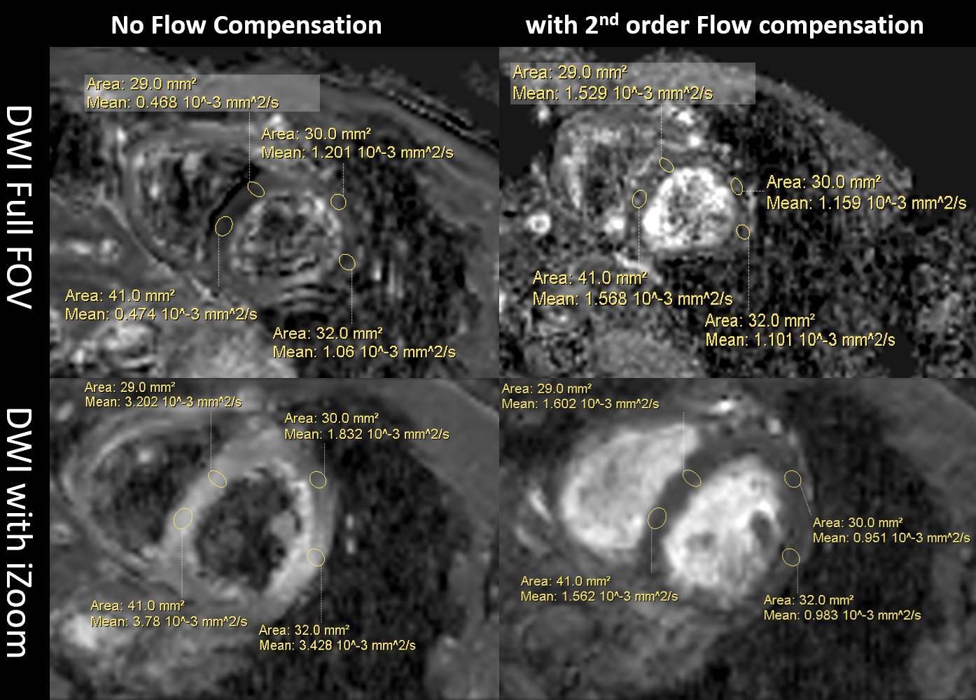
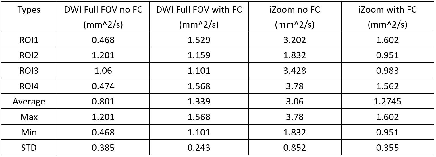
Fig. 2 shows the full FOV DWI images without FC2nd (A) has severe distortion and signal loss. Full FOV DWI with FC2nd (B) has less motion sensitivity, but it has much larger TE than without FC2nd (51ms to 101 ms). iZoom without FC2nd (C) has less distortion artifacts, but it still has a motion sensitivity iZoom with FC2nd has the best performance, which has shorter TE than full FOV DWI with FC2nd, but it still has a longer TE than iZoom without FC2nd (47ms to 74ms), which will reduce the SNR, to overcome this issue, we use average 10 vs original average 2 for it.Fig3 shows the ADC comparison for different schemes, it shows similar results, namely that full FOV DWI without FC2nd shows severe motion artifacts, full FOV DWI with FC2nd could reduce the motion artifacts, but has lower SNR and still has distortion and signal loss. iZoom without FC2nd shows non uniform ADC values, it may be caused by cardiac motion. ADC values by iZoom with FC2nd shows the best performance in these 4 schemes. In Table 2, the statistical comparison of 4 Region of Interest (ROI) also shows that ADC values with iZoom using SENSE and FC2nd has smaller standard variance. In conclusion, the ADC maps by iZoom with SENSE and FC2nd appear more robust.
Discussion and conclusions
The proposed method shows cardiac diffusion using the combination of iZoom, SENSE and 2nd order flow compensated diffusion gradient has better image quality, it is more robust for motion, and it has less distortion and signal loss than convention DWI with or without 2nd order flow compensated diffusion gradient. The preliminary results show that our proposed scheme could potentially further enhance the applicability of cardiac diffusion imaging. A clinical study should be performed to validate these results.Acknowledgements
No.References
1. Timothy GR, Marcel PJ, Himanshu B and David ES, Diffusion MRI in the heart, NBM 2017;30:e3426.
2. Sonia NV, Andrew S, Pedro F, Zohya K, Dudley P, David F, Cardiac Diffusion: Technique and Practical Applications. J. MAGN. RESON. IMAGING 2020;52(2):348-368;
3. Pruessmann KP, Weiger M, Scheidegger M, Boesiger P,SENSE: sensitivity encoding for fast MRI, Magn. Reason. Med., 1999;42(5):952-62.
4. Wu ZG, Zhang J, Fang WX, Huang F, B1 insensitive zoomed FOV imaging, ISMRM., 2015; 0953.
5. Christian TS, Constantin VD,Martin G,David A and Sebastian K, Second-Order Motion-Compensated Spin Echo Diffusion Tensor Imaging of the Human Heart, Magn Reson Med 2016;75:1669–1676.
6. Samo L,Filip S, Erica D, et. al, Motion‐compensated b‐tensor encoding for in vivo cardiac diffusion‐weighted imaging, NBM 2020;33:e4213.
7. Welsh CL, DiBella EVR, and Hsu EW, Higher-Order Motion-Compensation for In Vivo Cardiac Diffusion Tensor Imaging in Rats, IEEE Trans Med Imaging., 2015; 34(9): 1843–1853
Figures