4136
The application of REACT for central veins in patients with end-stage renal disease1The first affiliated hospital of Chongqing medical university, Chongqing, China, 2Philips Healthcare, Beijing, China
Synopsis
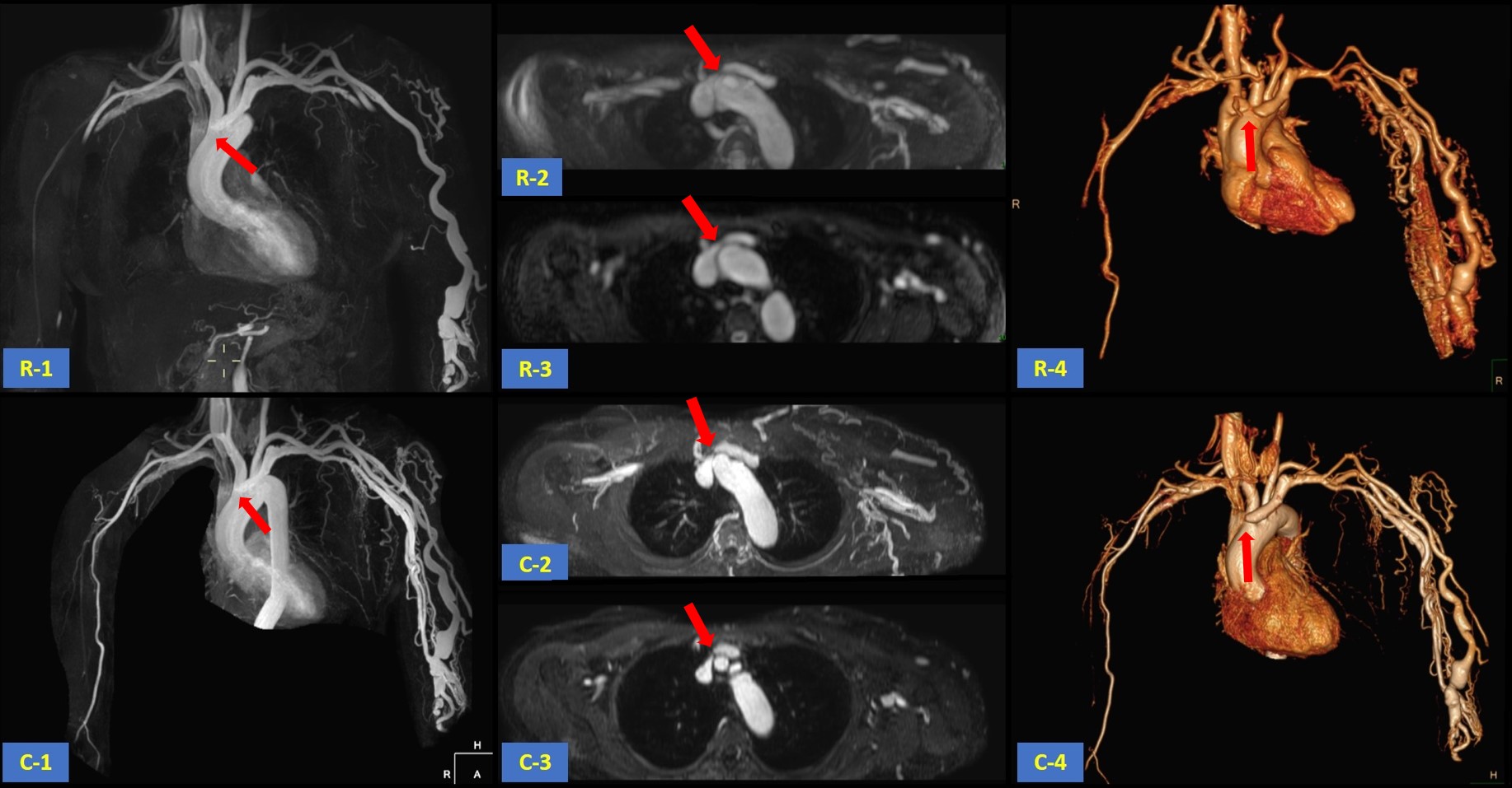
Relaxation-Enhanced Angiography without Contrast and Triggering (REACT) is a MR angiography technique without cardiac triggering, breath holding and contrast agent injection and is able to show robust blood-to-tissue contrast over multiple anatomies. This study aims to investigate the feasibility of REACT on central veins by comparing it with conventional CE-MRA and catheter angiography. Results showed that the image quality acquired from REACT was comparable to that from CE-MRA and there was no difference in central venous stenosis evaluated by REACT, CE-MRA and angiography.
Introduction
The central venous stenosis (CVS) of end-stage renal disease is a common complication of hemodialysis[1] and the gold standard for CVS imaging is catheter angiography, which is invasive. Contrast enhanced MR evaluation of the central veins has been reported to be extremely sensitive and specific in the detection of stenoses and occlusions[2]. But the gadolinium-based contrast agents as a causative factor may introduce nephrogenic systemic fibrosis, especially for ESRD patients. In recent years, MR venography with the blood pool specific agent acquired better image quality and can eliminate the risk of NSF, but the price is high. Relaxation-Enhanced Angiography without Contrast and Triggering (REACT) is a MRA technique without contrast and has been evaluated in pelvic venous vessels and pulmonary vasculature[3,4]. However, the evaluation of REACT in central venous stenosis has not been reported yet. Therefore, this study aims to assess the feasibility and diagnostic accuracy of REACT in the evaluation of central thoracic venous occlusive disease and compare it with CE-MRA and catheter angiography.Materials and Methods
The examinations were conducted on 3.0T MR system (Ingenia 3.0T, Philips Healthcare, the Netherlands). 40 consecutive patients (14 women and 26 men; age range, 30-85 years; mean age, 60.90years ± 12.81 [SD]) of end-stage renal disease with known or suspected central thoracic venous occlusive disease who had undergone REACT and CE-MRA imaging were absorbed in our group. Catheter angiography was performed within 1 week after MRA. REACT and CE-MRA images were evaluated in separate reading sessions by two independent radiologists for image quality and level of confidence and degree of venoocclusive disease. The accuracy of REACT and CE-MRA was assessed with catheter angiography as the standard of reference.Results
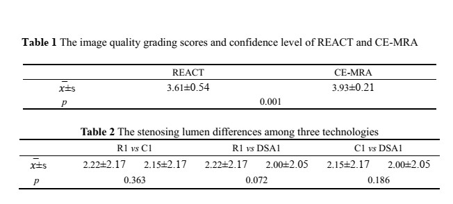
There was a high interobserver agreement for both methods of imaging at the central veins (ICC≥0.921). The image quality grading scores and confidence level of CE-MRA is a little better than that of REACT (Tabel1). The evaluation results in stenosis and occlusion of vein by REACT and CE -MRA had no difference from those by angiography (Table 2).Discussion
In our study, there is no statistical difference in the evaluation of central venous stenosis among REACT, CE-MRA and catheter angiography. The image quality acquired from REACT was not as well as CE-MRA, but was enough for clinical application. For some patients with implanting stent, REACT image also can be accurately assessed venous stenosis.Conclusion
Without any contrast media, REACT appears to be a feasible method for venous occlusive disease and to provide a high sensitivity comparable to CE-MRA.Acknowledgements
noneReferences
1. Krishna V N, Eason J B, Allon M. Central Venous Occlusion in the Hemodialysis Patient[J]. American Journal of Kidney Diseases the Official Journal of the National Kidney Foundation, 2016, 68(5):803-807.
2. Kim C Y, Mirza R A, Bryant J A, et al. Central veins of the chest: evaluation with time-resolved MR angiography[J]. Radiology, 2008, 247(2):558-66.
3. Terwolbeck M N, Zhang S, Bode M, et al. Relaxation-enhanced angiography without contrast and triggering (REACT) for pelvic MR venography in comparison to balanced gradient-Echo and T2-weighted spin-Echo techniques[J]. Clinical Imaging, 2021.
4. Pennig L, Wagner A, Weiss K, et al. Imaging of the pulmonary vasculature in congenital heart disease without gadolinium contrast: Intraindividual comparison of a novel Compressed SENSE accelerated 3D modified REACT with 4D contrast-enhanced magnetic resonance angiography[J]. Journal of Cardiovascular Magnetic Resonance, 2020, 22.
Figures