4134
Comparative analysis of the imaging quality in magnetic resonance coronary angiography with 3D mDIXON and B-TFE sequence on 3T
Gang Zhang1, Wei Xing2, Tingting Li2, Yan Zheng1, Ying Huang2, Junjing He2, and Xiuzheng Yue3
1The First Affiliated Hospital of Henan University of CM, Zhengzhou, China, 2Department of Magnetic Resonance, The First Affiliated Hospital of Henan University of CM, Zhengzhou, China, 3Department of Magnetic Resonance, Philips Healthcare, Zhengzhou, China
1The First Affiliated Hospital of Henan University of CM, Zhengzhou, China, 2Department of Magnetic Resonance, The First Affiliated Hospital of Henan University of CM, Zhengzhou, China, 3Department of Magnetic Resonance, Philips Healthcare, Zhengzhou, China
Synopsis
Due to the narrow diameter, tortuous route of coronary arteries, and the interference of respiration and heartbeats during scanning, MR coronary angiography (MRCA) remains challenge. In 3.0T MR systems, an improved 3D Balanced Turbo Field Echo (B-TFE) sequence is often used for MRCA[1,2], however, 3D mDIXON sequences have been applied for clinical application recently.[3,4] In this study, the subjective imaging quality, objective imaging quality and image authenticity indexes of MRCA with two imaging sequences were compared to analysis. Results showed that B-TFE had better subjective evaluation than mDixon, but mDIXON had better objective quality evaluation for SNR and CNR.
Introduction
Magnetic resonance angiography (MRA), as a non-invasive vascular imaging method, has been widely applied in clinical practice, providing rich imaging data for the diagnosis of vascular diseases. However, due to the narrow diameter, tortuous route of coronary arteries, and the interference of respiration and heartbeat during scanning, MRCA has always been a very difficult and challenging task. An improved 3D B-TFE sequence is often used for MRCA in 3.0T MR systems[1,2]. B-TFE sequence, which has high scanning speed, slight motion artifact and high blood signal[5], is suitable for MRCA and has become a common scanning method[6]. However, B-TFE is often not ideal in fat suppression, relatively sensitive to the uneven B0 field strength so that the image quality will be spoiled to a large extent. mDIXON has high signal to noise (SNR), uniform fat suppression, and high scanning speed combined with compressed SENSE technology. In recent years, mDIXON has been applied to MRCA more than before[3,4].Therefore, in order to choose more appropriate imaging techniques, we conducted a comparative study of MRCA with two different methods.Methods
12 healthy young volunteers(7 males and 5 females, aged range from 21 to 22) were scanned using B-TFE sequence and mDIXON sequence on 3.0T scanner (Ingenia CX, Philips, Netherlands) with a 32-channel coils in May 2021. Scaning parameters are recommended by Philips as shown in table 1.The coronary artery images were post-processed in ISP9 workstation (Philips, Netherlands) with surface reconstruction, maximum intensity projection, VR, etc, and evaluated with the original axial bitmap.Referring to the coronary artery segmentation method of the American College of Cardiology, this study mainly analyzed left main artery(LM), right coronary atery (RCA) including proximal, middle and distal segments,The left anterior descending branch(LAD) including proximal and middle segments, and the left circumflex branch (LCX) including proximal and middle segments.Subjective image quality was evaluated by referring to the Likert scoring method from 1point to 4 points. Among the above 8 study segments, 5 segments with scores greater than 3 were classified as qualified image quality.SNR= vascular signal intensity/background noise, CNR= (vascular signal intensity value - surrounding tissue signal intensity value)/background noise. Observe each segment of coronary artery, subjective diagnosis was divided into two types: no obvious stenosis, obvious stenosis.The measurement data were expressed as mean ± standard deviation.Wilcoxon rank sum test was used for subjective score data of coronary artery image quality, and T test was used for objective score data of SNR and CNR. P<0.05 was considered statistically significant.Results
11 healthy young volunteers was included with qualified MRCA image quality. One failed due to claustrophobia.The heart rate of 11 healthy young volunteers was 53-92 beats/min, with an average of 65 beats/min.The subjective evaluation mean value of B-TFE in coronary artery imaging was higher than that of mDixon sequence, but the difference was not statistically significant.Shown in table 2.SNR and CNR scores of mDIXON sequence were higher than those of B-TFE sequence, and the difference was statistically significant. As shown in table 3.None of the 11 healthy adults underwent DSA or CTA examination.Coronary MRA abnormalities were mainly manifested as vascular stenosis, the number of occurrences is shown in Table 4.Discussion
B-TFE can eliminate phase interference and retain magnetic resonance signal to the maximum extent[5].mDIXON can stably complete a wide range of water lipid separation and achieve good fat suppression[7].The results showed that the application of B-TFE and mDIXON sequences in MRCA can achieve satisfy imaging results, as shown in figure 1.11 from 12 healthy young volunteers complete the examination, with the subjective scores mostly above 3 points. mDIXON had blurred coronary artery edge and lower subjective quality score, while B-TFE had clear coronary artery edge and higher subjective quality score.B-TFE[5] utilized every TI interval to increase the ratio of T2/T1 relaxation time indicating that blood vessels have a very high contrast with soft tissue, and the image SNR is high. However, our examination showed that SNR and CNR of MRCA of mDixon sequence had relatively high absolute values, and the difference between the two sequences was statistically significant, which may be related to the characteristics of uniform fat suppression.MRCA sequences of mDIXON and B-TFE also showed a large number of vascular imaging abnormalities, and their manifestations were diverse, suggesting that the two examination methods need to be greatly improved.In addition, the sample size is small, and MRCA lacks the gold standard control.These may have had an impact on the resultsConclusion
3T MRCA using 3D B-TFE or mDIXON sequences can be used for coronary artery images. mDIXON sequence is a powerful alternative to B-TFE sequence. In addition, compressed SENSE technology can be used in mDIXON sequence and the scanning time is significantly shortened, suggesting that mDIXON sequence is potentially a promising and valuable MRCA imaging technique.Acknowledgements
No acknowledgement found.References
1. Nezafat M,Henningsson M, Ripley DP, et al. Coronary MR angiography at 3T: fat suppression versus water-fat separation.Magn Reson Med Sci. 2009;8:55-63. 2. Inoue K, Maeda M, Umino M, et al. Cervical carotid plaque evaluation using 3D T1-weighted black-blood magnetic resonance imaging: Comparison of turbo field-echo and turbo spin-echo sequences. Eur J Radiol. 2016;85:1035-1039. 3. Kourtidou S, Jones MR, Moore RA, et al. mDixon ECG-gated 3-dimensional cardiovascular magnetic resonance angiography in patients with congenital cardiovascular disease. J Cardiovasc Magn Reson.2019;21:52. 4. Mesropyan N, Isaak A, Dabir D, et al. Free-breathing high resolution modified Dixon steady-state angiography with compressed sensing for the assessment of the thoracic vasculature in pediatric patients with congenital heart disease. J Cardiovasc magn reson.2021;23:117. 5. Kawamitsu H, Kaji Y, Sugimura K. Magnetic resonance angiography of the renal arteries using three-dimensional balanced turbo field-echo sequence with progressive spin saturation. Magn Reson Mde Sci. 2005;4:43-46. 6. Kawada H, Goshima S, Sakurai K, et al. Utility of noncontrast magnetic resonance angiography for aneurysm follow-up and detection of endoleaks after endovascular aortic repair. Korean J Radiol. 2021;22:513-524. 7. Weiss KJ, Eggers H, Stehning C, et al. Feasibility and robustness of 3T magnetic resonance angiography using modified dixon fat suppression in patients with known or suspected peripheral artery disease. Front Cardiovasc Med.2020;7:549392.Figures
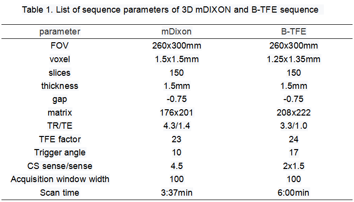
Table 1. List of sequence parameters of 3D mDIXON and B-TFE sequence
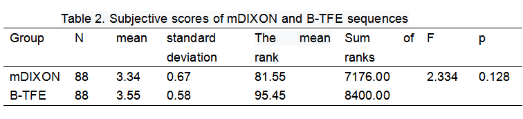
Table 2. Subjective scores of mDIXON and B-TFE sequences
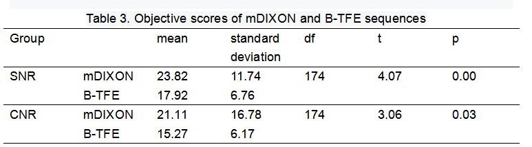
Table 3. Objective scores of mDIXON and B-TFE sequences
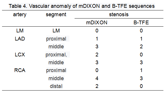
Table 4. Vascular anomaly of mDIXON and B-TFE sequences
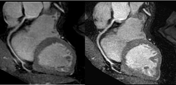
2. Figure 1. Title: angiography of RCA. Lgend: B-TFE image is displayed to the left and mDIXON image to the right. Both of the proximal segments of RCA appear abnormity with fuzzy edge and decreased signal which may be misdiagnosed as stenosis,mDIXON image has more fuzzy coronary artery edge and lower signal.
DOI: https://doi.org/10.58530/2022/4134