4132
PANDA: Simultaneous Phase-Contrast Bright and Dark-blood Angiography for Improvement of Carotid Plaque Assessment
Daichi Murayama1, Takayuki Sakai1, Masami Yoneyama2, Jihun Kwon2, and Shigehiro Ochi1
1Department of Radiology, Eastern Chiba Medical Center, Chiba, Japan, 2Philips Japan, Tokyo, Japan
1Department of Radiology, Eastern Chiba Medical Center, Chiba, Japan, 2Philips Japan, Tokyo, Japan
Synopsis
TOF-MRA sometimes cannot delineate the vessel clearly depending on the vessel orientation. Additionally, insufficient fat suppression can occur in FS-T1-VISTA, it may obscure the plaque depiction. we have developed a new sequence, termed Phase-contrast ANgiography with Dark-blood Application (PANDA), which can simultaneously acquire bright-blood image (MRA) and vessel-wall images (VWI) using the phase contrast method. There was no difference in the visualization ability of carotid plaque between PANDA-VWI and FS T1 VISTA. PANDA-MRA better visualized around the aorta compared to TOF-MRA. PANDA is useful method for simultaneous acquisition of MRA and VWI images.
Purpose
The current standard methods for carotid plaque assessment are time-of-flight (TOF) MRA and fat-suppressed T1-weighted 3D Turbo Spin-Echo (FS-T1-VISTA).1,2) However, TOF-MRA sometimes cannot delineate the vessel clearly depending on the vessel orientation. Additionally, insufficient fat suppression can occur in FS-T1-VISTA, it may obscure the plaque depiction. To overcome these problems of current techniques, we have developed a new sequence, termed Phase-contrast ANgiography with Dark-blood Application (PANDA), which can simultaneously acquire bright-blood image (MRA) and vessel-wall images (VWI) using the phase contrast method. [Fig.1] In this study, we investigated the usefulness of PANDA, simultaneous acquisition of MRA and VWI images, to improve the carotid plaque assessment.Methods
All subjects were examined with a 3.0T whole-body clinical system (Ingenia CX, Philips Healthcare). MRA (PANDA-MRA) was created from the phase image, and VWI (PANDA-VWI) was created by subtracting the phase image from the magnitude image. [Fig.2] We performed conventional methods (TOF-MRA and FS-T1-VISTA) and PANDA imaging in ten patients who had a carotid plaque in follow-up examination. To quantitatively evaluate carotid plaque visualization, we computed Contrast Ratio (CR) between the plaque and sternocleidomastoid muscle for both FS-T1-VISTA and PANDA-VWI. Imaging parameters for FS-T1-VISTA were; Coronal, voxel size=1.0×1.0×1.0mm3, FOV=250×250mm2, 150 slices, fat suppression=SPAIR, TR=600ms, TE=11ms, SENSE factor=2 and total acquisition time=4min30s. Imaging parameters for TOF-MRA were; Transverse, voxel size=1.0×1.0×1.0mm3, FOV=200×200mm2, 220 slices, TR=23ms, TE=3.5ms, Flip Angle=20, SENSE factor=2.5 and total acquisition time=4min32s. Imaging parameters for PANDA were; Coronal, voxel size=1.0×1.0×1.6mm3, FOV=250×250mm2, 150 slices, fat suppression=SPAIR, TFE prepulse = saturate, TR=8ms, TE=2.6ms, Flip Angle=10, TFE factor=6, SENSE factor=4 and total acquisition time=4min-7min(HR50-100).Results and Discussions
The CR of carotid plaque was not significantly different between PANDA-VWI and FS-T1-VISTA. [Fig.3] This suggests that there is no difference in the visualization ability of carotid plaque between PANDA-VWI and FS-T1-VISTA. In the clinical image, PANDA-MRA better visualized around the aorta compared to TOF-MRA. [Fig.4]Conclusion
PANDA improves the depiction of arteries especially around the aorta and is a useful method for simultaneous acquisition of MRA and VWI images.Acknowledgements
No acknowledgement found.References
1) Stroke. 2011; 42: 3132-3137
2) K inoue et al. EJR 2016; 85: 1035-1039
Figures
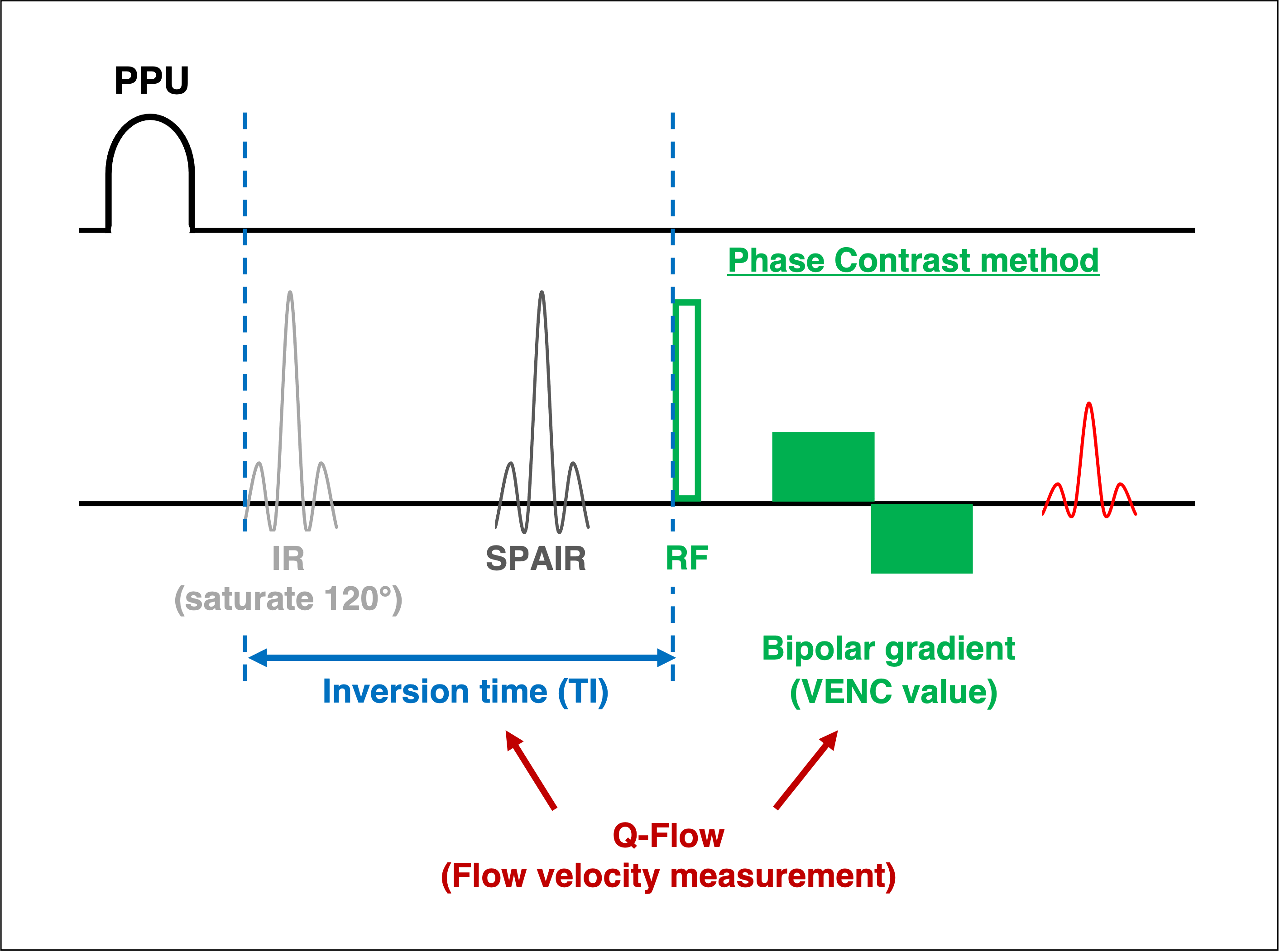
Fig.1 Acquisition scheme of PANDA (Phase-contrast ANgiography with Dark-blood Application). The basic sequence was the Phase-Contrast method using PPU synchronization. The prepulse using saturation recovery enhances T1 contrast. The Q-flow was used for analyzing the peak flow velocity and the maximum flow velocity of the common carotid arteries. The peak flow velocity was set as TFE prepulse delay and the maximum flow velocity was set as PC velocity (VENC).
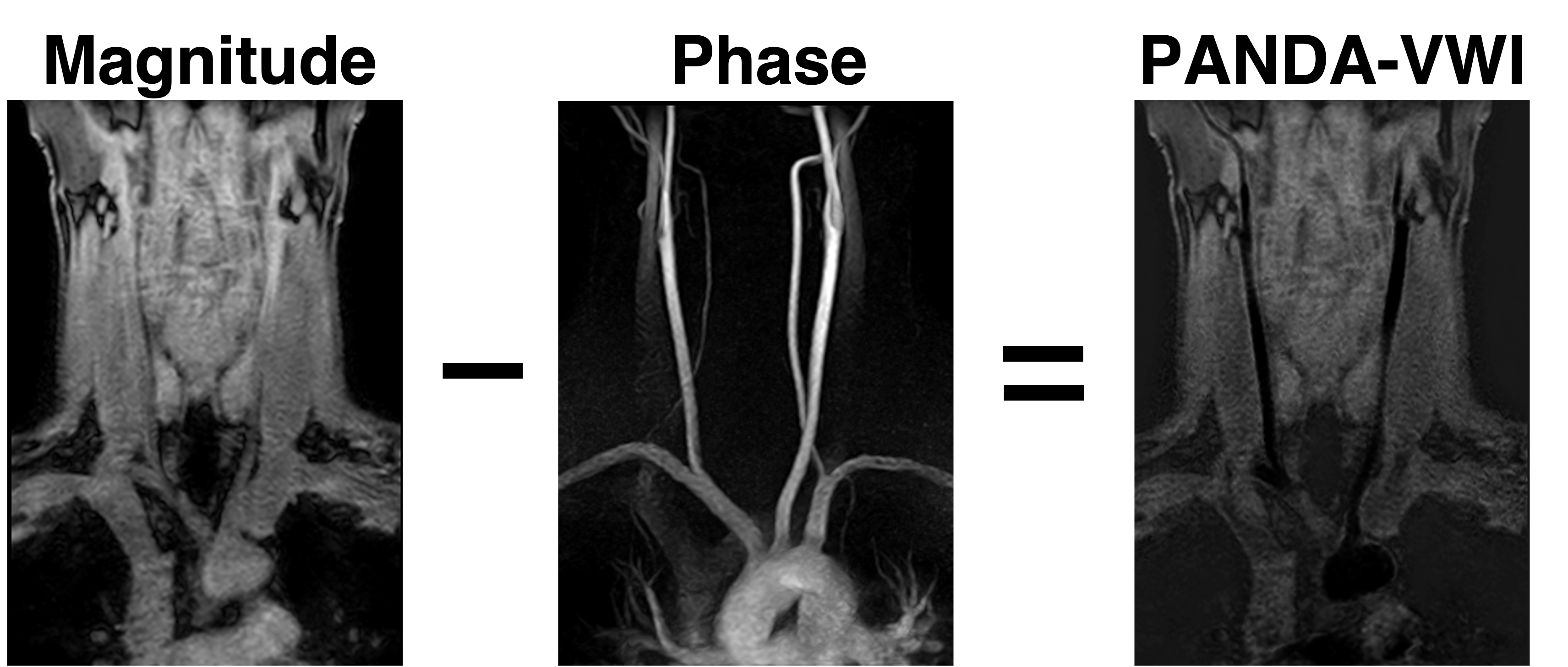
Fig.2 Vessel-wall images (PANDA-VWI) were created by subtracting the phase image from the magnitude image. In this process, weighting the phase images improved the accuracy of the subtraction.
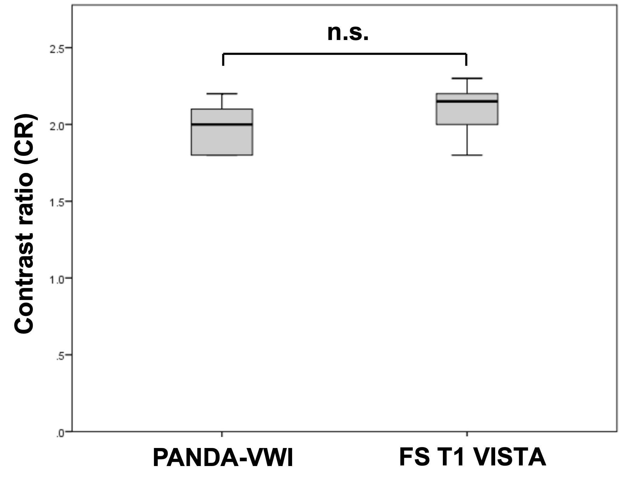
Fig.3 Contrast Ratio (CR) between the plaque and sternocleidomastoid muscle for PANDA-VWI and FS-T1-VISTA.

Fig.4 A 56-year-old woman who had a carotid plaque in follow-up examination. (A) Conventional methods(B) PANDA (Phase-contrast ANgiography with Dark-blood Application)
DOI: https://doi.org/10.58530/2022/4132