4124
Deep learning based whole heart T2-weighted dark blood imaging in a single breath hold1Department of Radiology, Tongji Hospital, Tongji Medical College, Huazhong University of Science and Technology, Wuhan, China, 2United Imaging Healthcare, Shanghai, China, 3UIH America, Inc., Houston, TX, United States
Synopsis
Cardiovascular magnetic resonance (CMR) T2-weighted dark blood (T2W-DB) imaging has great diagnostic value for detecting myocardial edema. In this study, a novel deep learning based acceleration framework (AI-assisted Compressed Sensing, ACS) was applied to a single-shot T2W-DB sequence for single breath-hold whole heart (9 slices) imaging. Both quantitative and qualitative assessment of the images suggested that the ACS T2-DB sequence offered better image quality with greatly reduced total scan time and the simplified scanning workflow.
Introduction
Conventional 2D dual inversion recovery (DIR) fast spin echo (FSE) sequence for cardiac T2W-DB morphological imaging acquires k-space data in segments across multiple cardiac cycles, which needs multiple breath holds. In clinical scenarios, irregular heart rate and breath holding difference tend to induce motion artifact. By incorporating an Artificial Intelligence (AI) module based on deep learning neural network for information recovery and artifact suppression 1, compressed sensing (CS) can achieve higher acceleration factor than parallel imaging while ensuring consistent image quality. In this study, a novel AI-assisted compressed sensing (ACS) 2 acceleration strategy was applied to the single-shot T2W-DB sequence. In addition, because long echo trains are required for single-shot acquisition, the variable flip angle strategy designed according to the myocardial T1 and T2 relaxation times is used for suppressing myocardial inhomogeneity and blurs. The purpose of this study was to investigate the clinical feasibility of the ACS single-shot T2W-DB sequence compared to conventional T2W-DB through quantitative and qualitative image assessment.Methods
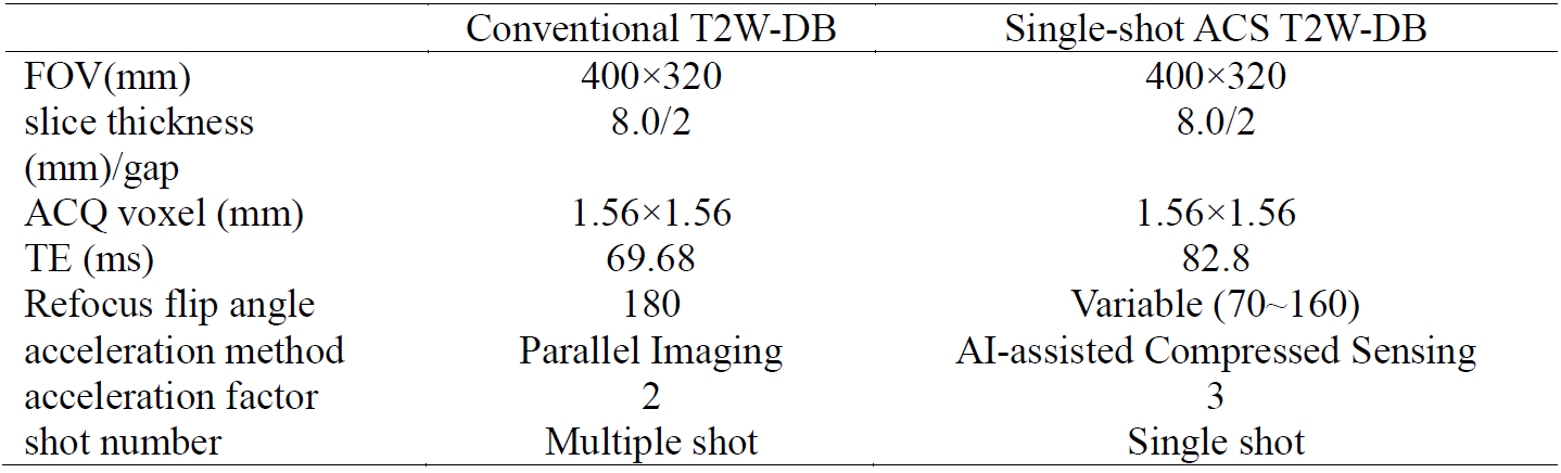
Subjects: 28 patients (21 males, 43±15 years) and 5 healthy volunteers (5 males, 29±4 years) were prospectively recruited with informed consent obtained in this study. Clinical indications included ischemic and non-ischemic cardiomyopathy, cardiac valve disease, myocarditis, and other diseases that were not classified.MRI scan: Cardiac Magnetic Resonance imaging (CMR) was performed on a 3.0-Tesla scanner (uMR790, United Imaging Healthcare, Shanghai, China), equipped with 12×2-channel cardiac coil. Conventional T2W-DB and single-shot ACS T2W-DB sequences were applied for heart imaging in short-axis view. The details of imaging parameters are shown in Table 1.
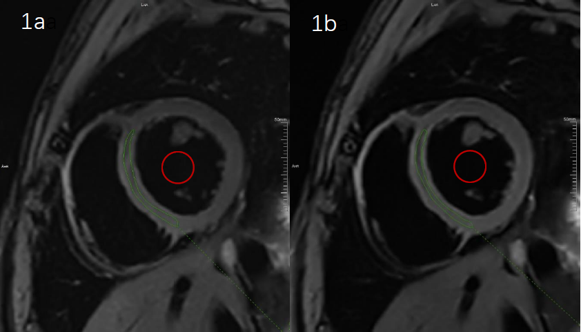
Image analysis: All imaging datasets were evaluated by Dicom format. The overall image quality and blood pool suppression were assessed by two cardiovascular radiologists using a 5-point scale of Likert score (1 = non-diagnostic, 2 = poor, 3 = satisfactory, 4 = good, 5 =excellent) independently. The visual quality of free wall of right ventricle (RV), free wall of left ventricle (LV) and interventricular septum were evaluated. Interventricular septum ROI and blood pool ROI were manually drawn by two radiographers on the middle slice of the heart, as Figure1 shows. Signal-to-noise ratio (SNR) and contrast-to-noise ratio (CNR) were then determined as follows:
$$SNR=\frac{Mean_{myocardium}}{SD_{myocardium}}$$
$$CNR=\frac{Mean_{myocardium}-Mean_{bloodpool}}{SD_{bloodpool}}$$
Sharpness Measurement: Image sharpness was measured by calculating the ratio of major frequency components in frequency domain 3.
Statistical analysis: Statistical analysis was performed using SPSS (version 23.0, Chicago, IL). The two observer Likert scores and all measurements were averaged prior to analysis, the measurement data and grade data between the conventional T2W-DB and the single-shot T2W-DB with ACS were assessed using paired wilcoxon signed-rank test and paired t-test, respectively. Kendall's W was used to assess inter-observer agreement of scoring data. ICC was used to assess inter-observer agreement of continual data. P<0.05 was considered as statistically significant.
Results
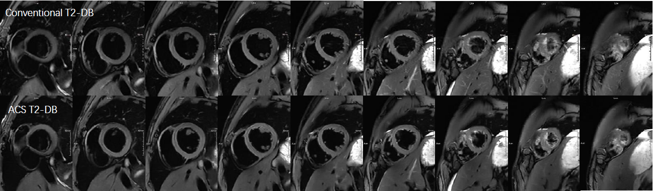
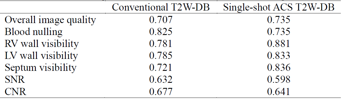
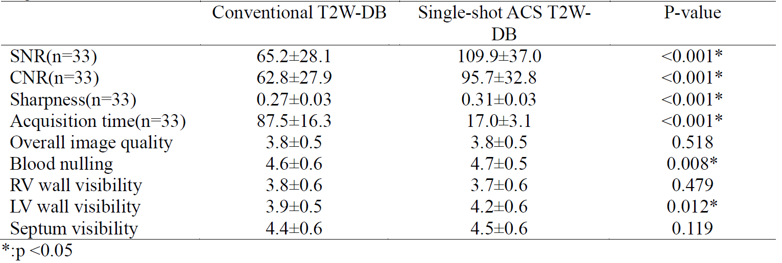
Compared to conventional T2W-DB, the acquisition time of single-shot ACS T2-DB was significantly reduced (87.5±16.3s vs 17.0±3.1s, p<0.001). Single-shot ACS T2-DB yielded significant higher SNR, CNR and sharpness (p<0.05 for all) than conventional T2-DB, while both methods showed no significant difference on overall image quality, RV and LV wall visibility as well as septal wall visibility (p> 0.05 for all). In terms of dark blood effect, single-shot ACS T2-DB was determined to have better performance than conventional T2-DB. The scores of the two cardiovascular radiologists and the results measured by the two radiographers were in good agreement (Table 2). Quantitative analysis and qualitative scoring data are shown in Table 3. The whole-heart images comparison of conventional T2-DB and single-shot ACS T2-DB are shown in Figure 2.Conclusion
Recently, deep learning-based acceleration methods with high acceleration factor have exhibited great potentials in MR imaging, however, few have been employed for CMR T2W-DB imaging. In this study, the single-shot ACS T2W-DB sequence acquires k-space data of one slice during one cardiac cycle, significantly reducing patient burden caused by repeated breath holding and the motion artifacts induced by irregular heart rate. The whole heart coverage (9 slices) T2W-DB imaging is completed within a single breath-hold, offering high quality images with greatly shortened acquisition time and simplified scanning workflow.Acknowledgements
No acknowledgement found.References
1.Knoll F, Murrell T, Sriram A, et al. Advancing machine learning for MR image reconstruction with an open competition: Overview of the 2019 fastMRI challenge. Magn Reson Med. 2020;(January):mrm.28338.
2.Renkuan Zhai, et al. Intelligent Incorporation of AI with Model Constraints for MRI Acceleration. Proc. Intl. Soc. Mag. Reson. Med. 27 (2021): 1760
3.De K, Masilamani V. Image Sharpness Measure for Blurred Images in Frequency Domain[J]. Procedia Engineering, 2013, 64:149-158
Figures