4110
A 24-channel Array for Awake NHP Imaging at 10.5 Tesla1Radiology, University of Minnesota, Minneapolis, MN, United States, 2Neuroscience, University of Minnesota, Minneapolis, MN, United States
Synopsis
A 24-channel receiver – dual channel transmitter array for awake monkey imaging at 10.5 Tesla is presented. This was achieved by adding an 8-channel extension to a previously built 16-channel receive array. The new layout does not require any user adjustments and fully supports monkey imaging.
Introduction
Gains in parallel imaging are made possible by increases in signal-to-noise ratio (SNR) at ultra-high fields (UHF) and high density coil arrays. For primate imaging where the imaging volume is significantly smaller these gains become increasingly important. In support of this, several innovative coil designs have been presented, predominantly for anesthetized animal imaging1-4. Experimental difficulty increases significantly at UHF, particularly for awake animal imaging. In recent years fewer designs have been presented for these awake UHF imaging studies, which in addition to the receivers requires integration of transmitters. Here the principle of integrated loop transmitters with receive arrays5-7 is a particularly promising compromise to achieve the required combination of transmission and reception with high channel count. Utilizing this principle in related prior work, we presented a 16 channel8 awake primate array and developed miniaturized front-end technology for up to 128 channel reception9.Methods
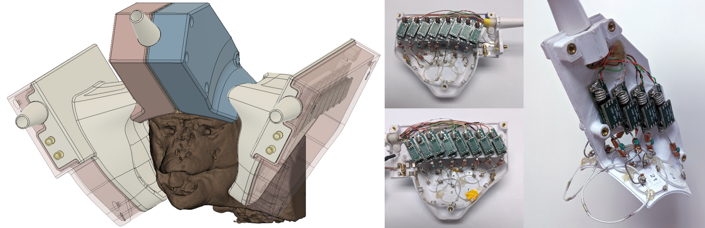
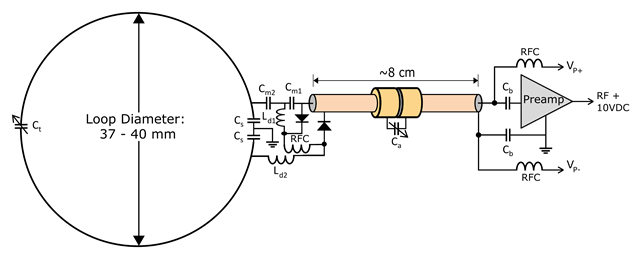
The complete array assembly is composed of three form-fitted housings each containing eight integrated receivers made of loop coils with diameters ranging from 30 to 40 mm. The two side housings each contain one 13 cm x 8.5 cm transmitter loop. The receive channels follow the circuit topology shown in figure 2. All receive loops include a 5 mm x 5 mm matching circuit board, are capacitor segmented twice and are overlap decoupled with neighboring loops. The exceptions here are two 40 mm diameter, non overlapped eye loops which are capacitor segmented four times. For improved decoupling and overall SNR each coil was connected via semi-rigid coaxial cables of average length 6 cm to WMA447D preamplifiers (WanTcom, Chanhassen, MN, USA) tuned to 447 MHz. Cable traps in each receiver path suppressed common mode currents and were needed for overall stable operation. All coil housings are composed of PETG and 3D printed in-house using FDM (Fusion3 F410, Greensboro, NC, USA). The coil housings were designed to be fully enclosed, allow for visual presentation, and easily repositionable for subjects of different sizes.Results and Discussion
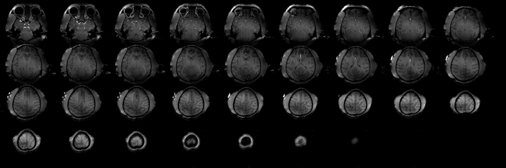
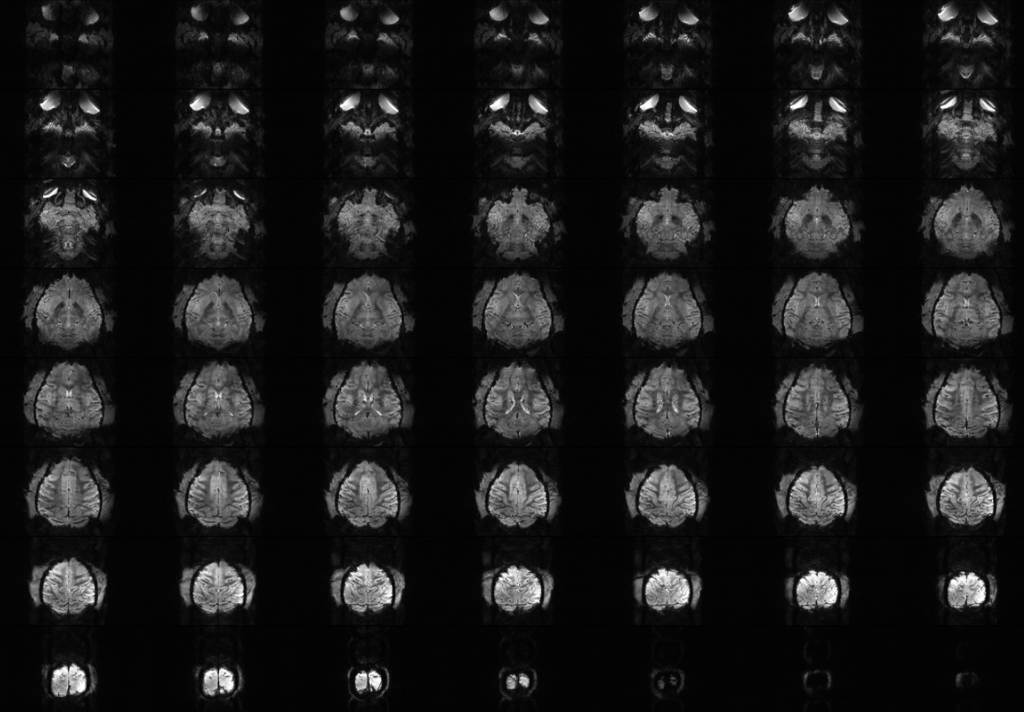
The matching for all receiver loops averaged about -14 dB and was -8 dB in the worst case loading situation. Coupling between nearest neighbor receive loops within each housing was around -9.5 dB at worst prior to preamplifier decoupling. Coupling between receive loops in different housings was around -14.5 dB at worst and the coupling of the closest receive loops in different housings averaged -21 dB. Imaging results from the coil are shown in figures 3 and 4 and indicate that excellent brain coverage is achievable at 10.5T even when using only two transmitter loops. In future work we will evaluate possible advantages of a third transmitter loop integrated into the middle shell this in greater detail.Acknowledgements
This research was funded by NIH BTRC P41 EB027061, U01 EB025144, P30 NS076408, NIH S10 RR029672 grants.References
1. Janssens, T., Keil, B., Serano, P., Mareyam, A., McNab, J. A., Wald, L. L. & Vanduffel, W. A 22-channel receive array with Helmholtz transmit coil for anesthetized macaque MRI at 3 T. (2013) NMR Biomed 26, 1431-1440.
2. Gilbert, K. M., Gati, J. S., Barker, K., Everling, S. & Menon, R. S. Optimized parallel transmit and receive radiofrequency coil for ultrahigh-field MRI of monkeys. (2016) Neuroimage 125, 153-161.
3. Li, M., Li, Y., Jin, J., Yang, Z., Zhang, B., Liu, Y., Song, M., Freakly, C., Weber, E., Liu, F., Jiang, T. & Crozier, S. A dedicated eight-channel receive RF coil array for monkey brain MRI at 9.4 T. (2020) NMR Biomed 33, e4369.
4. Lagore, R. L., Moeller, S., Zimmermann, J., DelaBarre, L., Radder, J., Grant, A., Ugurbil, K., Yacoub, E., Harel, N. & Adriany, G. An 8-dipole transceive and 24-loop receive array for non-human primate head imaging at 10.5 T. (2021) NMR Biomed 34, e4472.
5. Adriany, G., Schillak, S., Waks, M., Tramm, B., Roebroeck, A., Formisano, E., DeMartino, F., Yacoub, E. S. & Vaughan, J. T. A Flexible 4 Ch. Transmit / 16ch. Receive Auditory Cortex Array for HiRes fMRI at 7 Tesla. in Proc. Int. Soc. Reson. Med. (2013). 2734.
6. Gilbert, K. M., Schaeffer, D. J., Zeman, P., Diedrichsen, J., Everling, S., Martinez-Trujillo, J. C., Pruszynski, J. A. & Menon, R. S. Concentric radiofrequency arrays to increase the statistical power of resting-state maps in monkeys. (2018) Neuroimage 178, 287-294.
7. Zhang, X., Zhang, J., Gao, Y., Qian, M., Qu, S., Quan, Z., Yu, M., Chen, X., Wang, Y., Pan, G., Adriany, G. & Roe, A. W. A 16-Channel Dense Array for In Vivo Animal Cortical MRI/fMRI on 7T Human Scanners. (2021) IEEE Trans Biomed Eng 68, 1611-1618.
8. Jungst, S., Lagore, R., Metha, P., Grier, M. D., Heilbronner, S. R., Adriany, G., Hayden, B. Y. & Zimmermann, J. Awake rhesus macaque imaging at ultra high field strength (10.5T). in Society of Neurosience. 174.
9. Lagore, R., Jungst, S., Radder, J., Auerbach, E., Moeller, S., Grant, A., DelaBarre, L., Waks, M., Van de Moortele, P.-F., Adriany, G. & Ugurbil, K. A 128-channel receive array for 10.5T human head imaging. in Proc Intl Soc Mag Reson Med 29. 177.
Figures