4106
Rendering MRI Coils Less Visible in MR images1Department of Radiology, Weill Cornell Medicine, New York, NY, United States, 2Department of Radiology and Imaging, Hospital for Special Surgery, New York, NY, United States, 3Department of Psychology, New York University, New York, NY, United States, 4GE Healthcare, Aurora, OH, United States
Synopsis
It is commonly undesirable to see an RF coil in MR images. Novel research into innovative coil designs often involves the use of new materials that can be visible in MR images. In this abstract, we address this limitation by doping the coil substrate with Magnevist, drawing from a little-known phenomenon of signal hypointensity via T2-shortening for large concentrations of gadolinium-based (Gd) contrast agents that is especially applicable to hydrogen-rich materials. We show successful reduction of MR signal of >60% using our previously developed stretchable, liquid-metal based Ecoflex coil (6) using relaxometry measurements and in vitro experiments.
Introduction
In MR imaging, it is commonly undesirable to see the outline of the RF coil used because the expectation is for the focus to be on the anatomy. Novel research into innovative coil designs often involves the use of new materials that can be visible on MR images. It would be a clear impediment to limit the selection of coil materials to ensure their invisibility and could severely limit the potential of these novel designs. An example of such innovative coil technology is in the arena of stretchable/flexible designs: liquid metal-based radiofrequency (RF) coils1-6 build on the inherent stretchability of liquid conductors but also require a suitable stretchable substrate. In our previous work (6), we used pure Ecoflex and observed strong MR signal from the polymer, which motivated the quest for a universal solution to reduce signal intensity in arbitrary coil materials. In this abstract, we address this limitation by doping the coil substrate with Magnevist, drawing from a little-known phenomenon of signal hypointensity stemming from T2 shortening for large concentrations of gadolinium-based (Gd) contrast agents. We show successful reduction of MR signal using the Ecoflex coil example6.Methods
Theory. For a gradient-recalled-echo (GRE) sequence with given flip angle $$$\alpha$$$, repetition time TR, and echo time TE, signal intensity depends on T1 and T2 relaxation times7$$SI_{GRE}\sim sin(\alpha)\cdot \frac{1-e^{-TR/T_1}}{1-cos\alpha\cdot e^{-TR/T_1}}$$ [Eq.1].
Gd contrast agents facilitate relaxation of nearby hydrogen protons and shorten both T1 and T2. For T1-shortening, increased signal intensity occurs, but for T2-shortening, and especially at high concentrations of gadolinium, decreased signal intensity occurs7. Our goal is to utilize T2-shortening to reduce the appearance of Ecoflex polymer on MR images.
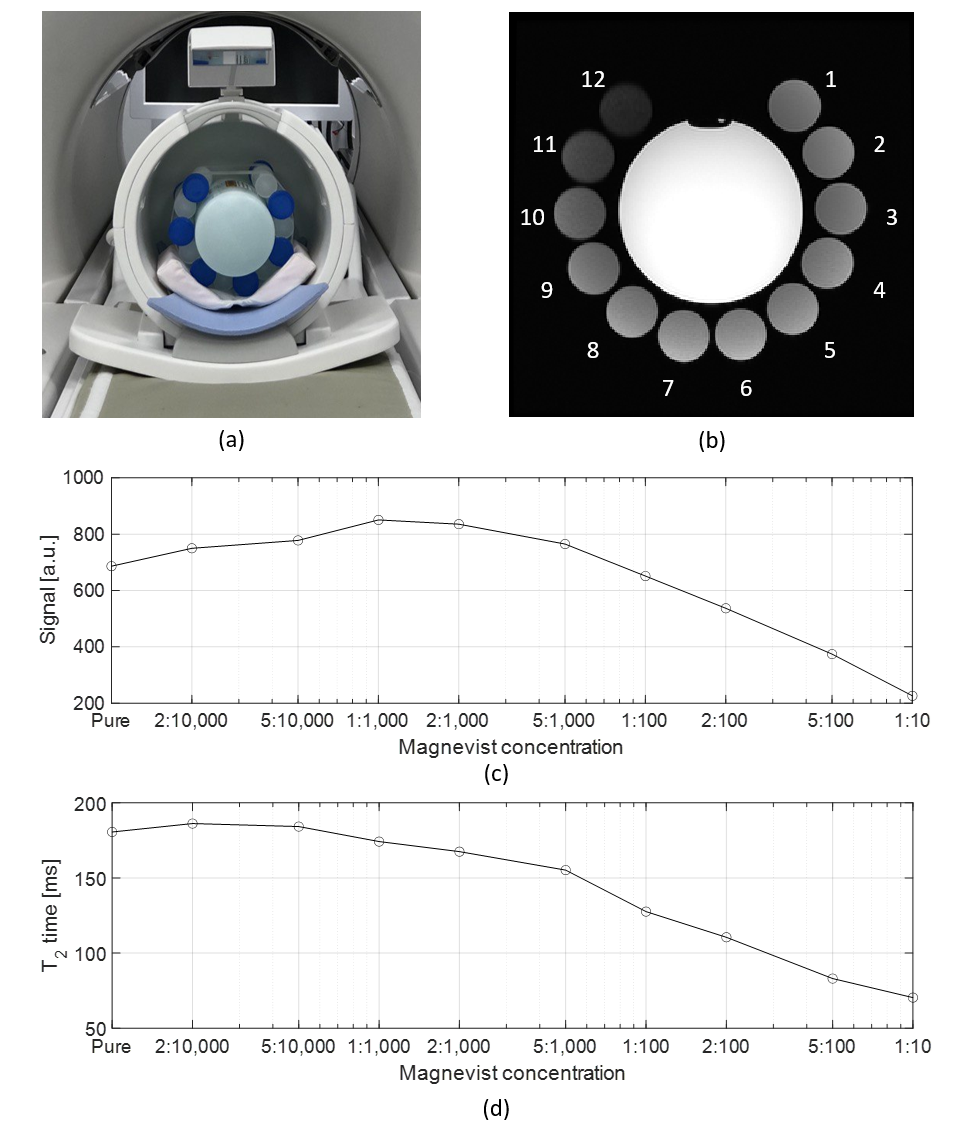
Sample preparation. Three samples of commonly used Ecoflex polymer (00-10, 00-30, and 00-50) were prepared by mixing the corresponding crosslinker and prepolymer at a ratio of 1:1. For the doped samples, Ecoflex 00-30 was mixed with different volume ratios of a Gd contrast agent (Magnevist) (Figure 1a). After mixing, all samples were degassed in a vacuum chamber to remove air bubbles, transferred to graduated plastic test tubes, and cured at room temperature (Figure 1b).
Relaxometry measurements. Samples were equidistantly arranged around a standard homogeneous cylindrical phantom (OD=9.5cm, L=30cm, Figure 2a), positioned centrally inside a 32-channel head coil, and scanned on a 3T MRI scanner (GE Healthcare, MR750). T2 mapping was performed by acquiring a single axial slice with a spin-echo (SE) sequence (TR=1500ms, slice thickness 5mm, 32 TEs from 8.3-133ms). Post-processing was performed using Matlab, with circular, manually drawn, regions of interest to determine the average signal intensity. The T2 value of each sample was found by fitting the average signal intensity to the following equation $$SI_{avg.}=S_0\cdot e^{-TE/T_2}$$ [Eq.2].
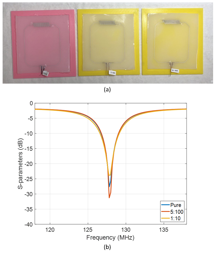
Coil fabrication and bench tests. Three geometrically identical coils were fabricated as outlined previously6. The polymer substrate was fabricated using i) pure Ecoflex 00-30 polymer and Ecoflex 00-30 mixed with Magnevist at a ratio of ii) 5:100 and iii) 1:10 (Figure 3a). S11 parameters (Figure 3b) and unloaded quality factor were measured using a vector network analyzer (Keysight, E5071C).
Phantom imaging. The coils were placed on a standard rectangular silicone phantom (W=22cm, L=33cm, H=16cm) for imaging also at 3T using an FSE sequence (TR=315ms, TE=25.5ms, FOV=24cm, pixel size 0.9x0.9mm, slice thickness=3mm, ETL=2).
Results
Relaxometry measurements. Figure 2b shows one of the SE images acquired with an intermediate echo time (TE) of 33.2ms. The signal is decreased in samples with high Magnevist content (samples #10-12). With the addition of Magnevist, the signal increases by up to 24% at 1:1,000 concentration (Figure 2c). As concentration is increased further, the signal gradually drops to only 30% of its initial value at a concentration of 1:10. Figure 2d shows T2 with respect to the Magnevist concentration to remain relatively stable around 180ms (up to 1:10,000) and then to drop to 39% (70.35ms) of its original value for higher concentrations.Coil fabrication and bench tests. The three coil prototypes exhibited similar S11 parameters (-27+/-3dB) and Q factors (22.2+/-1.5) indicating maintained frequency stability and unloaded losses.
Phantom imaging. Figures 4a-c show FSE images of a homogenous phantom acquired with the 3 coils. The coils doped with Magnevist were less visible on the MR scans, yielding an imaging signal reduction of up to 83% and 92% for 5:100 and 1:10 ratios, respectively. Figures 4d-f show the corresponding SNR maps calculated by dividing the signal maps (a)-(c) by the standard deviation of the background noise. The SNR remains stable for all coils (426±5). Figures 4g-I show the signal intensity measured inside the phantom along the vertical line going through the center of each coil, confirming that doping of the polymer with Magnevist does not deteriorate coil sensitivity.
Conclusions
This study demonstrates that doping coil materials with Gd-based contrast agents (Magnevist) can reduce their appearance in MR images without sacrificing coil sensitivity. Signal intensity reduction depends on sequence parameters, but even for a SE sequence with a relatively short TE=33.2ms, signal reduction is greater than 60% in an Ecoflex substrate. This method could also prove interesting for other applications, such as the design of phantoms with low MR signal intensity, e.g. for the mimicking of lung tissue or bone.Acknowledgements
This work was supported by the National Institutes of Health under NIH R00EB024341, and GE Healthcare. The authors would like to acknowledge Muc Chu, Jojo Borja, and Jonathan Dyke of the Citigroup Biomedical Imaging Center for the helpful technical discussions, and Dr. Hollis Potter of the Hospital for Special Surgery for the guidance and research support of the project.References
1. Varga M, Mehmann A, Marjanovic J, Reber J, Vogt C, Pruessmann KP, et al. Adsorbed eutectic GaIn structures on a neoprene foam for stretchable MRI coils. Advanced Materials. 2017;29(44):1703744.
2. Mehmann A, Varga M, Vogt C, Port A, Reber J, Marjanovic J, et al. On the bending and stretching of liquid metal receive coils for magnetic resonance imaging. IEEE Transactions on Biomedical Engineering. 2018;66(6):1542-8.
3. Mehmann A, Vogt C, Varga M, Port A, Reber J, Marjanovic J, et al. Automatic resonance frequency retuning of stretchable liquid metal receive coil for magnetic resonance imaging. IEEE transactions on medical imaging. 2018;38(6):1420-6.
4. Port A, Luechinger R, Albisetti L, Varga M, Marjanovic J, Reber J, et al. Detector clothes for MRi: A wearable array receiver based on liquid metal in elastic tubes. Scientific Reports. 2020;10(1):1-10.
5. Port A, Luechinger R, Brunner DO, Pruessmann KP. Elastomer coils for wearable MR detection. Magnetic Resonance in Medicine. 2021;85(5):2882-91.
6. Motovilova E, Tan ET, Taracila V, Vincent JM, Grafendorfer T, Shin J, et al. Stretchable self-tuning MRI receive coils based on liquid metal technology (LiquiTune). Scientific reports. 2021;11(1):1-10.
7. Rohrer M, Bauer H, Mintorovitch J, Requardt M, Weinmann H-J. Comparison of magnetic properties of MRI contrast media solutions at different magnetic field strengths. Investigative radiology. 2005;40(11):715-24.
Figures