4101
Improved TSE imaging at Ultra-High Field using Non-localized Efficiency Shimming and Acquisition of Modes Optimized for Refocused Echo (AMORE)
Xiaoxuan He1, Simon Schmidt1, Stefan Zbyn1, Tobey Haluptzok1, Steen Moeller1, and Gregory J. Metzger1
1Center for Magnetic Resonance Research, University of Minnesota, Minneapolis, MN, United States
1Center for Magnetic Resonance Research, University of Minnesota, Minneapolis, MN, United States
Synopsis
To mitigate B1+ inhomogeneity at ultra-high field (UHF), we propose a novel non-localized efficiency shim and utilize this to optimize for T2W TSE contrast in a technique termed Acquisitions of Modes Optimized for Refocused Echo (AMORE). A non-localized efficiency shim is ideal for turbo spin echo (TSE) or fast spin echo (FSE) imaging of smaller anatomical targets such as brain and knee, while AMORE is an optimized TIAMO for improved TSE imaging in the torso by ensuring the desired B1+ is achieved. With AMORE two phase-only modes appear sufficient to mitigate the B1+ shadings and nulls in the interior torso.
Introduction
One major challenge for TSE imaging at UHF is B1+ inhomogeneity. This is typically addressed by RF shimming that trades efficiency for homogeneity accompanied by an inevitable loss of SAR efficiency (i.e. $$$B_1^+/\sqrt{SAR}$$$ ). These tradeoffs make it more difficult to perform TSE imaging in the torso due to larger fields of view (FOV) over which B1+ homogeneity is optimized and limitations in peak available B1+. While the dynamic RF shimming approach using time interleaved acquisition of modes (TIAMO) [1] is a promising solution to address such challenge in the torso, the optimal RF shim or “mode” design for TSE imaging remained to be investigated. To address these issues, we propose a novel non-localized efficiency shim combined with an optimized TIAMO strategy for TSE which we referred to as Acquisitions of Modes Optimized for Refocused Echo (AMORE). AMORE provides improved TSE imaging in the torso at UHF with demonstrated advantages in B1+ efficiency, image contrast and uniformity.Methods
- Non-localized efficiency shimming:We define a novel non-localized efficiency shimming cost function as: $$$f(\mathbf{x})=\sum_{i=1}^{N_s}\frac{r(\mathbf{s}_i\mathbf{x})^Hr(\mathbf{s}_i\mathbf{x})}{\mathbf{x}^H\mathbf{x}}$$$, where $$$r(\alpha)=\begin{cases}\alpha_{min}-\alpha, & \alpha < \alpha_{min}\\0, & \alpha \geq \alpha_{min} \end{cases}$$$, $$$\alpha=\left|s_ix\right|$$$ is the voxel-wise flip angle under the given transmit sensitivity $$$\mathbf{s}_i$$$ and shim $$$\mathbf{x}$$$, and where $$$\alpha_{min}$$$ is the minimum flip angle threshold to be used in optimization. By penalizing only voxels with under-flipping, this above cost function is expected to produce solutions that are “efficient” in the sense that B1+ nulls are minimized within the entire imaging FOV without explicitly constraining over-flipping.
- In-vivo RF shimming study:
To illustrate the proposed cost function, a standard T2W TSE brain imaging using the non-localized efficiency shimming was acquired in 1 subject demonstrating improved field profiles compared to a standard/default circularly polarized (CP) mode. The B1+ maps and the corresponding images were shown in Figure 1.
- Cost function of AMORE:
The proposed AMORE approach extends the above cost function to multiple shims where the voxel-wise maximum flip angle across all modes is evaluated by: $$$f(\mathbf{x})=\sum_{i=1}^{N_s}\frac{r(\alpha_M)^Hr(\alpha_M)}{\mathbf{x}_M^H\mathbf{x}_M}$$$, where $$$\alpha_M=\max_{m\in{\{1,2,...,N_m\}}}{|\mathbf{s}_i\mathbf{x}_m|}$$$. In contrast to the previously presented TIAMO optimization strategy using the least-squares optimization of modes [5], referred to as LS-TIAMO, AMORE optimizes T2W contrast by improving the maximum flip angle across modes to achieve the nominal flip angle. Based on our observations, at least one of the modes needs to achieve the nominal flip angle to maintain the desired T2W contrast.
- Phantom AMORE study:
To quantitatively evaluate the contrast performance in T2W TSE TIAMO acquisitions, a custom contrast phantom of human torso size was constructed and imaged using different mode designs including LS-TIAMO and AMORE, where the contrast to noise ratio (CNR) was calculated for every tube contained in the phantom with respect to its local surrounding polyvinylpyrrolidone (PVP). Per-mode B1+ maps were acquired for each design to compare the transmit efficiency. A summary of the results was shown in Figure 2.
- In-vivo AMORE study:
To compare the performance between LS-TIAMO and AMORE, T2W TSE TIAMO acquisitions were performed in the pelvis of 4 subjects using a 16-channel loop-dipole transceiver array at 7T[4]. Both LS-TIAMO and AMORE were designed and used the same imaging acquisition parameters. Power calibration was based on the prostate, where an additional standard T2W TSE acquisition with phase shimming was performed to determine the transmitter voltage and provided a reference contrast in the prostate. All TIAMO modes were constrained to be phase-only (i.e. equal magnitude across all channels). An example dataset with B1+ maps was shown in Figure 3, where the other in-vivo images acquired are shown in Figure 4.
Results
The proposed non-localized efficiency shimming produced a CP-like shim with less imperfections in the brain (Fig. 1). For the phantom AMORE study, AMORE increased overall B1+ by 35% and could achieve a similar contrast to LS-TIAMO at a significantly lower transmitter voltage. For the in-vivo AMORE study, AMORE reduced signal dropout in the torso and manifested overall higher signal intensity and improved contrast including in the prostate when compared with LS-TIAMO, with a mild over-flipping in the subcutaneous lipid near the coil elements.Discussion
We demonstrated the improvements in TSE imaging when using the proposed non-localized efficiency shimming and its extension AMORE. The proposed cost functions underlying these approaches are inspired by the observation that TSE imaging is more prone to under-flipping than over-flipping. While LS-TIAMO produces a more homogeneous B1+ field, it comes at the cost of lower transmit efficiencies and possible signal dropout, the latter arising from the fact that its solution space contains regions where neither of the modes provides the desired flip angle unless a higher transmit power is used. Therefore, LS-TIAMO is less desirable for torso TSE imaging at UHF and the proposed AMORE is a more efficient and robust approach. In addition, our results demonstrated that with AMORE two phase-only modes appear sufficient to mitigate the B1+ shadings and nulls in the interior torso, although phase-magnitude modes may potentially perform better given the increased degrees of freedom.Conclusion
The proposed non-localized efficiency shimming and AMORE improves TSE acquisitions at UHF in multiple anatomies by increasing overall B1+ and reducing signal dropout, which may serve as a clinically feasible routine TSE acquisition with improved image uniformity than existing approaches.Acknowledgements
We would like to thank Dr. Maria Giovanna Di Trani for help with the AMORE acronym and funding sources: P41 EB027061 & R01 EB029985.References
1. Orzada, S., et al., RF excitation using time interleaved acquisition of modes (TIAMO) to address B1 inhomogeneity in high-field MRI. Magn Reson Med, 2010. 64(2): p. 327-33.2. Deniz, C.M., et al., Maximum efficiency radiofrequency shimming: Theory and initial application for hip imaging at 7 tesla. Magn Reson Med, 2013. 69(5): p. 1379-88.
3. Eichfelder, G. and M. Gebhardt, Local specific absorption rate control for parallel transmission by virtual observation points. Magn Reson Med, 2011. 66(5): p. 1468-76.
4. Erturk, M.A., et al., A 16-channel combined loop-dipole transceiver array for 7 Tesla body MRI. Magn Reson Med, 2017. 77(2): p. 884-894.
5. Orzada, S., et al., Time-interleaved acquisition of modes: an analysis of SAR and image contrast implications. Magn Reson Med, 2012. 67(4): p. 1033-41.
Figures
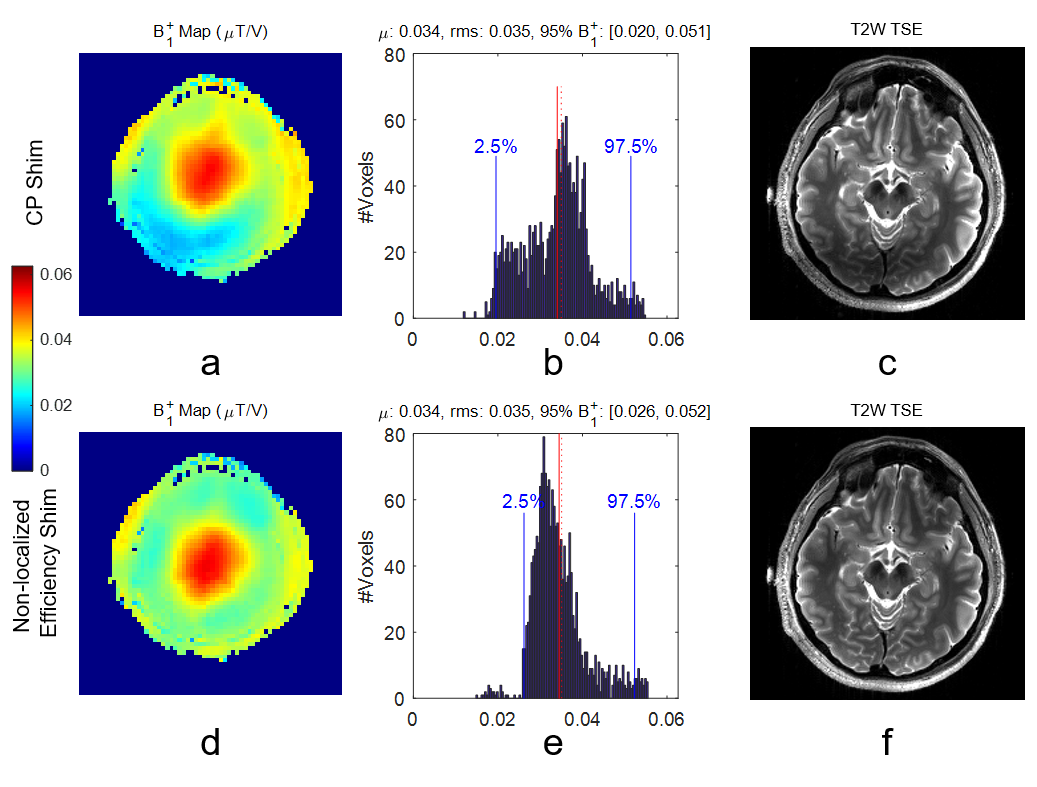
Figure 1. Comparisons of default CP shim
(a – c) and proposed non-localized efficiency shim (d – f) illustrated by the
predicted B1+ maps (a, d), histograms of voxel-wise B1+ (b, e) and T2w TSE
images (c, f). The proposed shimming reduced imperfections of the CP shim, as
demonstrated by an improved B1+ in the posterior part of the brain, increased
minimum B1+ in the voxel-wise distribution, and reduced signal dropout in the
TSE images. µ: mean value; rms: root of mean squares.
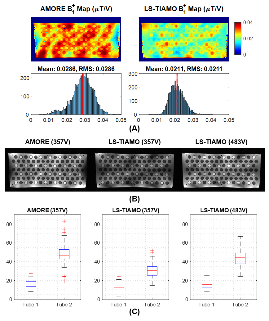
Figure 2. The comparison between LS-TIAMO
and AMORE in the phantom study is demonstrated by: (A) the maximum B1+ map and
its histogram, (B) TIAMO combined T2W TSE images, (C) Contrast to noise ratio
(CNR) of the tubes. Compared with LS-TIAMO, the proposed AMORE increases
average B1+ by 35% and achieves a similar contrast at a significantly lower
transmitter voltage.
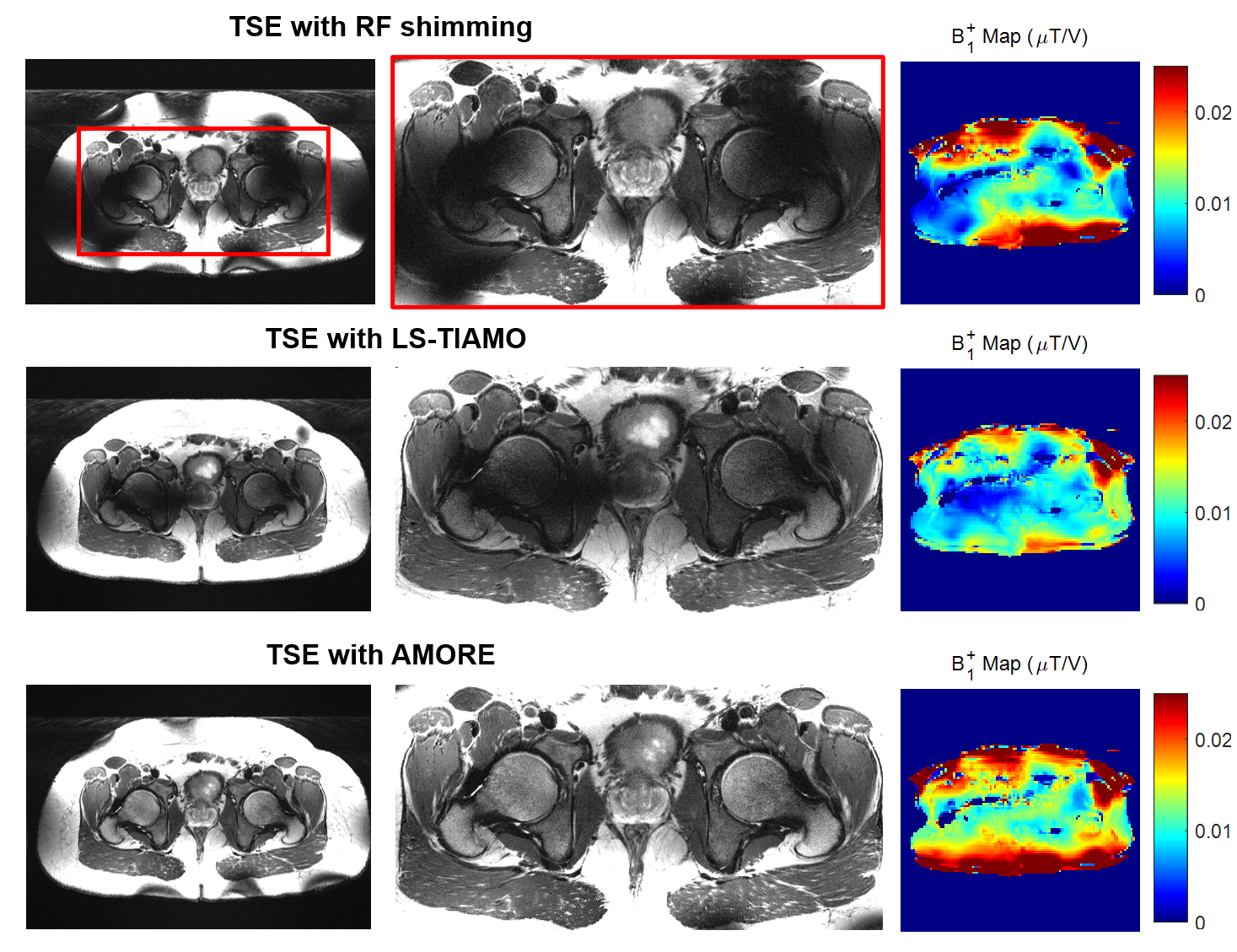
Figure 3. An example dataset of TSE
images acquired in the torso using a phase shimming in the prostate (upper),
LS-TIAMO (middle) and AMORE (lower). The full-size images and zoomed-in
versions demonstrated an improved image uniformity and contrast in the interior
torso when using AMORE due to an overall higher transmit efficiency as
indicated by the B1+ maps. Acquisition parameters: TR/TE: 7000/65 ms,
flip angle: 120°, voxel-size: 0.7x0.7x3 mm3, matrix size: 672 x 672, turbo-factor:
13, acquisition time: 06:06 (phase-shimming) and 06:34 (LS-TIAMO / AMORE, iPAT 2).
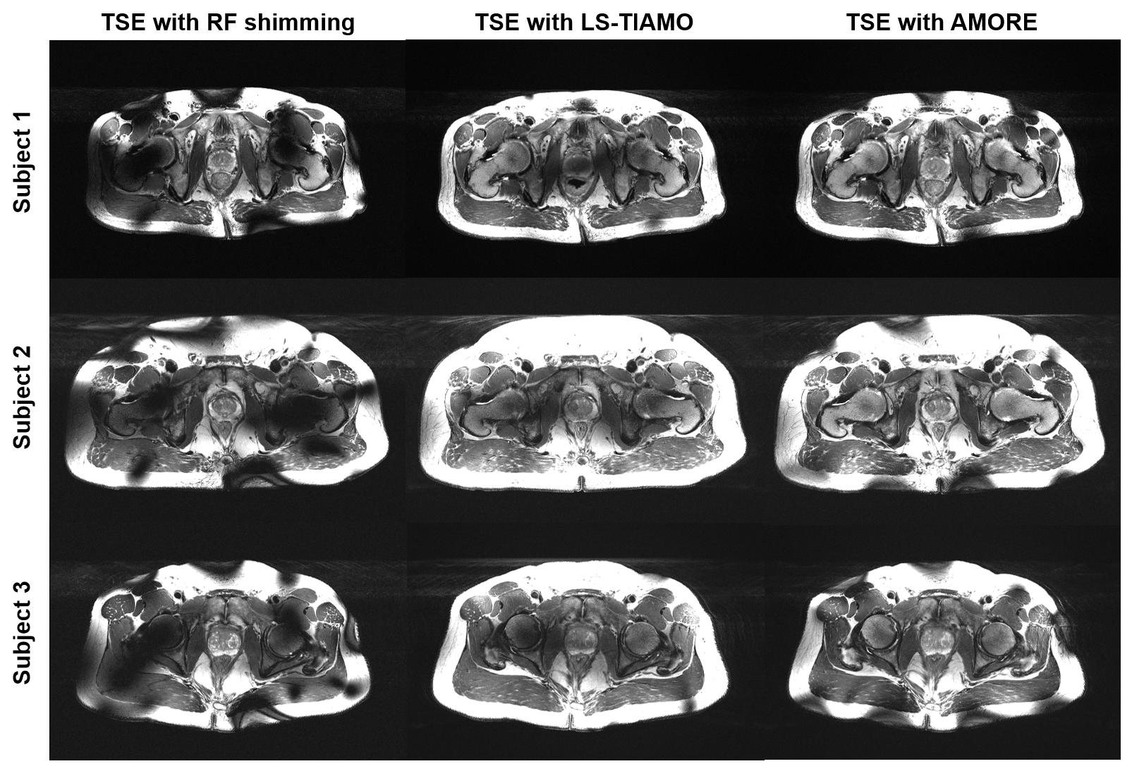
Figure 4. Additional in-vivo TSE images
acquired in the torso using phase shimming in the prostate, LS-TIAMO and AMORE.
Despite subject-specific variabilities, AMORE shows a consistently less shading
and improved contrast in the interior torso, with a slightly increased
over-flipping near the subcutaneous lipid near the coil elements.
DOI: https://doi.org/10.58530/2022/4101