4090
Visualizing and generating a quantitative map of short T2* tissues and producing CT-like imaging of bony structures using multi-echo UTE1Canon Medical Systems USA, Inc., Tustin, CA, United States
Synopsis
Two-dimensional Fast Spin Echo is a workhorse of clinical MRI. However, its minimum TE is in the order of milliseconds or tens of milliseconds, which limits its ability to image tissues with short T2*. This work aims to optimize the multi-echo ultra-short echo time (UTE) sequence to (i) visualize, (ii) generate an associated quantitative map of tissues with short T2*, and (ii) to produce CT-like images of bony structures, which often are not visible in clinical images routinely acquired using FSE sequence.
Introduction
Fast Spin Echo (FSE) is the most widely used sequence in clinical protocols due to its speed, SNR, and robustness to field inhomogeneities. However, its minimum TE is in order of tens milliseconds, which hinders its ability to image short T2*/T2 tissues such as tendon, meniscus, and bone. On the other hand, ultra-short echo-time (UTE) sequence can achieve minimum TE in the range of 0.1 ms by utilizing center-out radial sampling (and other modifications such as minimum phase excitation, ramp sampling, etc.). In this work, we demonstrate the use of multi-echo UTE to (i) visualize, (ii) generate associated T2* map of tissues with short T2*, and (iii) to produce CT-like imaging of bony structures.Methods
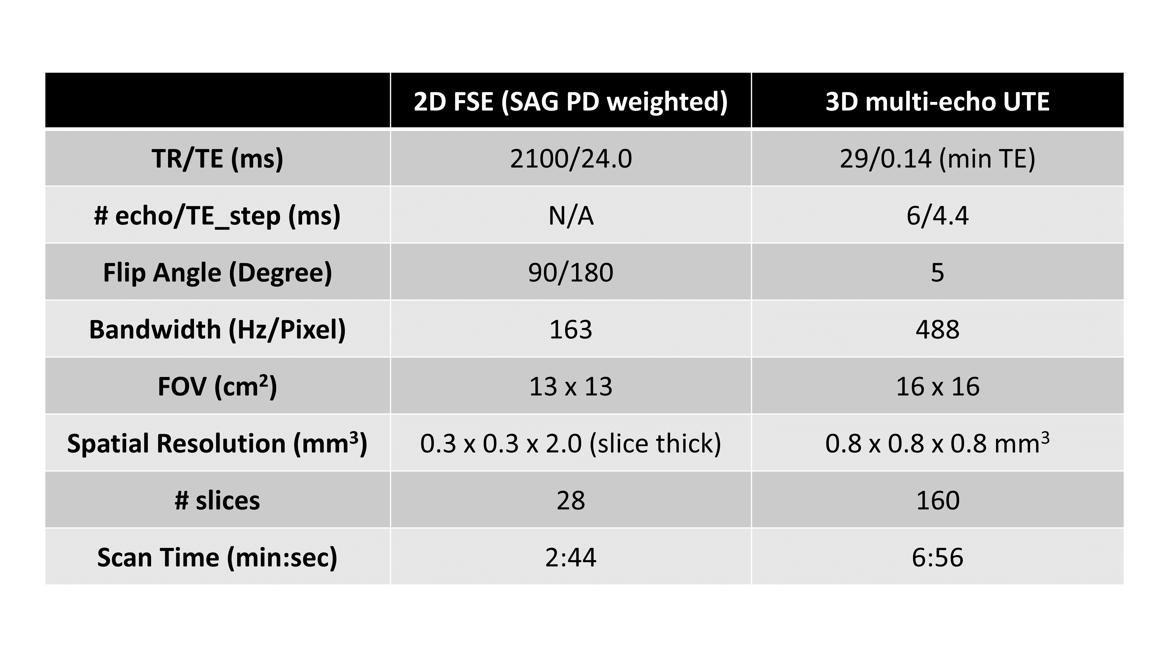
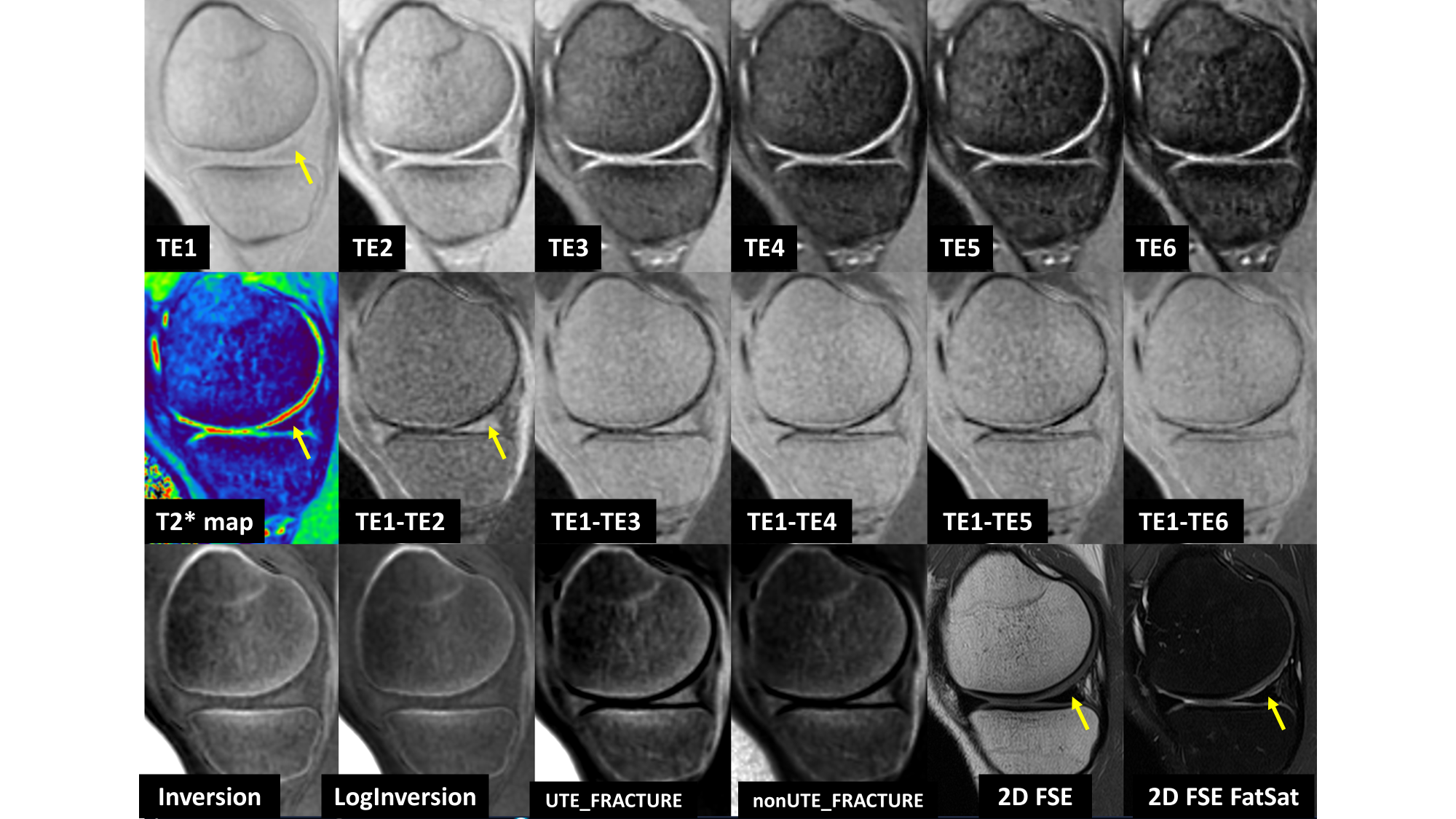
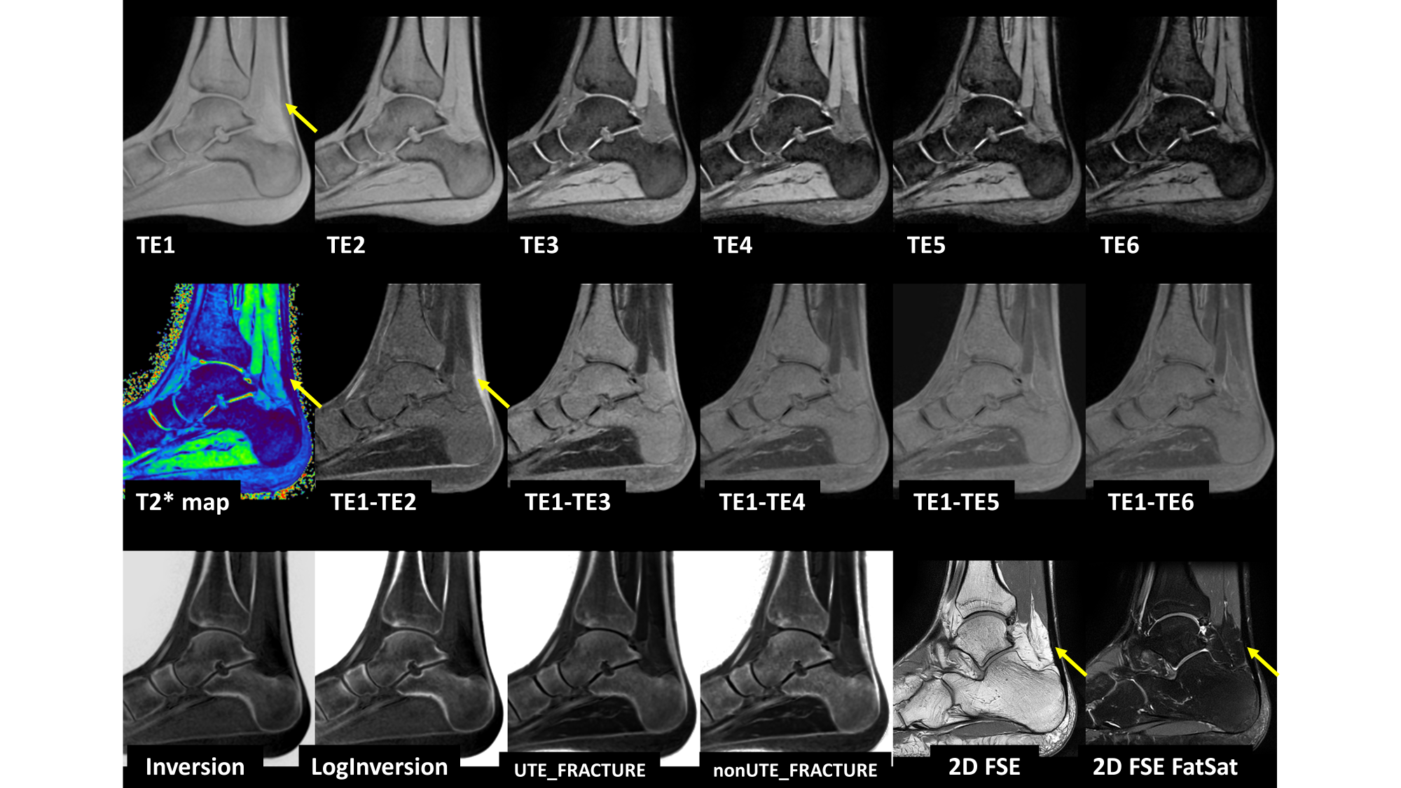
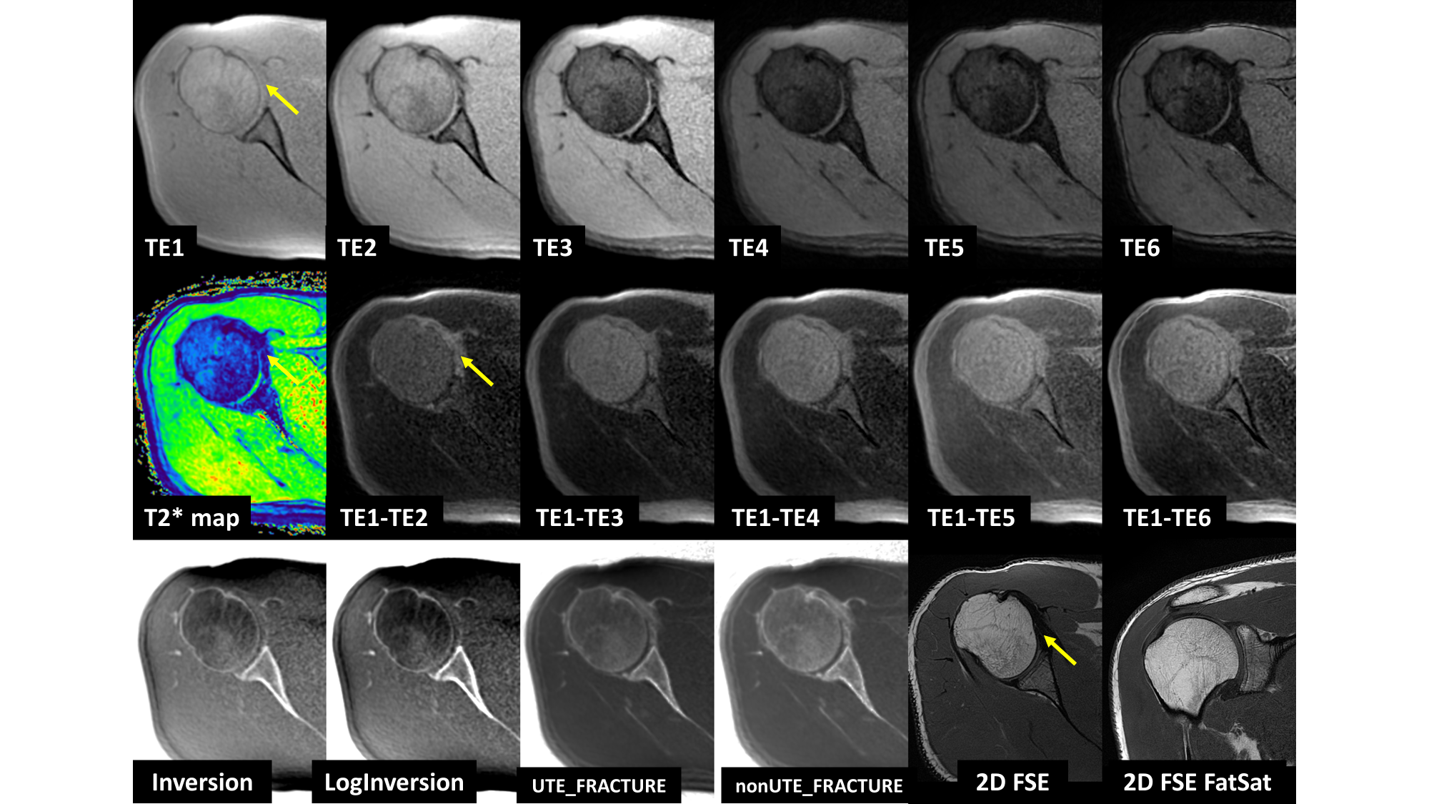
Images of ankle, knee, and shoulder were acquired on volunteers’ ankle, knee, and shoulder using a 1.5T scanner (Canon, Tochigi, Japan). In addition to routine clinical protocol using 2D FSE sequences with standard sequence parameters, multi-echo UTE sequences were performed. Parameters of a representative FSE sequence and multi-echo UTE sequence are shown in Table 1.To enhance visualization of short T2* tissues, higher TE images were subtracted from the minimum TE image. To generate the associated T2* map, multi-echo UTE images were fitted to an exponential decay model. To produce CT-like imaging of bony structures, four different methods were performed on the intensity corrected multiple UTE images. First method: Simple inversion was applied on the first echo image (refer to as Inversion). Second method: Logarithmic inversion was applied on the first echo image (refer to as LogInversion) [1]. Third method: Logarithmic inversion was applied to the sum of six-echo images minus the longest one similar to the FRACTURE method [2] (refer to as UTE_FRACTURE). Note that, the original FRACTURE paper uses a conventional multi-echo gradient echo sequence with minimum TE of 2.3 ms. Therefore, the fourth method to generate CT-like images is in the same way as the third method but the first echo from multi-echo UTE was excluded (refer to as nonUTE_FRACTURE).
Results
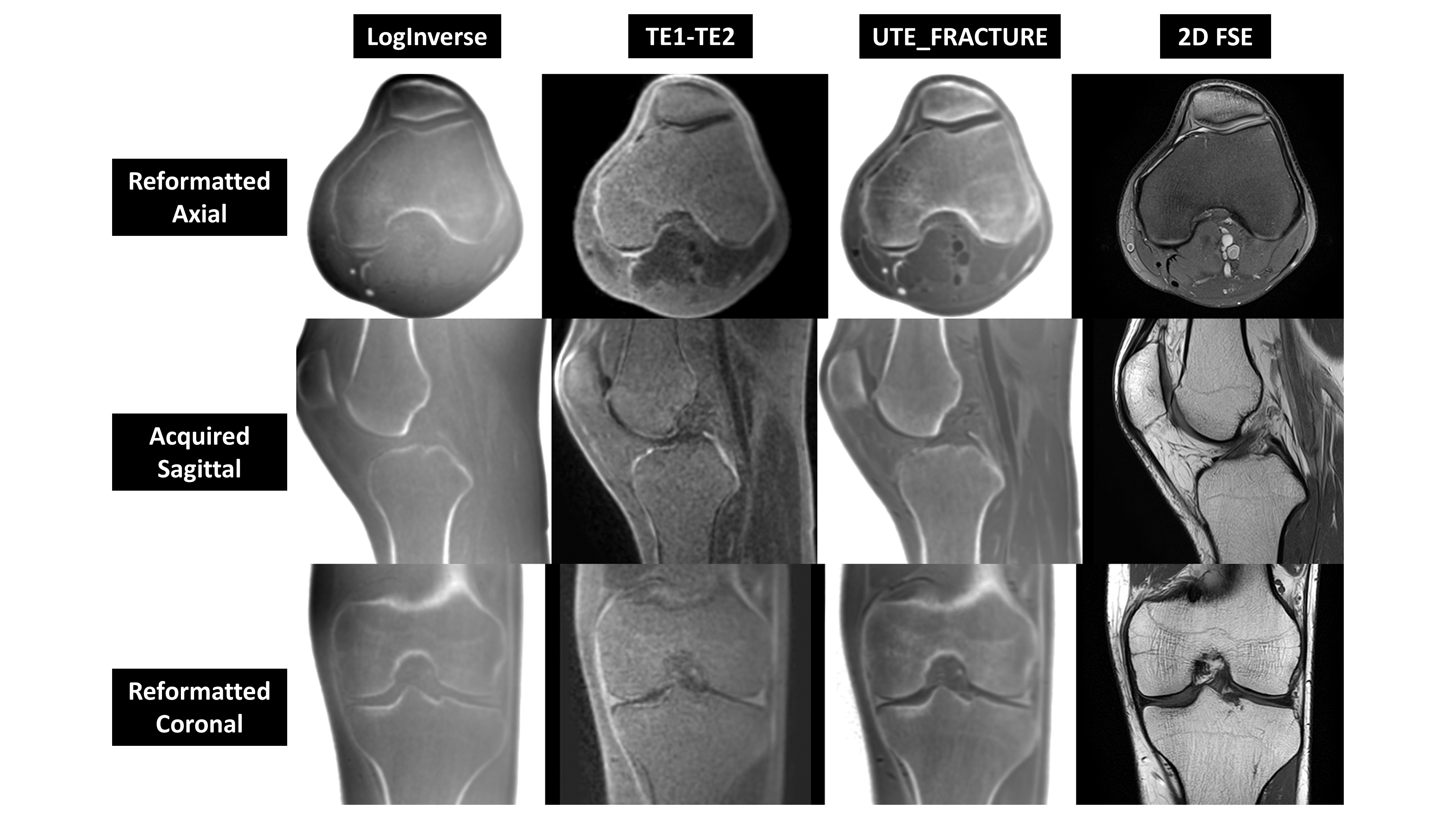
Figure 1 shows knee images with arrows pointing to the meniscus. Figure 2 shows ankle images with arrows pointing to the Achilles tendon. Figure 3 shows shoulder images with arrows pointing to the subscapularis tendon. In Figures 1, 2, and 3, the top row shows raw six-echo UTE images, the middle row shows the T2* map and subtraction images, and the bottom row shows CT-like images created from the four different methods and corresponding routine 2D FSE images.Since the multi-echo data was acquired with 0.8 mm3 isotropic resolution, it can be reformatted in any plane or orientation. Figure 4 shows UTE reformatting knee data in three planes and corresponding 2D FSE images.
Discussion and Conclusion
Multi-echo UTE can simultaneously provide enhanced visualization of short T2* tissues and the associated T2* map and CT-like images of bony structures. This potentially could provide additional information to complement the routine FSE imaging for clinical diagnosis, especially of the tissues with short T2*, which are often not visible in routine FSE-acquired images.Acknowledgements
No acknowledgement found.References
[1] Wiesinger F, Sacolick LI, Menini A, Kaushik SS, Ahn S, Veit‐Haibach P, Delso G, Shanbhag DD. Zero TE MR bone imaging in the head. Magnetic resonance in medicine. 2016 Jan;75(1):107-14.
[2] Johnson B, Alizai H, Dempsey M. Fast field echo resembling a CT using restricted echo-spacing (FRACTURE): a novel MRI technique with superior bone contrast. Skeletal Radiology. 2021 Aug;50(8):1705-13.
Figures