4059
Inversion Recovery for Resolving the Olefinic Resonance in Spinal Bone Marrow at 3 T1Department of Oncology, University of Alberta, Edmonton, AB, Canada, 2Department of Medical Physics, Cross Cancer Institute, Edmonton, AB, Canada
Synopsis
Magnetic resonance spectroscopy measures of the olefinic resonance (≈ 5.3 ppm) enables estimation of fat unsaturation levels. In spinal bone marrow, the olefinic peak is significantly overlapped by that of water (≈ 4.7 ppm). Previously, STEAM with a TE of 100 ms was demonstrated to resolve the olefinic resonance from that of water in spinal bone marrow at 3 T. In this work, we show that an inversion recovery with short-TE STEAM (20 ms) suppresses the water signal, resolving the olefinic resonance and yielding a signal to noise ratio 3.3x larger than that obtained with long-TE STEAM.
Introduction
Magnetic resonance spectroscopy (MRS) measures of the fat olefinic resonance (≈ 5.3 ppm) enables assessment of fat unsaturation levels (1-3). In anatomical regions where water is present, such as in spinal bone marrow, the water resonance (≈ 4.7) overlaps that of the olefinic, rendering its quantification challenging. Previously, long echo time (TE) Point RESolved Spectroscopy (PRESS) and Stimulated Echo Acquisition Mode (STEAM) were optimized to resolve the olefinic resonance from that of water in spinal bone marrow at 3 T (4,5). The long TE of 200 ms for PRESS and 100 ms for STEAM resulted in adequate olefinic signal with water signal significantly reduced by T2 relaxation (4,5). STEAM with a TE of 100 ms resulted in higher signal to noise ratio (SNR) for the olefinic resonance compared to PRESS with a TE of 200 ms due to less olefinic T2 signal loss (4). However, a TE of 100 ms is also relatively long and results in significant T2 signal loss for the olefinic protons. The objective of this work is to investigate the feasibility of employing inversion recovery with a short-TE STEAM sequence to suppress water signal in spinal bone marrow at 3 T.Methods
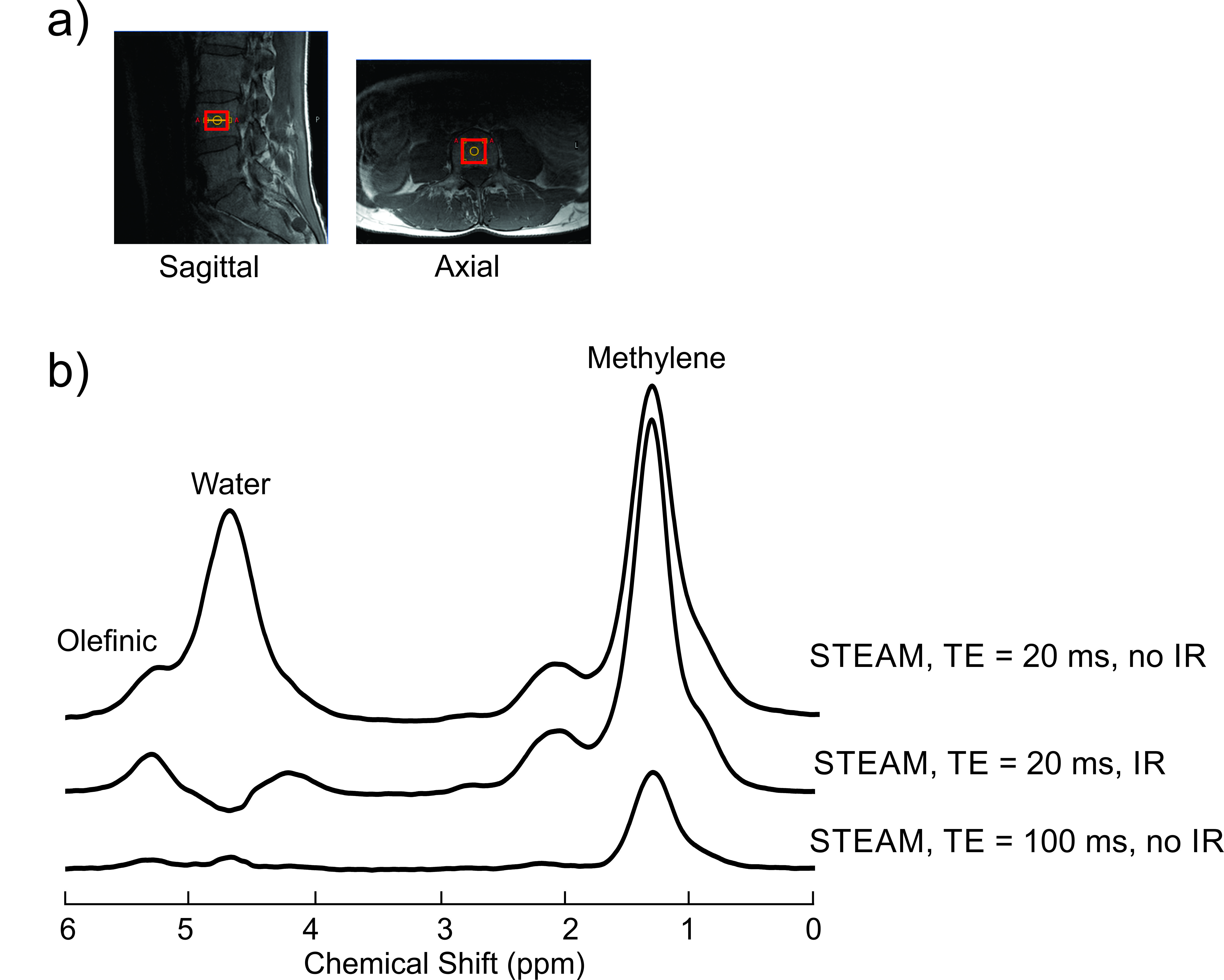
Experiments were performed with a 3 T Philips Intera MRI scanner. The body coil was used for excitation and the Philips spine coil was employed for signal reception. Spectra were acquired from the L4 vertebra of a 46 year old healthy male volunteer. Three spectra were acquired from a 20 x 20 x 15 mm3 voxel with a STEAM sequence (mixing time, TM = 20 ms) with the following parameters: averages = 32, samples = 2048 and sampling frequency = 2000 Hz. Two spectra were acquired with a short TE of 20 ms, one without inversion recovery and one with inversion recovery preceding the sequence. The inversion recovery pulse had a bandwidth of 100 Hz and the inversion recovery time used was 300 ms. A third spectrum was obtained with a TE of 100 ms and no inversion recovery pulse.Results
Figure 1(b) show the spectra acquired from the voxel shown in (a). Significant overlap of water and olefinic signal can be seen in the short TE spectrum acquired with no inversion recovery. Application of the inversion recovery pulse suppressed the water signal, resolving the olefinic resonance. The SNR of the olefinic peak was calculated to be about 83. This is approximately 3.3x larger than that (about 25) obtained with STEAM with a TE of 100 ms (previously optimized to resolve the olefinic resonance in spinal bone marrow).Discussion
Preceding a short-TE STEAM sequence with an inversion recovery pulse to suppress water signal resolved the olefinic signal from that of water. The shorter TE reduces losses due to T2 relaxation resulting in an SNR at least three times higher than that obtained by applying the previously optimized long TE sequence. Water signal suppression with inversion recovery can be improved by performing measurements to obtain an estimate of the water T1. In addition, reducing the olefinic and water linedwidths would improve performance. This can be done by improving shimming and potentially by individual spectral corrections prior to averaging (6,7).Conclusion
Inversion recovery with a short-TE STEAM sequence is feasible for resolving the olefinic resonance from that of water in spinal bone marrow at 3 T.Acknowledgements
The Natural Sciences and Engineering Research Council of Canada (NSERC) is gratefully acknowledged for funding.References
1.Johnson NA, Walton DW, Sachinwalla T, et al. Noninvasive assessment of hepatic lipid composition: Advancing understanding and management of fatty liver disorders. Hepatology 2008;47:1513-1523.
2.Lundbom J, Hakkarainen A, Söderlund S, Westerbacka J, Lundbom N, Taskinen M-R. 1. 1. Long-TE 1H MRS suggests that liver fat is more saturated than subcutaneous and visceral fat. NMR Biomed 2011;24:238-245.
3.Mosconi E, Fontanella M, Sima DM, et al. Investigation of adipose tissues in Zucker rats using in vivo and ex vivo magnetic resonance spectroscopy. J Lipid Res 2011;52:330-336.
4.Bingölbali A, Fallone BG, Yahya A. Comparison of optimized long echo time STEAM and PRESS proton MR spectroscopy of lipid olefinic protons at 3 Tesla. J Magn Reson Imaging 2015;41:481-486.
5.Troitskaia A, Fallone BG, Yahya A. Long echo time proton magnetic resonance spectroscopy for estimating relative measures of lipid unsaturation at 3 T. J Magn Reson Imaging 2013;37:944-949.
6.Gajdošík M, Chadzynski GL, Hangel G, et al. Ultrashort-TE stimulated echo acquisition mode (STEAM) improves the quantification of lipids and fatty acid chain unsaturation in the human liver at 7 T. NMR Biomed 2015;28:1283-1293.
7.Roumans KHM, Lindeboom L, Veeraiah P, et al. Hepatic saturated fatty acid fraction is associated with de novo lipogenesis and hepatic insulin resistance. Nat Commun 2020;11:1891.
Figures