4057
Two-Step Semi-Supervised Denoising for Low-field Diffusion MRI1Hyperfine, Guilford, CT, United States, 2New York University, New York City, NY, United States, 3Yale University, New Haven, CT, United States, 4Massachusetts General Hospital, Boston, MA, United States
Synopsis
In clinical low-field MRI, prolonged data acquisition is impractical, limiting the achievable SNR during imaging. In the absence of ground truth, unsupervised denoising is desirable, but many of them underperform on correlated noise structure of reconstructed MR images. In this work, we present an effective two step training framework for removing correlated MR noise without ground truth. We demonstrate that the proposed approach outperforms the existing denoising methods when applied to the low-field (64mT) diffusion-weighted images and demonstrate that significant noise reduction is possible. 82.5% of processed images were expertly rated clearly/far better overall.
Background
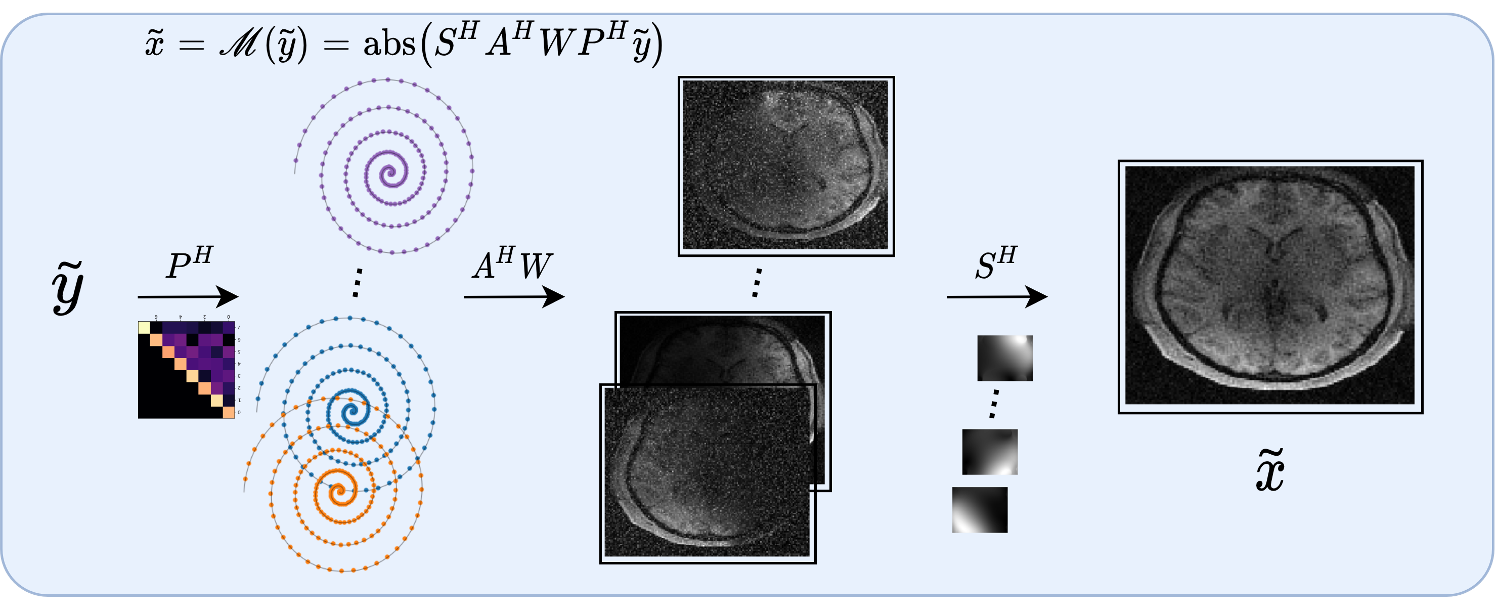
In clinical low-field MRI (64mT)[1,2], MR sequences are designed such that a reasonable SNR can be achieved within an acceptable scan time. Therefore, image denoising techniques are necessary for further improving the SNR, which at the same time has the potential for improving the accuracy of the downstream tasks or further reducing the scan time.Currently, deep learning based image denoising methods offer state-of-the-art performance in a variety of image denoising tasks[3-8]. However, they typically require a sufficiently large dataset of clean images, which are difficult to obtain for MRI in clinical settings. To solve this problem, unsupervised denoising was proposed for training deep learning-based denoisers without requiring clean data.[9-16] Yet, most methods require the noise to be independent and identically distributed (i.i.d) in the image. Nevertheless, The noise distribution in the images obtained through complex MR reconstruction pipeline is often non-i.i.d.[17,18] See Figure 1 for the example of a typical modern MR reconstruction pipeline. In this work, we propose a two-step training approach for effectively removing correlated MR noise without requiring clean images from the target domain.
Materials and Methods
Imaging: A portable 64mT MRI scanner (Hyperfine SwoopTM) was deployed to 6 different acquisition sites and patients were recruited and provided consent under a protocol approved by an institutional review board. The patients underwent a DWI imaging protocol (TE/TR = 24ms/34ms, 2x2x6mm3 resolution, b=890 s/mm2, scan time = 8 min, followed by b=0 s/mm2, scan time=1.5 min) using a 1Tx/8Rx coil. In total, nearly 600 DWI images were collected, including 289 and 308 images at b-value of 890s/mm2 and 0 s/mm2.Proposed Method: The network architecture consists of a bias-free[19] DNCNN[8] with 20 convolutional layers. 2D 64x64 axial patches are used for training. During the inference, the entire 3D image is fed slice-by-slice to the network and the output DWI is generated.
Training: The denoiser was trained in the following two steps process (Figure 2). In the first step, a supervised denoising is performed on a large-scale dataset with clean references. For the source dataset, a total of 400 cases of T1w and T2w images from the HCP projects[20] and 400 cases of T1w, T2w and FLAIR images acquired at 64mT were used. Once the denoiser is trained, it is applied to 600 DWI images. Data augmentation (such as image sharpening) is applied to DWI images and are then added to the training dataset. In the second step, the denoiser is re-trained from scratch on the extended dataset. The denoiser from the first and second steps are referred to as Sup (supervised) and SeqSSL (sequential semi-supervised learning). We used L1+SSIM loss with Adam optimizer and each training took approximately 17 hours. All methods were implemented in Tensorflow 2.3.
Evaluation:
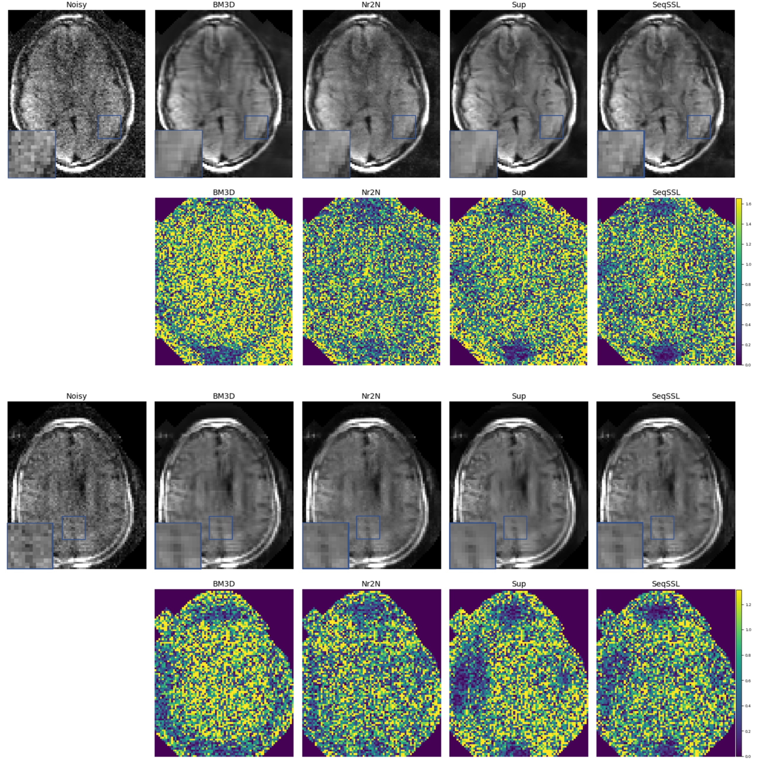
In order to test the efficacy of our proposed approach, we apply the proposed framework to denoise diffusion-weighted MR images at 64 mT acquired using the portable MR system. The proposed method was qualitatively compared to the competing methods: BM3D[21], Noisier2Noise (Nr2N)[13], and supervised learning. For BM3D, hyper-parameters were manually tuned for each slice. For training Nr2N, we trained the model to predict images at $$$\sigma=0.05$$$ from an input with noise level $$$\sigma=0.1$$$.
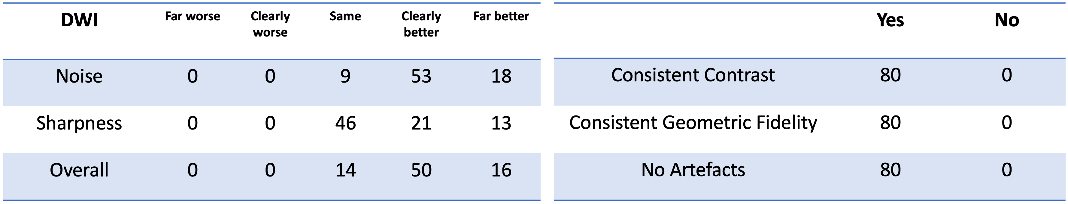
The proposed denoising framework was then qualitatively evaluated by four expert graders with background in either MR physics, clinical science, and/or radiology. Images before and after denoising were shown to the raters as a side-by-side comparison. The users were asked to rate if the denoised image was “Far better”, “Clearly Better”, “Same”, “Clearly Worse”, or “Far Worse”, in terms of noise, sharpness and overall quality. The raters were also asked if the denoised image had consistent image features as the input in terms of contrast, geometric fidelity, and whether artifacts were introduced.
Results
In Figure 3, examples of the denoised DWI images are shown. BM3D results were competitive; however, it required careful parameter tuning. Nr2N resulted in inconsistent levels of denoising. This may be due to the fact that in practice, the noise variance present in the images are variable, whereas Nr2N requires training at a fixed noise level. Some degree of over-smoothing can be observed in Sup. SeqSSL alleviated this issue of over-smoothing and behaved more consistently across all images.Results of the expert grading are summarized in Figure 4. In particular, 100% of the graders indicated “Same”, “Clearly Better”, “Far Better” for all categories. At least 88.8%, 42.5%, 82.5% voted “Clearly Better”, “Far better” for reduced noise, sharpness and overall quality, respectively. The raters also scored “Yes” for all consistency questions.
Discussion and Conclusions
In this work, we proposed a two-step training framework that can be applied to data with correlated noise without access to ground truth. The proposed framework not only yielded competitive performance to existing unsupervised approaches, it was also more robust across different noise levels. Our future work will consider more extensive sets of experiments to better understand the behavior of the proposed model under different source datasets. The proposed framework new possibilities beyond denoising and we plan to investigate its application to other inverse problems such as MR image reconstruction.Acknowledgements
No acknowledgement found.References
Sheth, Kevin N., et al. "Assessment of brain injury using portable, low-field magnetic resonance imaging at the bedside of critically ill patients." JAMA neurology. 2020.”
O’Halloran, Rafael, et al., “Bedside Stroke Imaging at 64mT”, ISMRM2020, Abstract #1799
Burger, H.C., et al.: Image denoising: Can plain neural networks compete with bm3d? In: 2012 IEEE conference on computer vision and pattern recognition. pp. 2392–2399. IEEE (2012)
Jifara, W., et al.: Medical image denoising using convolutional neural network: a residual learning approach. The Journal of Supercomputing 75(2), 704–718 (2019)
Kong, Z., et al.: A comprehensive comparison of multi-dimensional image denoising methods. arXiv preprint arXiv:2011.03462 (2020)
Tian, C., et al.: Deep learning on image denoising: An overview. Neural Networks (2020)
Weigert, M., et al.: Content-aware image restoration: pushing the limits of fluorescence microscopy. Nature methods 15(12), 1090–1097 (2018)
Zhang, K., et al.: Beyond a gaussian denoiser: Residual learning of deep cnn for image denoising. IEEE transactions on image processing 26(7), 3142–3155 (2017)
Batson, J., Royer, L.: Noise2self: Blind denoising by self-supervision. In: International Conference on Machine Learning. pp. 524–533. PMLR (2019)
Krull, A., et al.: Noise2void-learning denoising from single noisy images. In: Proceedings of the IEEE/CVF Conference on Computer Vision and Pattern Recognition. pp. 2129–2137 (2019)
Lehtinen, J., et al.: Noise2noise: Learning image restoration without clean data. arXiv preprint arXiv:1803.04189 (2018)
Metzler, C.A., et al.: Unsupervised learning with stein’s unbiased risk estimator. arXiv preprint arXiv:1805.10531 (2018)
Moran, N., et al.: Noisier2noise: Learning to denoise from unpaired noisy data. In: Proceedings of the IEEE/CVF Conference on Computer Vision and Pattern Recognition. pp. 12064–12072 (2020)
Schlemper J, Dey N, Salehi SSM, Lazarus C, O’Halloran R, Kundu P, Sofka M. Unsupervised Denoising for Low-field Diffusion MRI. Proceedings of the 2021 ISMRM & SMRT Annual Meeting & Exhibition. Abstract Number 2190.
Soltanayev, S., Chun, S.Y.: Training and refining deep learning based denoisers without ground truth data. arXiv preprint arXiv:1803.01314 (2018)
Yan, H., et al.: Unsupervised image noise modeling with self-consistent gan. arXiv preprint arXiv:1906.05762 (2019)
Brooks, T., et al.: Unprocessing images for learned raw denoising. In: Proceedings of the IEEE/CVF Conference on Computer Vision and Pattern Recognition. Pp. 11036–11045 (2019)
Guo, S., et al.: Toward convolutional blind denoising of real photographs. In: Proceedings of the IEEE/CVF Conference on Computer Vision and Pattern Recognition. pp. 1712–1722 (2019)
Mohan, S., et al.: Robust and interpretable blind image denoising via bias-free convolutional neural networks. arXiv preprint arXiv:1906.05478 (2019)
Van Essen, D.C., et al: The wu-minn human connectome project: an overview. Neuroimage 80, 62-79 (2013).
Dabov, K., et al.: Image denoising by sparse 3-d transform-domain collaborative filtering. IEEE Transactions on image processing 16(8), 2080–2095 (2007)
Figures
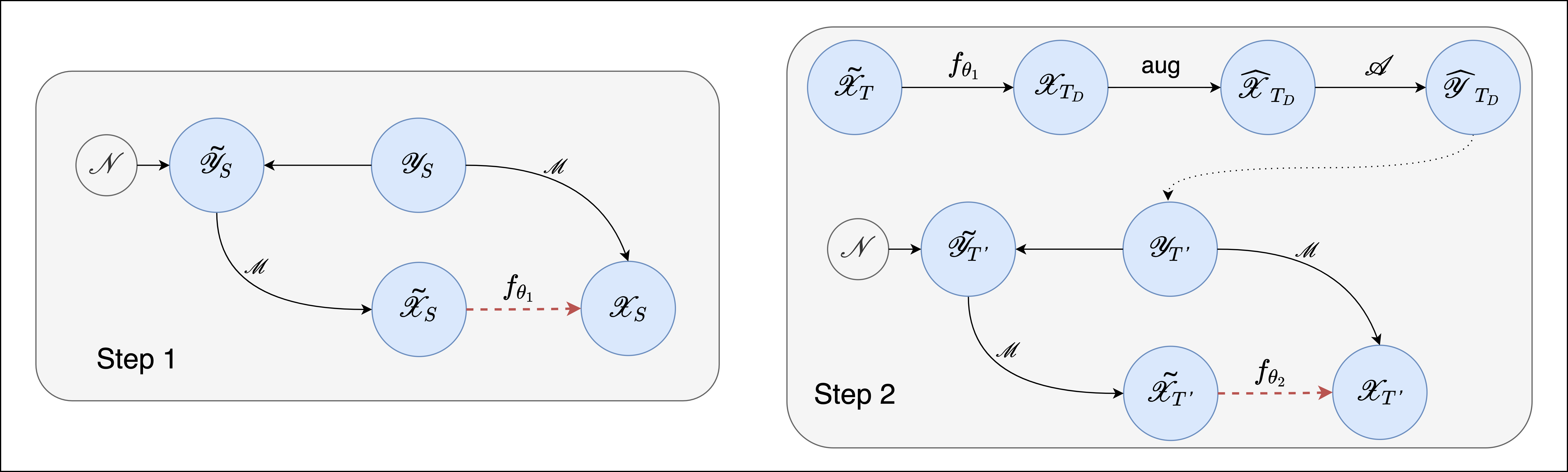
The proposed two step training: Step 1: the supervised network is trained on the available dataset in domain S (red dashed arrow). The structured noise is modelled by adding Gaussian noise to simulated raw data $$$y$$$ and is propagated through the reconstruction pipeline ($$$\tilde{x}$$$. In the second step, the trained model $$$f_{\theta_1}$$$ is used to denoise the noisy images $$$\tilde{x}_T$$$ from the target domain $$$T$$$, creating new clean reference images for the second stage supervised training. The final model is given by $$$f_{\theta_1}$$$.