4043
Evaluation of the Effects of Capsinoids on Brown Adipose Tissue Recruitment and Activation in Obesity Using PET/MRI
Jie Deng1, Lisa M Neff2, and Bin Zhang3
1Radiation Oncology, UT Southwestern Medical Center, Dallas, TX, United States, 2Chicago, IL, United States, 3Department of Pediatrics, Cincinnati Children's Hospital Medical Center, Cincinnati, OH, United States
1Radiation Oncology, UT Southwestern Medical Center, Dallas, TX, United States, 2Chicago, IL, United States, 3Department of Pediatrics, Cincinnati Children's Hospital Medical Center, Cincinnati, OH, United States
Synopsis
In this randomized, placebo-controlled trial in subjects with obesity, we implemented multi-parametric MRI including fat fraction (FF), R2*, and T1 values on PET-MRI in both thermoneutral and cold-activated non-shivering thermogenesis conditions to investigate whether brown adipose tissue property changes measured by MRI can reflect its activation secondary to cold-activated stimulation and chronic capsinoids ingestion before and after weight loss. In subjects taking capsinoids, FF decreased and R2* increased significantly after weight loss as compared with baseline, and these changes were significantly larger than the changes seen after weight loss in the group taking placebo.
INTRODUCTION
Human brown adipose tissue (BAT), when present, plays a role in cold- and diet-induced thermogenesis, potentially providing some protection against the development of obesity. Capsinoids are nunpungent analogs of capsaicin which may activate BAT by stimulating sensory neurons in the gastrointestinal tract. A study of young, lean men with little or no detectable BAT prior to treatment has shown that chronic ingestion of capsinoids increases cold-induced thermogenesis, suggesting that capsinoids may also facilitate recruitment of BAT [1]. The purpose of this study was to implement multi-parametric MRI in both thermoneutral and cold-activated non-shivering thermogenesis (NST) conditions to investigate whether MRI measures of BAT tissue property changes can reflect BAT activation secondary to cold-activated stimulation and chronic capsinoids ingestion more sensitively than PET.METHODS
Males with obesity, ages 18-50, were recruited for a randomized, placebo-controlled trial of capsinoids supplementation. The study consisted of two main phases: the first in which subjects maintained their usual diet and activity for 8 weeks, and the second in which subjects consumed a low-calorie diet for 12 weeks followed by 2 weeks of weight stability. Imaging studies were performed on a hybrid PET/MRI scanner (Fig. 1) before and after each phase (3 visits in total), although the final scan was not conducted for all subjects due to the COVID19 pandemic. MRI images were acquired under both thermoneutral and NST conditions after cold exposure, while 18F-FDG PET images were acquired simultaneously with MRI under NST condition [2]. Fat fraction (FF), R2*, and T1 values were measured on MRI using dual flip angle (5° and 20°) 6-echo in/out-of-phase gradient echo sequence [3]. Standardized uptake value (SUV) of FDG was measured on PET images to calculate the volume and activity of metabolically active BAT, defined as any neck fat voxel with SUV>1.5. Linear mixed model was used to assess the correlation between PET/MRI measurements and various factors (group, cold exposure, visit, and weight). Spearman’s correlation was used to assess the correlation between the MRI measurement changes due to cold exposure and PET measurements.RESULTS
A total of 19 subjects were included in this study and randomized into a capsinoids group (N=10) and placebo group (N=9). In PET imaging, active BAT was detected in 11/19 subjects on visit 1, 11/15 subjects on visit 2, and 3/9 subjects on visit 3. Using a linear mixed model analysis, PET measures of BAT activity were not significantly different between treatment groups or visits. In contrast, MRI-based FF and R2* was significantly associated with an interaction term between treatment group and visit (p < 0.0001 and p = 0.04, respectively), adjusted for cold-activation condition and weight. MRI-based measurements between two groups were dependent on visits, and MRI measurements at each visit were dependent on treatment conditions as well. Specifically, compared with visit 1, FF of the group of subjects taking placebo (FFplacebo) decreased by 0.4% on visit 2 and 4.9% on visit 3, while FF of the group of subjects taking capsinoids (FFcapsinoids) deceased by 0.2% on visit 2 and 7.4% on visit 3 (Fig. 1) (p <0.0001). Compared with visit 1, R2*placebo increased by 3.4 s-1 on visit 2 and 9.8 s-1 on visit 3, while R2*capsinoids increased by 4.4 s-1on visit 2 and 14.4 s-1 on visit 3 (Fig. 2) (p = 0.037). Independent of visit and group, FF decreased by 0.5% after cold exposure compared with pre-cold (p = 0.01). MRI FF changes after cold exposure (post-pre: ΔFF) were correlated with percentage of body fat (r = 0.56, p = 0.01) measured by DXA on visit 1. In combined group analysis, T1 measured at visit 3 was significantly higher than visit 1 by 845ms (p = 0.003) and but not significantly higher than visit 2 by 185ms (p = 0.4). From the Spearman’s correlation analysis, active BAT volume measured by PET was significantly correlated with T1 changes after cold exposure (post-pre: ΔT1) (r = 0.76, p = 0.03). However, due to the small sample sizes on visit 2 and 3, there was not sufficient data to evaluate the correlation between these measurements.DISCUSSION
Our study used hybrid PET-MRI to assess BAT response to chronic capsinoids treatment to achieve more accurate tissue characterization and functionality of BAT than either method alone. It is one of the first studies to assess the effect of chronic capsinoids ingestion on BAT in subjects with obesity. The two-phase design allowed for separation of the metabolic effects of capsinoids from those of weight loss. Simultaneous acquisitions of FDG-PET and MRI not only shortened the imaging exam time, but more importantly, enabled more accurate localization and analysis of FDG-vivid areas overlaid on fat tissues identified by MRI. FF and R2* measured by MRI are sensitive quantitative MRI features for detecting BAT tissue properties secondary to cold induced thermogenesis and capsinoids BAT recruitment.CONCLUSION
PET may not detect quiescent BAT with activity under certain threshold. MRI is sensitive to subtle changes of the inherent tissue properties during thermogenic stimulation and chronic activation process by capsinoids ingestion.Acknowledgements
The study was supported by a grant from NIDDK (R01DK112281) to Northwestern University (Neff/Deng co-PI), where the trial was conducted. Dr. Neff is currently an employee of Eli Lilly and Company, which has no affiliation with this work. At the onset of the study, she was employed by Northwestern University Feinberg School of Medicine, Chicago, IL. Dr. Neff has previously received research funding from Amryt Pharmaceuticals and served as a research investigator for Novo Nordisk.References
[1] Yoneshiro, T., et al., Recruited brown adipose tissue as an antiobesity agent in humans. J Clin Invest, 2013. 123(8): p. 3404-3408.[2] Gariani K, et al, Hybrid PET/MRI as a tool to detect brown adipose tissue: Proof of principle, Obesity Research & Clinical Practice, Volume 9, Issue 6, 2015, Pages 613-617, https://doi.org/10.1016/j.orcp.2015.05.004.[3] Deng J, Neff LM, Rubert NC, et al. MRI characterization of brown adipose tissue under thermal challenges in normal weight, overweight, and obese young men. J Magn Reson Imaging. 2018;47(4):936-947. doi:10.1002/jmri.25836Figures
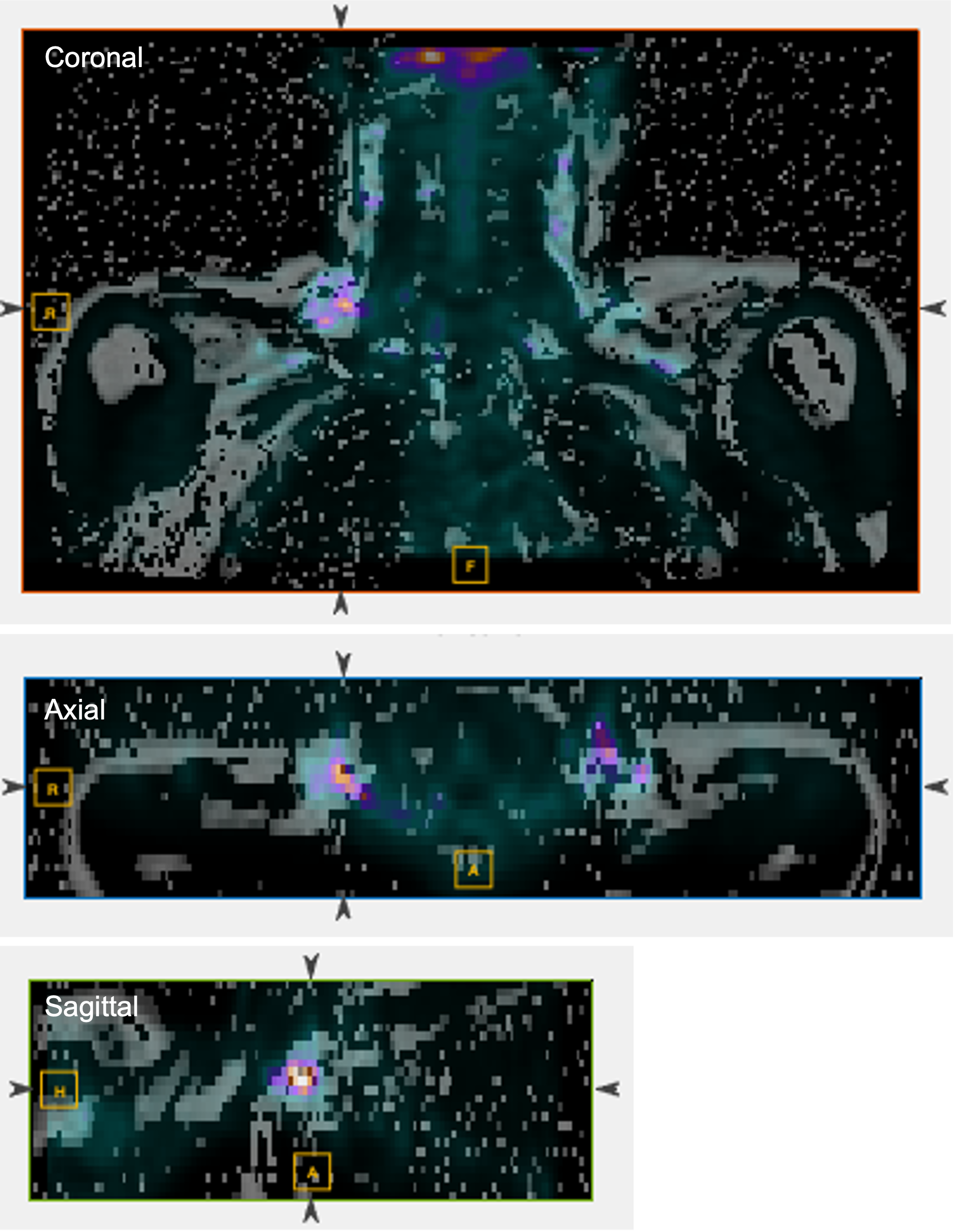
Figure 1. MRI-based fat fraction (FF) map overlaid with PET image acquired simultaneously with MRI. Areas with SUV >1.5 on FDG-PET represent active brown fat in supraclavicular regions.
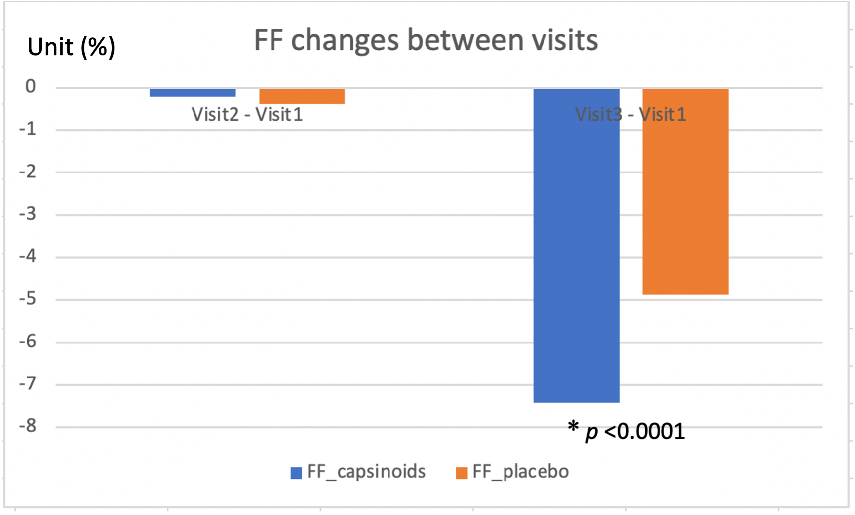
Figure 2. The decrease of FFcapsinoids at visit 3 relative to visit 1 were significantly larger than that of FFplacebo. There was no obvious FF change between visit 2 and visit 1.
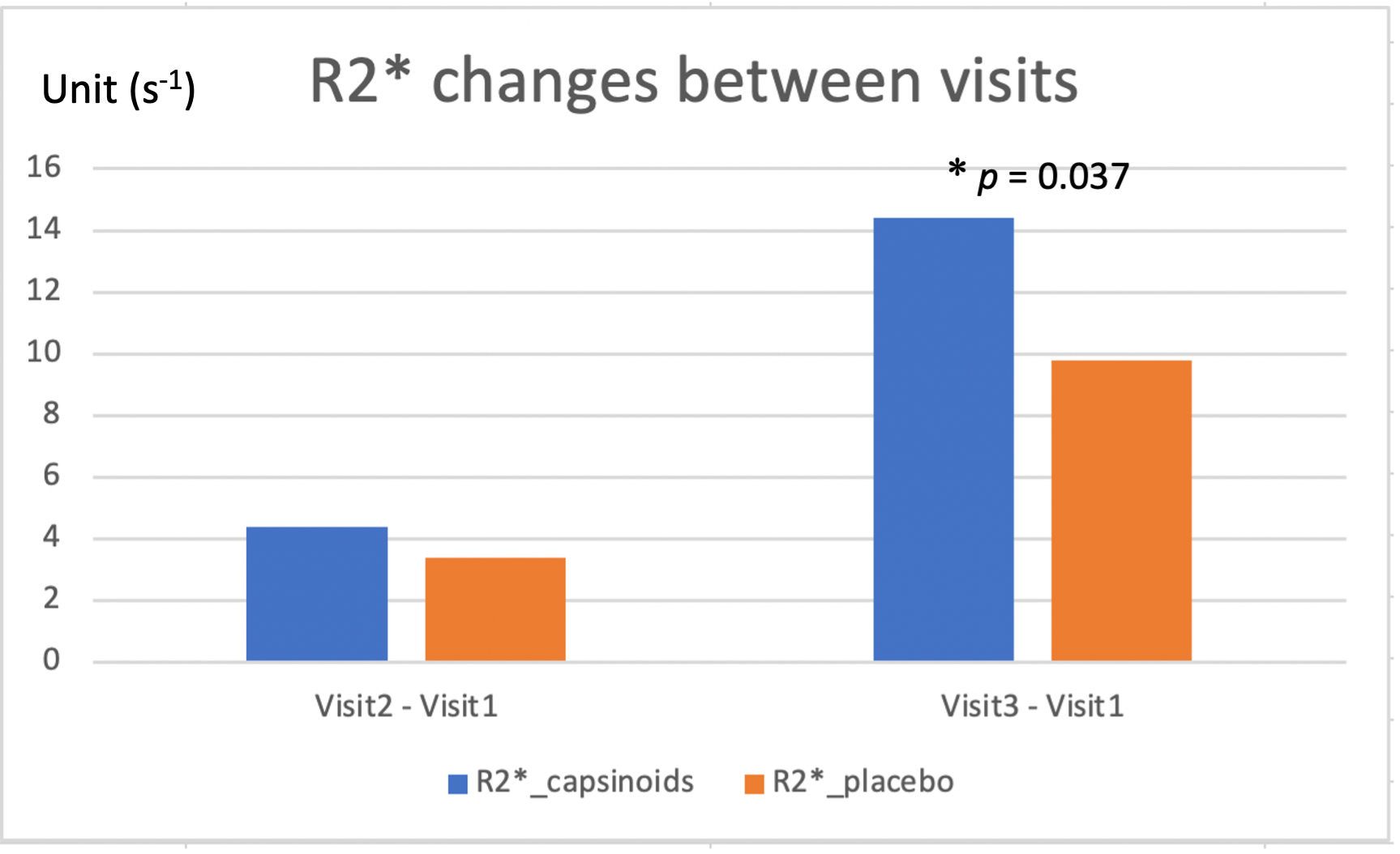
Figure 3. The increase of R2*capsinoids at visit 3 relative to visit 1 were significantly larger than that of R2*placebo. In comparison, there was less R2* change between visit 2 and visit 1.
DOI: https://doi.org/10.58530/2022/4043