3967
Development of a High-Speed MRI Protocol with Deep Learning Reconstruction Method for Brain Imaging in a Clinical Setting1GE Healthcare, Brooklyn, NY, United States
Synopsis
The purpose of this study was to develop a High-Speed MRI (hsMRI) protocol with deep learning reconstruction (DL Recon) for adult and pediatric brain imaging to provide consistent, stout image quality in less than 1/4 the scan time of standard protocols. This was achieved utilizing FSE T1, SS T2w, Gradient Echo (GRE) T2*, and Diffusion Weighted & FLAIR Echo Plane Imaging (EPI) sequences. Evaluation was performed on of 20 patients comparing current protocol to hsMRI with results indicating imaging using hsMRI protocol was of clinically diagnostic quality allowing significant reduction in scan times compared to industry standard routine brain imaging protocols.
Purpose
Historically, MRI has proven valuable when imaging the brain to assist in visualizing and diagnosing many abnormalities around the world. When performing this type of exam, like any other MR scan, it’s essential that data is free of motion/ghosting artifacts to make an accurate clinical diagnosis. Time in the bore acquiring data can have direct contribution for causing and preventing the above-mentioned artifacts. The increase of the number of MR systems located in the emergency department (ED) provides quicker access to MR imaging. Many of these ED patients pushes the needs for more rapid imaging protocols while still providing highest potential image quality. Many facilities have also made significant strides to improve system/fleet utilization while trying to reduce overall exam duration across the board. Because brain imaging is still the one of the most frequent exams completed on an MR system, the desire to reduce exam duration is directly impacted by brain exams. As a result, the aim of this study is the assess the benefit non-research clinical sequences with DL Recon to develop a reduced scan-time rapid brain protocol with no significant compromise of diagnostic capability and quality.Method
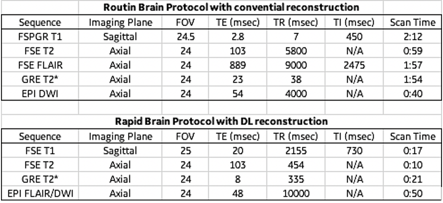
In this study, we acquired standard routine brain and the newly develop express brain protocol for FSE sagittal T1, axial FLAIR, T2w, GRE T2* and DW-EPI sequences in 20 patients. The study was performed on, 3T 70cm bore MR scanners (Discovery MR750w, GE Healthcare, USA) using GE Signa MRI Brain Array Coil (8 channel, High Resolution). Imaging parameters for deep-learning reconstruction imaging data set described in table (fig. 1). were developed by the authors to produce signal-to-noise ratios similar standard brain sequences in a fraction of the time. The sequences collected with current clinical voxel size were reconstructed using conventional reconstruction method and the rapid brain (excluding DWI/FLAIR EPI) sequences were reconstructed with the DLRecon method for removal of image noise. All data was collected without gadolinium contrast focusing on the core imaging sequences prior to the administration of contrast.The Deep-Learning Reconstruction (DLRecon) method comprised a deep convolutional residual encoder network trained from a database of over 10,000 images to reconstruct images with high signal-to-noise ratio and high spatial resolution. The network offered a tunable noise reduction factor to accommodate user preference. The DLRecon network was embedded into a new reconstruction pathway that operated in parallel with the conventional reconstruction such that two sets of image series were generated from a single set of raw MR data. All deep learning reconstruction sequences used a 75% noise reduction calculation.
Results
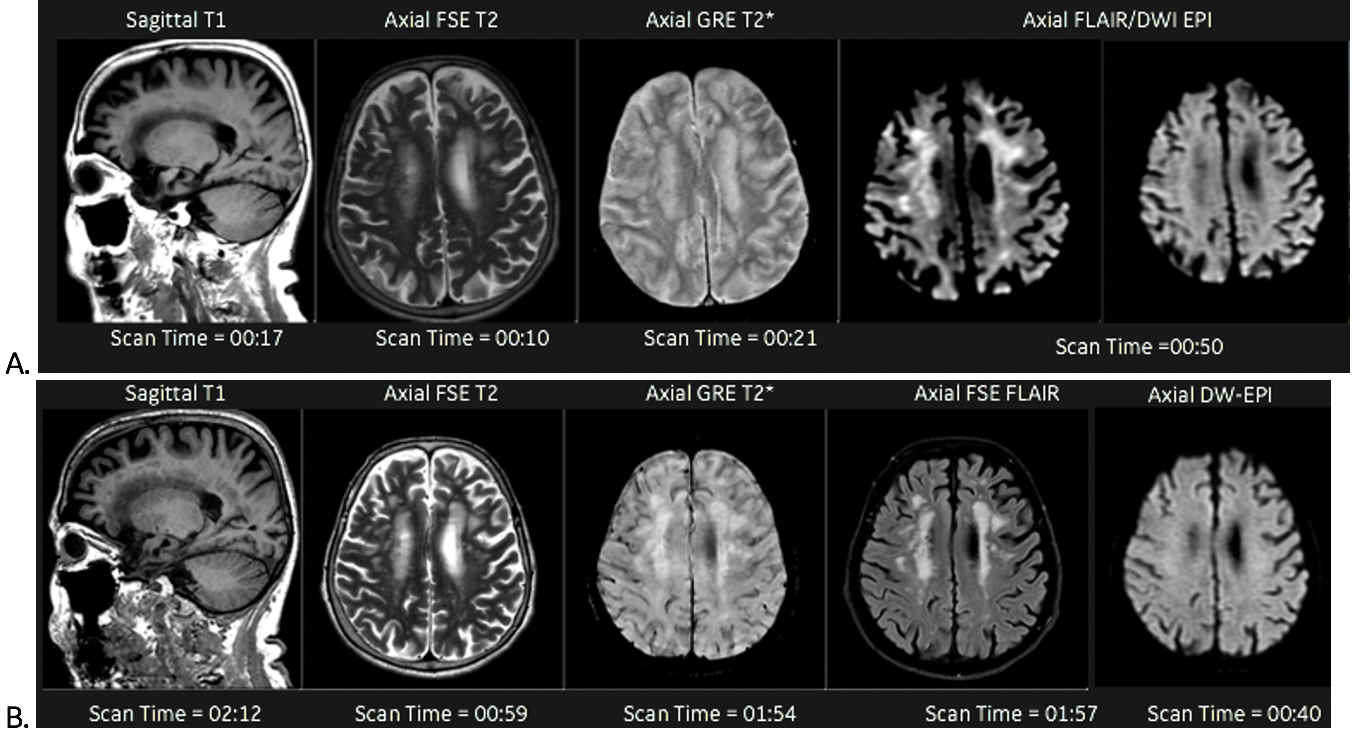
By employing the DL reconstruction combined with the hsMRI, image quality was maintained while significantly reducing overall exam duration. This shift from exam time of 10 – 15 to 1.5 – 2 minutes resulted in quality images, reduction in the need to repeat due to artifacts and a overall improved patient experiences (fig 2.).Discussion
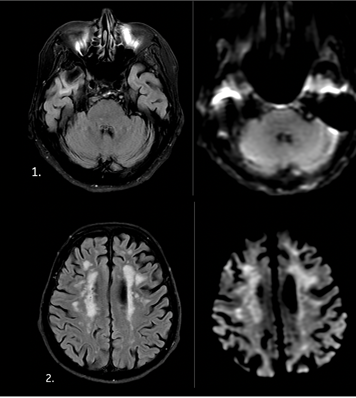
Our results from this abstract provides preliminary data on the use of implementing DLRecon to hsMRI for significant exam/sequence time reduction while preserving quality similar to standard brain protocols. The concept of fasting imaging in MR has been on the forefront of many users for a variety of reason such as reducing the use of CT, improve utilization to free up the scanner for increase patient throughput, reduce the need of sedation for pediatrics and with the evolution of MR systems for ED patients. Even with advances in MR gradient and RF coil technology, the ability to have consistent quality fast MR scans have proven difficult. The introduction of Deep-Learning bridges this gap, making it possible to achieve both speed and quality. Nonetheless, T2 FLAIR contrast image quality is still questionable with this high-speed approach due to low spatial resolution and distortion artifacts. FLAIR FSE requires 2 – 3 acquisitions in order to properly play out TR and TI, so using FSE approach was not a viable option in achieving such fast exam times. This was done by replacing the T2 weight B0 that is typically acquired with DWI with T2 FLAIR weighted B0 and using those reconstructed images to provide FLAIR imaging & DWI in one pass. A detail review shows similar sensitivity to FSE FLAIR when comparing to the hybrid FLAIR-DWI, but also reveals poor spatial resolution comparison and increase artifacts at the skull base because it is EPI (fig 3). The T2* GRE is also lower in quality & resolution compared to 3D SWI, but it’s still highly sensitive to small disturbances in the magnetic field making it useful for detecting blood products.Conclusion
In this work, we demonstrated that high-speed brain protocols can provide sufficient diagnostic capabilities compared to routine standard protocols with less than one-fourth of the scan time without significant changes to image quality. This reduction allows for significant increase in scanner utilization, operational efficiency and overall time the patient may need to be sedated. The fast nature of this protocol also provides reduction typical imaging related artifacts such as motion and image ghosting. More work is required on FLAIR contrast or finding an alternative to FSE FLAIR that can be acquired in less than 1 minute.Acknowledgements
No acknowledgement found.References
[1] Rozovsky, Katya et al. Fast-brain MRI in children is quick, without sedation, and radiation-free, but beware of limitations. Journal of Clinical Neuroscience
[2] Ramgopal, S., Karim, S.A., Subramanian, S. et al. Rapid brain MRI protocols reduce head computerized tomography use in the pediatric emergency department. BMC Pediatr 20, 14 (2020).
Figures