3966
High-resolution Diffusion Tensor Imaging at 7T with Multi-band Multi-shot EPI acquisition and Deep Learning Reconstruction1GE Healthcare, Houston, TX, United States, 2GE Healthcare, Waukesha, WI, United States, 3GE Healthcare, Calgary, AB, Canada, 4Center for Magnetic Resonance Research, University of Minnesota, Minneapolis, MN, United States, 5GE Healthcare, Menlo Park, CA, United States
Synopsis
Diffusion tensor imaging (DTI) is a well-established tool for providing insights into brain network connectivity and detecting brain microstructure but suffers from artifacts, low SNR, low spatial resolution, and long scan times. High-resolution DTI at 7T with multiband MUSE (MB-MUSE) and noise reduction methods have shown many potentials for mitigating these challenges. In this study, we combine a deep learning reconstruction method with MB-MUSE to overcome the image quality challenges and demonstrate improved quantification of high-resolution DTI at 7T compared with MB-MUSE and MB-MUSE with low-rank denoising.
Introduction
Diffusion tensor imaging (DTI) is a well-established tool for providing insights into brain network connectivity and detecting brain microstructure. However, DTI suffers from artifacts (distortion, Gibbs ringing, etc.), low SNR, and low spatial resolution, leading to errors in tensor estimation and increased acquisition times. While the high sensitivity at 7T enables higher spatial resolution, it also brings additional challenges of increased distortion and rapid T2* decay, which necessitates a segmented DTI acquisition approach. Acceleration strategies are also needed to shorten the prohibitively long acquisition time of a thin slice whole brain multi-shot DTI with high number of diffusion directions. High-resolution DTI at 7T with multiband multi-shot EPI acquisition and noise reduction methods1,2 have shown many potentials for mitigating these challenges. Multi-shot EPI can reduce image distortion; multiband can accelerate the acquisition; noise reduction reconstruction methods improve the SNR. However, the performance of the low-rank or PCA-based noise reduction methods is dependent on the setting of reconstruction parameters. Moreover, Gibbs ringing artifacts are still present or enhanced in the processed images, affecting the measures derived from dMRI3,4. In this study we aim to combine a deep learning-based reconstruction method with multi-band multi-shot diffusion acquisitions to improve the image quality and quantification of high-resolution DTI at 7T by 1) improving SNR and in-plane resolution and 2) reducing Gibbs ringing artifacts.Methods
Reconstruction: The proposed reconstruction included the conventional Multi-band multiplexed sensitivity-encoding (MB-MUSE)5 and a deep learning-based MR image reconstruction (DLRecon) pipeline6, which removes both noise and ringing artifacts. For comparison, a recently proposed Noise reduction with Distribution Corrected (NORDIC) PCA was applied2,7 to the original complex valued MB-MUSE images to remove the noise.Data Acquisition: DTI images were acquired on a GE 7T SIGNA MRI scanner with a 32-channel head coil and an ultra-high performance gradient (max. strength 100mT/m, max. slew rate=200T/m/s). The images were acquired at two resolutions: 1.2x1.2x1.2mm3 and 1.0x1.0x1.0mm3. For 1.2mm3 isotropic acquisition, TE=58ms, TR = 4 s, b = 1000 s/mm2, directions = 45. For 1-mm isotropic acquisition, TE = 61 ms, TR = 4.5s, b=800s/mm2, directions = 32. Other acquisition parameters are same: FOV = 256X256X78mm3, partial Fourier = 0.75, bands = 2, shots = 4.
Results and Discussion
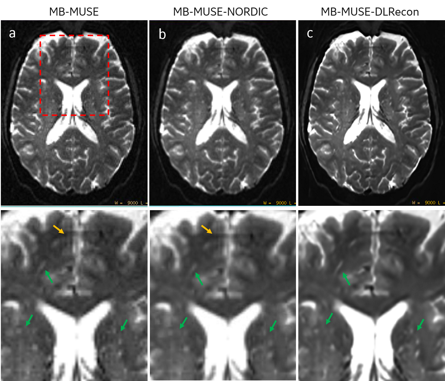
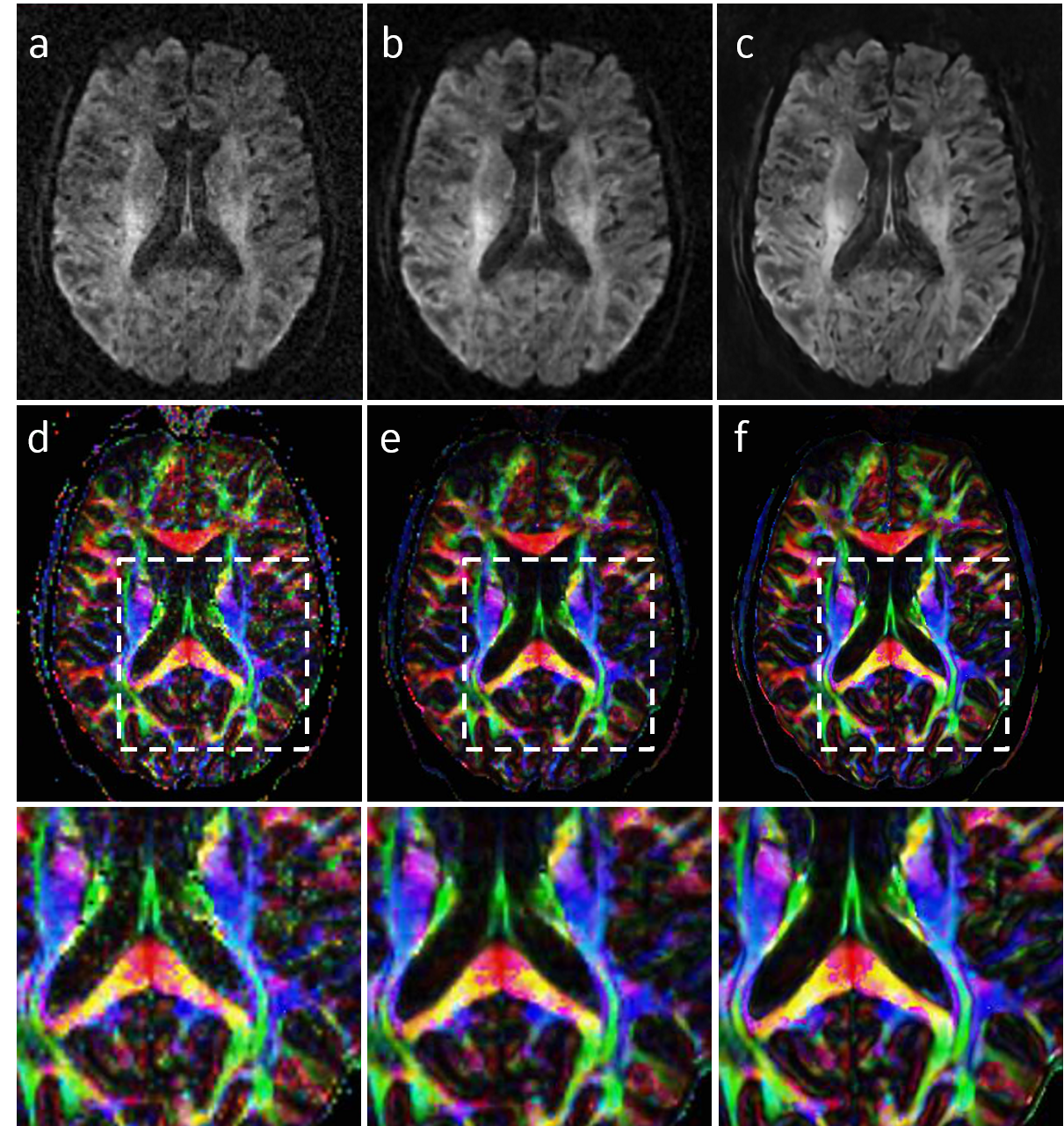
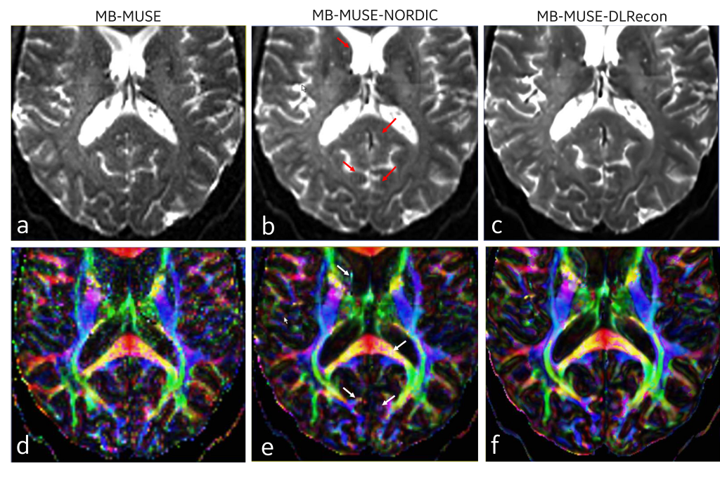
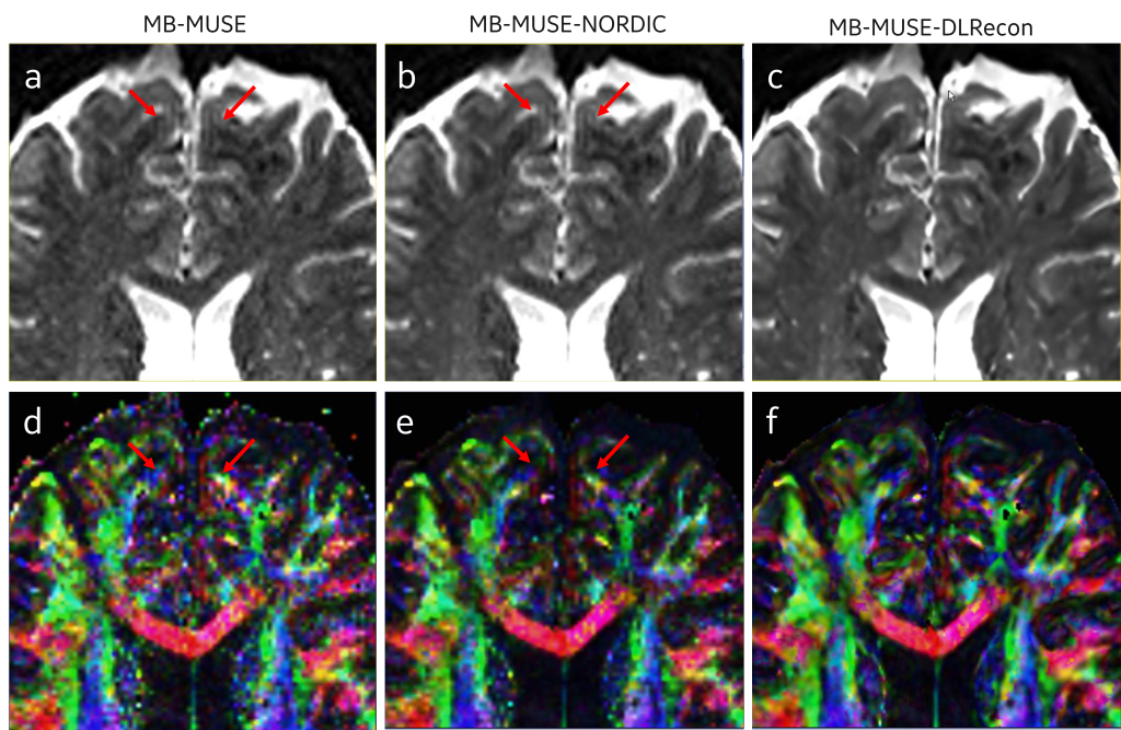
Figure 1 showed the b0 images acquired at 1.2-mm isotropic resolution and reconstructed using the original MB-MUSE, MB-MUSE with NORDIC and MB-MUSE with DLRecon. Compared to the original MB-MUSE, both NORDIC and DLRecon improved the SNR. However, the small vessels in DLRecon images are clearer than those in NORDIC images. The edges of other tissues (i.e. white/grey matter, ventricle) in DLRecon images are sharper than those in NORDIC images because DLRecon can also improve the in-plane resolution. Since NORDIC can only remove the noise, Gibbs ringing artifacts are present in both the original MB-MUSE and NORDIC images. However, Gibbs ringing artifacts were well removed by DLRecon. A similar result was found with b1000 images (Figure 2): both NORDIC and DLRecon improved the SNR, but the DLRecon image (Fig. 2c) is sharper than the NORDIC image (Fig. 2b). The derived color-coded fractional anisotropy (FA) map from DLRecon images (Fig. 2f) is also sharper than that from NORDIC images (Fig. 2e).Moreover, DLRecon removed Gibbs ringing artifacts in the source images and reduced the quantification error in the derived FA map, as shown in Figure 3. As discussed in the previous study3,4, Gibbs ringing can affect the derived measurements in DTI. When the noise is removed, the effect of Gibbs ringing artifacts becomes more obvious. The lines in the FA map (Figure 3e) resulting from the Gibbs ringing artifacts are more obvious in the NORDIC image, indicating the over-estimate or under-estimate of FA. However, DLRecon can simultaneously remove noise and ringing artifacts, improving SNR and the measurements, as shown in Figure 3f.
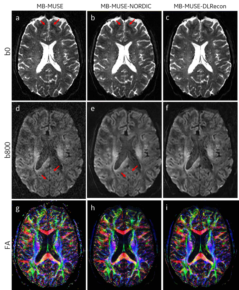
The same results were observed in images acquired at 1-mm isotropic resolution. As shown in Figure 4, both NORDIC and DLRecon achieved efficient denoising, but the DLRecon image is sharper than the NORDIC image because of the in-plane resolution improvement. The Gibbs ringing artifacts are present in b0 and b800 MB-MUSE images with and without NORDIC, resulting in lines in the derived FA maps. However, DLRecon efficiently removed these Gibbs ringing artifacts. The color-coded direction map derived from DLRecon images is also sharper than that derived from NORDIC images.
The effect of Gibbs rining on diffusion metric estimation has been previously demonstrated3 in the literature and postprocessing tools for ringing suppression have been researched. In this work we explored the benefits of the DL reconstruction algorithm that operates on complex MR data, without the use of any postprocessing tools.
Conclusion
Both NORDIC and DLRecon provide effective denoising, but DLRecon can effectively remove Gibbs ringing, improve the in-plane resolution and image sharpness in addition to noise removal, resulting in better SNR, FA measurements, and in-plane resolution.Acknowledgements
Development of NORDIC was partly supported by research funds: P41 EB027061 and U01 EB025144References
1. Dai E, Bruns B, Yang B, et al. Ultra-high-resolution diffusion MRI on 7T with multiband multishot EPI acquisition and joint reconstruction. CMRR ultra high field workshop, 2021
2. Moeller S, Pisharady PK, Ramanna S, et al. NOise Reduction with DIstribution Corrected (NORDIC) PCA in dMRI with complex-valued parameter-free locally low-rank processing. NeuroImage. 2021;226:117539.
3. Veraart J, Fieremans E, Jelescu IO, et al. Gibbs ringing in diffusion MRI. Magnetic resonance in medicine. 2016;76(1):301-14.
4. Perrone D, Aelterman J, Pižurica A, et al. The effect of Gibbs ringing artifacts on measures derived from diffusion MRI. Neuroimage. 2015;120:441-55.
5. Hing-Chiu Chang, etc. Interleaved EPI based fMRI improved by Multiplexed Sensitivity Encoding (MUSE) and Simultaneous Multi-Band Imaging
6. Lebel, R.M. Performance characterization of a novel deep learning-based MR image reconstruction pipeline. August 2020, http://arxiv.org/abs/2008.06559
7. https://github.com/SteenMoeller/NORDIC_Raw
Figures