3950
8ch multi transmit with ultrasonic dual axis gradient and 32ch receive setup for fast spatial encoding at 7T
Mark Gosselink1, Aris van Ieperen1, Wout Schuth2, Martino Borgo2, Dimitri Welting1, Edwin Versteeg1, and Dennis Klomp1
1Department of Radiology, University Medical Center Utrecht, Utrecht, Netherlands, 2Futura composites, Heerhugowaard, Netherlands
1Department of Radiology, University Medical Center Utrecht, Utrecht, Netherlands, 2Futura composites, Heerhugowaard, Netherlands
Synopsis
We propose a setup to boost spatiotemporal encoding for MRI in the human brain using a conventional receiver array (32ch) and fast gradients (6100T/m/s). Moreover, we demonstrate that the operation frequency of a high-end gradient-amplifier can be increased to ultrasonic frequencies so to avoid unpleasant acoustic noise. By using this amplifier with 8 RF transmitters, 32 receivers and a 2-channel gradient coil, a light-weight setup is constructed for operation in a 7 Tesla MRI system. The setup is demonstrated for operation in the human brain showing good FLAIR data and no peripheral nerve stimulation at the slew rate of 6100T/m/s.
Introduction
Silent MRI can be obtained when using an extra gradient insert that can drive an EPI readout above the human hearing frequency and can be operated in conjunction with the conventional body gradients [1]. When driving gradients in EPI mode, the selwrate can be increased by an order of magnitude before causing peripheral nerve stimulation. Here we propose a light-weight design that includes a second gradient axis, so to facilitate spiral readouts above 20kHz, and investigate if human brain MRI performance is maintained and peripheral nerve stimulation remains absent.Methods
The gradient amplifier used is a NG500 1.1 (Prodrive Technologies, Son, Netherlands). The FPGA firmware of the gradient amplifier has been modified to allow for a higher frequency range output. The new firmware modifies the output driver to allow the output of a harmonic of the base frequency, increasing the output frequency of one amplifier to 23kHz and t a second amplifier to 27kHz. The internal end-stage filter was removed and brought to the gradient coil as a resonant circuit. A dual axis gradient insert was built (Futura, the Netherlands) that has been made with windings in the Y and Z axes, (Fig 1). The high driving frequency of the insert allowed for minimal use of epoxy, as minimal movement is expected due the inertia of the coil above 20 kHz. Therefore, the entire setup weighs less than 25kg. Capacitors have been added to the inductive windings to filter unwanted amplifier currents while enhancing gradient field efficiency. To minimize eddy currents, no RF shield was used between the insert gradient and the RF coils. Eight ultrathin (2mm) 7T 1H dipoles transmitter, matched to ~50 ohm at 298MHz, are integrated into the design so to provide sufficient space for a standard 32 channel receiver array (NOVA, USA). To measure the efficacy of the ultrasonic gradient setup a field camera (Skope, Swiss) has been placed inside the insert gradient and RF setup. In vivo MRI scans were obtained with the entire setup. First, constructive B1+ phase shimming was applied, followed by a B1 map to verify flip angle setting. Then a FLAIR sequence was obtained using the 32 receivers and 8ch transmitters in the presence of the insert gradient. Finally, the 6100mT/m/s (for Z) and 3040mT/m/s (for Y) were applied for 64 times in 10ms shots to the human brain of a healthy volunteer to verify absence of peripheral nerve stimulation.Results
With the gradient amplifier driven at less than 30% of its range, already 6100T/m/s at 42mT/m (Z) and 3040mT/m/s at 18mT/m (Y) was obtained, resulting in the expected operating frequency of 23 and 27kHz. In the presence of the gradient insert, the 8 channel dipole array could obtain 15.6uT in the human brain (Fig 2), while the 32 receivers provided excellent FLAIR images of the human brain (Fig 3). Using the identical gradient waves as measured by fieldcameras, repeated 64 times, no peripheral nerve stimulation was observed by the healthy volunteer.Discussion
While a fixed resonance above 20kHz is pleasant for sound perception and simplifies cable management, it coincides with limitations in flexibilities to setup the MRI experiment. Furthermore, no standard MR-sequences exist for 5 gradient channels. Nonetheless, the setup demonstrates similar performance as a state-of-the-art 7T 8ch-32ch head coil, even in the presence of a close fitting two channel gradient insert. Moreover, as the third axis is not implemented, full visual view is maintained.Conclusion
We successfully increased the operation frequency of a high-end gradient amplifier by altering the firmware and bringing the end filter to the coil. Moreover, a low weight setup was constructed that houses 8 transceivers, 32 receivers, 2 channel cooled gradient coil for operation in a 7T MRI system for MRI experiments in the human brain, even providing access for visual stimulation. The operation of the setup was confirmed using field cameras and MRI images from the human brain, while no PNS was observed when operating spirals at 6100 and 3040T/m/s in the brainAcknowledgements
No acknowledgement found.References
Versteeg, E. et al. MRM2021Figures
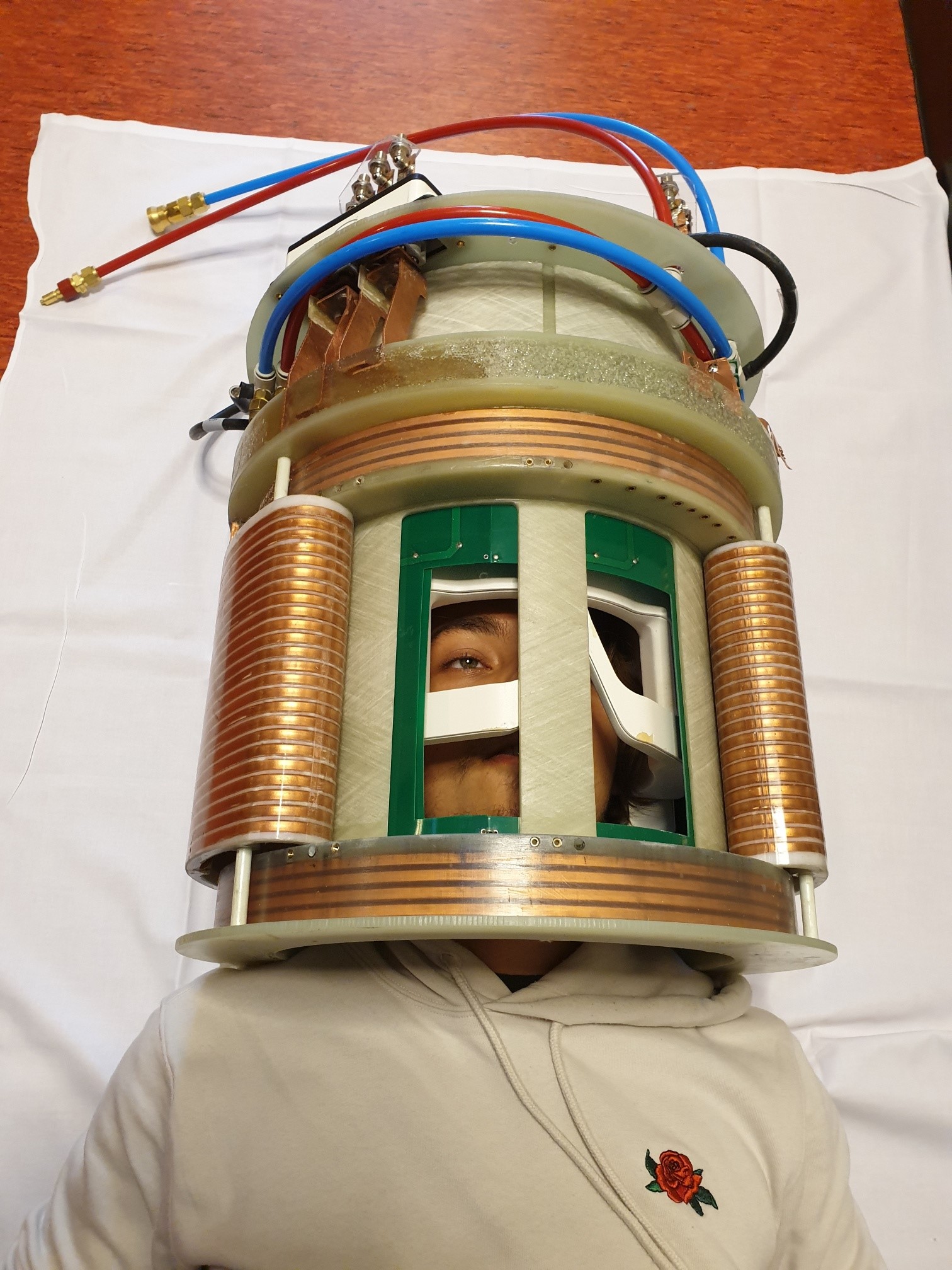
Figure 1: Photo of the fully integrated setup, that
comprises of the 32ch receiver array (closest to the head), the 8 ch dipole
array (dark green), the Y and Z gradients, and cooling tubes..
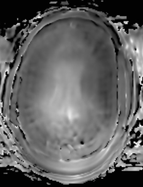
Figure 2: B1 map obtained with
the 8 dipoles in the presence of the gradient insert, the 32ch receiver array
and the human brain. Note the reference B1 was set to 12uT, and the map
demonstrates 130% of B, so the actual B1 was 15.6uT..
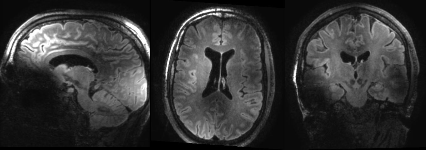
Figure 3: B1 map obtained with
the 8 dipoles in the presence of the gradient insert, the 32ch receiver array
and the human brain. Note the reference B1 was set to 12uT, and the map
demonstrates 130% of B, so the actual B1 was 15.6uT..
DOI: https://doi.org/10.58530/2022/3950