3939
Spin Echo-Echo Planar Imaging Sequence Validation for MR Elastography of Intervertebral Discs
Megan Co1, Huiming Dong2, Daniel J. Boulter2, Xuan V. Nguyen2, Safdar N. Khan2, Brian Raterman2, Brett Klamer3, Arunark Kolipaka1,2, and Benjamin A. Walter1,2,4
1Biomedical Engineering, The Ohio State University, Columbus, OH, United States, 2Radiology, The Ohio State University, Columbus, OH, United States, 3Center for Biostatistics, The Ohio State University, Columbus, OH, United States, 4Spine Research Institute, The Ohio State University, Columbus, OH, United States
1Biomedical Engineering, The Ohio State University, Columbus, OH, United States, 2Radiology, The Ohio State University, Columbus, OH, United States, 3Center for Biostatistics, The Ohio State University, Columbus, OH, United States, 4Spine Research Institute, The Ohio State University, Columbus, OH, United States
Synopsis
Magnetic resonance elastography (MRE) is an imaging technique that can non-invasively assess shear properties of intervertebral discs (IVD), a potential biomarker for disease. This study validated the use of a spin echo-echo planar imaging (SE-EPI) sequence for MRE of the IVD compared against the standard gradient recalled echo (GRE) sequence. Volunteers were scanned with the GRE and two variants of the SE-EPI sequence: SE-EPI with breath holds (SE-EPI-BH) and SE-EPI with free breathing (SE-EPI-FB). SE-EPI-based MRE-derived stiffnesses are highly reproducible and repeatable and correlate with current standard GRE MRE-derived stiffness estimates while reducing scan times and potentially improving patient compliance.
Introduction
Non-invasively assessing the mechanical function of spinal soft-tissues may allow earlier diagnosis of injury and disease, improve patient stratification, and facilitate in-vivo evaluation of regenerative interventions for low back pain (LBP). Magnetic resonance elastography (MRE) is a non-invasive imaging technique that allows the assessment of the shear properties of soft tissues. We have previously demonstrated that MRE-derived shear stiffness measurements acquired using a conventional 2D gradient recalled echo (GRE) sequence increase with IVD degeneration, potentially serving as a biomarker for disease1. However, previous studies have demonstrated that 2D stiffness measurements acquired from a single slice can overestimate tissue stiffness and that 3D measurements acquired from multiple slices are more accurate2,3. However, 3D GRE acquires slices sequentially leading to long scan times. Spin echo-echo planar imaging (SE-EPI) MRE sequences can rapidly acquire multiple slices within the effective repetition, potentially reducing scan time. In addition, GRE sequences require breath holds while SE-EPI sequences allow for scans with or without breath holds, potentially increasing patient compliance. Therefore, the objective of this study was to validate a SE-EPI MRE sequence for application within the IVD by (1) comparing the SE-EPI and GRE derived stiffnesses and (2) by determining the reproducibility and repeatability of the breath hold (SE-EPI-BH) and free breathing (SE-EPI-FB) version of the SE-EPI sequence.Methods
All protocols were approved by The Ohio State University Institutional Review Board and all images were acquired on a 3T MR scanner (Prisma, Siemens Healthcare, Germany). At least one lumbar IVD (L34-L45) from twenty-eight subjects (ages: 19-55) was scanned. 80 Hz vibrations were applied to the subject’s lower back via a pneumatic driver system. GRE scans consisting of three transverse slices through each IVD were acquired using a previously described GRE sequence1. This was immediately followed by a second MRE scan of the same IVD consisting of three transverse slices using two variants of a previously described SE-EPI sequence4: SE-EPI-BH and SE-EPI-FB (scan 1). The MRE-derived shear stiffness measurements from the GRE and these first SE-EPI scans were used to evaluate the comparability between GRE and SE-EPI sequences. The subject was then repositioned and the same IVD was scanned for a second time using the SE-EPI sequences (scan 2). The first and second sets of SE-EPI scans were used to assess reproducibility of the SE-EPI sequences. The same IVD was immediately scanned again using the SE-EPI sequences without repositioning the subject in between scans (scan 3). The second and third sets of SE-EPI scans of the subject in the same position were used to assess repeatability of the SE-EPI sequences. Shear stiffness measurements were calculated via the principal frequency analysis (PFA) method1. Octahedral shear strain – signal to noise ratio (OSS-SNR) was calculated for all scans as described elsewhere5. Linear mixed models were used to estimate the mean difference of OSS-SNR between all sequences. The comparison between stiffness measurements derived from the GRE and SE-EPI sequences as well as SE-EPI reproducibility and repeatability were evaluated using Lin’s concordance correlation coefficients (CCC) and Bland-Altman tests. For the comparison of GRE versus SE-EPI sequences, SE-EPI stiffness values were adjusted by each group’s observed mean bias before estimating Lin’s CCC.Results and Discussion
Good correlation was observed between shear stiffnesses derived from the SE-EPI sequences with those derived from the GRE sequence after adjusting for the observed mean difference with CCC values greater than 0.73 and 0.78 for the nucleus pulposus (NP) and annulus fibrosus (AF) region, respectively (Figure 1). OSS-SNR was not significantly different between GRE and SE-EPI sequences. SE-EPI sequences generated highly reproducible and repeatable stiffness measurements with CCC values greater than 0.97 in the NP and AF regions, respectively, (Figure 2) and reduced scan time by at least 51% compared to GRE. SE-EPI-BH and SE-EPI-FB stiffness measurements were similar with CCC values greater than 0.98 for both regions (Figure 3).Conclusion
This study demonstrated the feasibility of utilizing a SE-EPI MRE sequence within IVDs across the degenerative spectrum. Results showed that SE-EPI sequences provided good correlation to GRE MRE-derived shear stiffness estimates with slight and consistent underestimation and similar OSS-SNR. SE-EPI derived stiffness estimates were highly reproducible and repeatable. Further, the flexibility of the SE-EPI sequences to occur under both free breathing and breath hold configurations creates more options to address patient compliance and facilitates the scanning process to obtain viable data. Overall, the reduced scan times, increase in patient compliance, and high reproducibility and repeatability of measurements comparable to GRE suggest that SE-EPI-based MRE of the IVD has greater potential to be utilized in a clinical setting for imaging of patients suffering from low back pain.Acknowledgements
Funded by the Department of Biomedical Engineering, the National Institute of Arthritis and Musculoskeletal and Skin Diseases R01AR075062, and the National Heart, Lung and Blood Institute of the National Institutes of Health R01HL124096. The content is solely the responsibility of the authors and does not necessarily represent the official views of the National Institutes of Health. Special thanks to Prateek Karla and Sreyas Pillai for assistance with image post-processing.References
- Walter BA, Mageswaran P, Mo X, et al. MR elastography-derived stiffness: A biomarker for intervertebral disc degeneration. Radiology. 2017. doi:10.1148/radiol.2017162287
- Kenyhercz WE, Raterman B, Illapani VSP, et al. Quantification of aortic stiffness using magnetic resonance elastography: Measurement reproducibility, pulse wave velocity comparison, changes over cardiac cycle, and relationship with age. Magn Reson Med. 2016. doi:10.1002/mrm.25719
- Wassenaar PA, Eleswarpu CN, Schroeder SA, et al. Measuring age-dependent myocardial stiffness across the cardiac cycle using MR elastography: A reproducibility study. Magn Reson Med. 2016. doi:10.1002/mrm.25760
- Dong H, Jin N, Kannengiesser S, Raterman B, White RD, Kolipaka A. Magnetic resonance elastography for estimating in vivo stiffness of the abdominal aorta using cardiac-gated spin-echo echo-planar imaging: a feasibility study. NMR Biomed. 2020;(September):1-14. doi:10.1002/nbm.4420
- McGarry MDJ, Van Houten EEW, Perrĩez PR, Pattison AJ, Weaver JB, Paulsen KD. An octahedral shear strain-based measure of SNR for 3D MR elastography. Phys Med Biol. 2011;56(13). doi:10.1088/0031-9155/56/13/N02
Figures
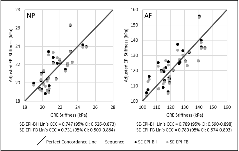
Figure 1 – Comparison between GRE and SE-EPI derived
shear stiffness: Lin’s concordance correlations based on the mean shifted
data between MRE-derived shear stiffness values acquired from GRE and SE-EPI
sequences for the nucleus pulposus (NP) and annulus fibrosus (AF) region.
SE-EPI-Breath Hold (SE-EPI-BH) shown in black and SE-EPI-Free Breathing
(SE-EPI-FB) shown in gray.
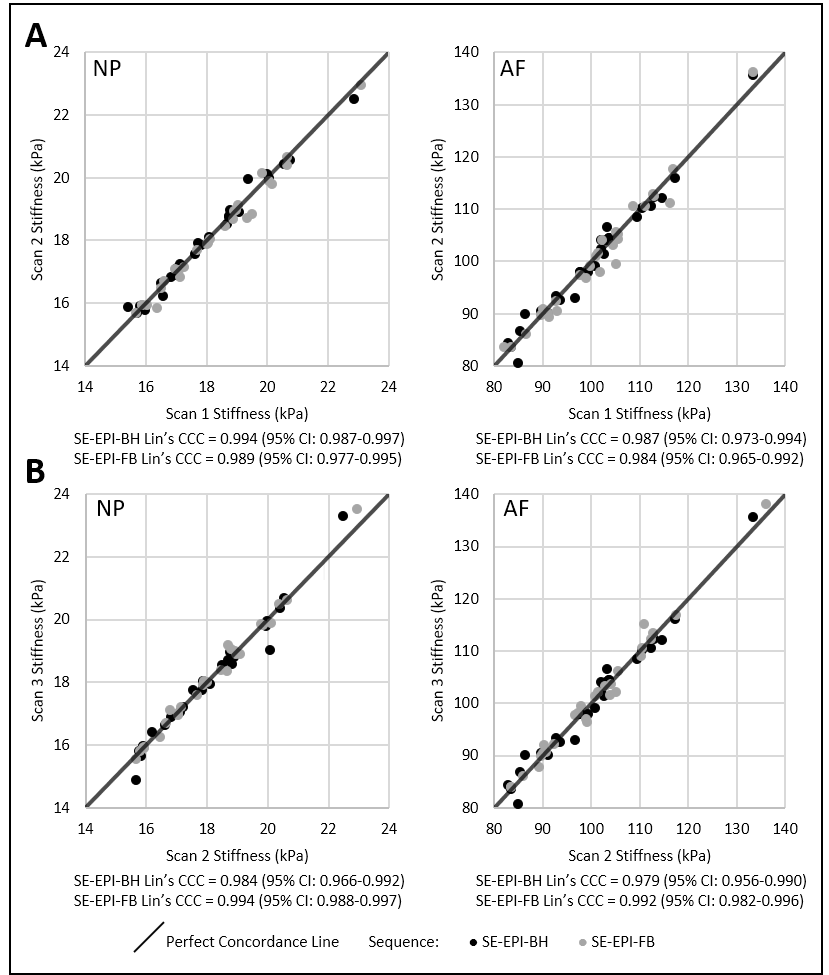
Figure 2 –
Reproducibility & repeatability analysis of SE-EPI derived shear
stiffness: Lin’s concordance correlations demonstrating the agreement
between (A) scan 1 and scan 2 (reproducibility) and between (B) scan 2 and scan
3 (repeatability) of SE-EPI sequences for the nucleus pulposus (NP) and annulus
fibrosus (AF) region; SE-EPI-Breath Hold (SE-EPI-BH) shown in black and
SE-EPI-Free Breathing (SE-EPI-FB) shown in gray.
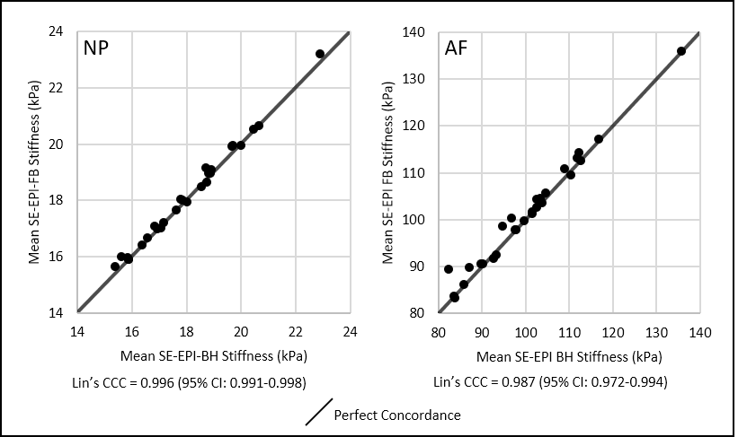
Figure 3 – Comparison between SE-EPI sequences: Lin’s
concordance correlations between MRE-derived shear stiffness values acquired
from SE-EPI-Breath Hold (SE-EPI-BH) and SE-EPI-Free Breathing (SE-EPI-FB) sequences
for the nucleus pulposus (NP) and annulus fibrosus (AF) region.
DOI: https://doi.org/10.58530/2022/3939