3938
A 3D SPACE-DIXON Technique for Fat Suppression in Knee Imaging1MR R&D Collaborations, Siemens Medical Solutions USA, New York, NY, United States, 2MR R&D Collaborations, Siemens Medical Solutions USA, Los Angeles, CA, United States, 3Department of Radiology, New York University Grossman School of Medicine, New York, NY, United States, 4Department of Radiology, NYU Grossman School of Medicine, New York, NY, United States
Synopsis
We present a 3D SPACE-DIXON technique with improved fat suppression performance for musculoskeletal 3D SPACE MRI. Phantom experiments were used to evaluate the fat suppression efficiency. Preliminary in vivo results indicate the clinical utility of this technique for proton density-weighted 3D MRI.
Introduction
In musculoskeletal (MSK) MRI, chemical shift and inversion recovery-based fat suppression techniques are employed to improve the conspicuity of various acute and chronic abnormalities[1]. However, both techniques are affected by B0 and B1 field variations, which result in inhomogeneous fat suppression[2].DIXON techniques acquire data at different echo times (TE) to encode the chemical shift between fat and water, which is more robust to B0 heterogeneities and permits more uniform fat suppression[3,4]. In addition, compared to the fat suppressed (FS) images and the non-FS images in separate acquisitions, Dixon techniques can provide various image contrasts, including water-only images and in-phase images equivalent to FS and non-FS images respectively, in a single acquisition for better efficiency.
The aim of this work was to develop a 3D SPACE based 2-Point DIXON technique for efficient and uniform fat suppression and evaluate it in proton density fat fraction (PDFF) phantoms and in vivo acquisitions.
Methods
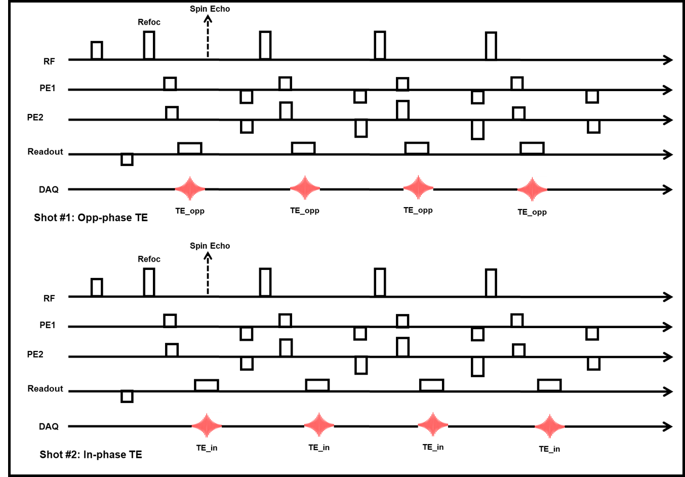
Data AcquisitionFigure 1 shows a prototype pulse sequence diagram for the SPACE-DIXON acquisition. 3D k-space data is acquired at the opposed-phase and in-phase echo times in separate shots, interleaved across TRs to minimize motion effects. The in-phase echo is acquired at the spin echo, corresponding to the conventional non-fat suppressed SPACE acquisition. This DIXON scheme is integrated with the SPACE variable refocusing flip angle approach to enable SAR efficient sampling.
The acquired in- and opposed-phase data are processed retrospectively offline using a prototype DIXON algorithm to generate the fat-only and water-only images. The contrast in the computed water image can be qualitatively enhanced using a weighted combination of fat and water signal: $$$Water_{enhanced} = Water + \lambda Fat$$$ Choice of the scaling factor $$$\lambda$$$controls the amount of fat suppression.
Phantom MRI
The prototype sequence was tested on a 3T MRI system (MAGNETOM Prisma, Siemens Healthcare, Erlangen, Germany). Performance of the SPACE DIXON technique was evaluated using a set of commercial PDFF phantoms (Calimetrix, Madison, WI USA) with PDFF values of 0%, 5%, 10%, 20%, 30%, 40%, and 100%. Reference PDFF estimates were obtained using a product multi-echo VIBE-DIXON sequence with the following parameters: resolution: 1.7mm3, TE=1.24–7.4 ms, sup>, ETL=40, TE=108ms, TR=1000ms, Acceleration factor=CAIPIRINHA 2x2, Acq Time= 5min. 3D SPACE-DIXON data were acquired with parameters matched to the non-FS protocol except acceleration factor = GRAPPA 2x2 and Acq Time = 7min34sec. Additionally, a 2D TSE-DIXON sequence was acquired to generate reference water-only images with the following parameters: resolution=0.47mmx0.47mmx3mm, ETL=15, TE=31ms.
In-vivo MRI
Data were acquired on volunteers after IRB approval and informed consent on a 3T MRI system (MAGNETOM Prisma, Siemens Healthcare, Erlangen, Germany). Non-fat suppressed 3D PD SPACE data were acquired with the following parameters: resolution=0.68mm3,ETL=60,TE=33ms,TR=900ms, Acceleration factor=CAIPIRINHA 2x2, Acq Time= 4min10sec. SPAIR fat suppressed 3D T2 SPACE data were acquired with the following parameters: resolution=0.68mm3,ETL=40,TE=108ms,TR=1000ms, Acceleration factor=CAIPIRINHA 2x2, Acq Time= 5min. 3D SPACE-DIXON data were acquired with parameters matched to the non-FS protocol except acceleration factor=GRAPPA 2x2 and AcqTime = 7min34sec. Additionally, a 2D TSE-DIXON sequence was acquired to generate reference water-only images with the following parameters: resolution=0.47mmx0.47mmx3mm,ETL=15,TE=31ms.
Results
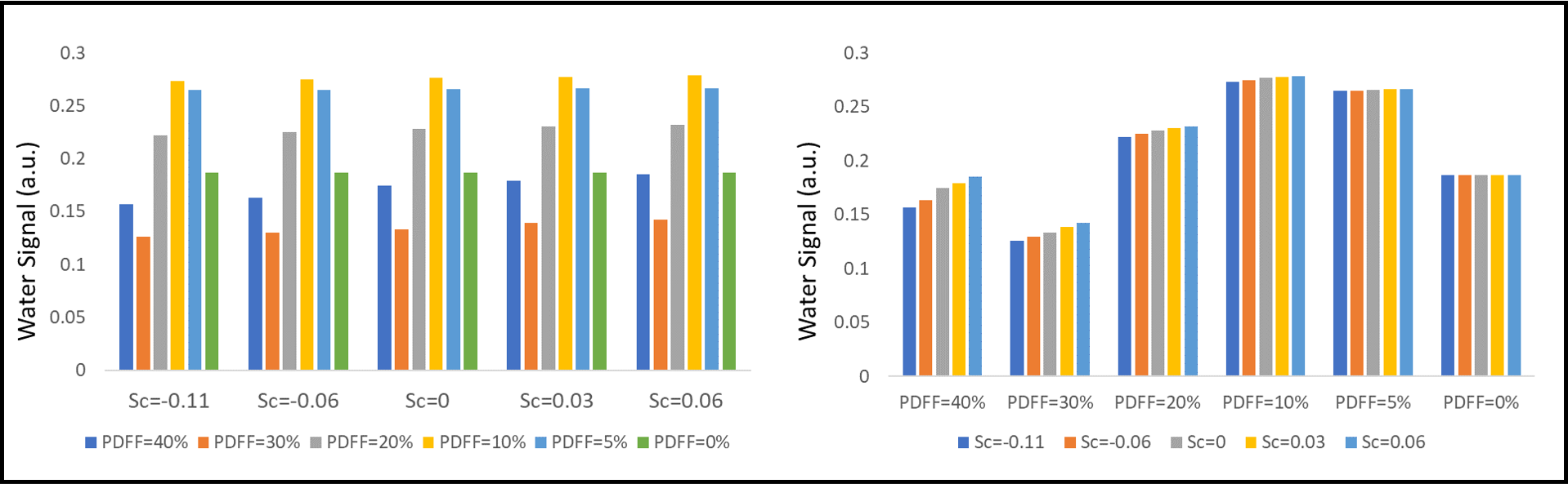
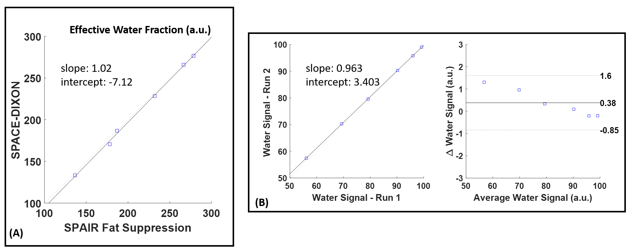
The fat and water images generated by the 3D SPACE-DIXON technique were used to compute an enhanced water image using the following scaling factors $$$\lambda = -0.11, -0.06, 0, 0.03, 0.06$$$. Figure 2 shows a plot of the water-signal values for the different enhancement scaling factors. Note that there is an increase in the water signal with higher scaling factors especially for the high PDFF phantoms.Figure 3A shows a linear correlation plot of the residual water signal from a SPAIR fat-suppressed 3D SPACE acquisition and the proposed 3D SPACE-DIXON sequence for the different phantoms, demonstrating very good correlation (r=0.98). Figure 3B shows results from the phantom reproducibility study. The linear correlation plot and Bland-Altman plot comparing the water signal values obtained from two separate measurements are shown. The mean difference in water signal between the measurements was 0.38 with the 95% limits of agreement being [-0.85, 1.6]. A correlation analysis indicated very strong correlation between the two measurements (Pearson r = 0.99). A Bland-Altman analysis was performed between the two measurements and the estimated water signal has a coefficient of reproducibility of 1.22 and a coefficient of variation of 0.76%.
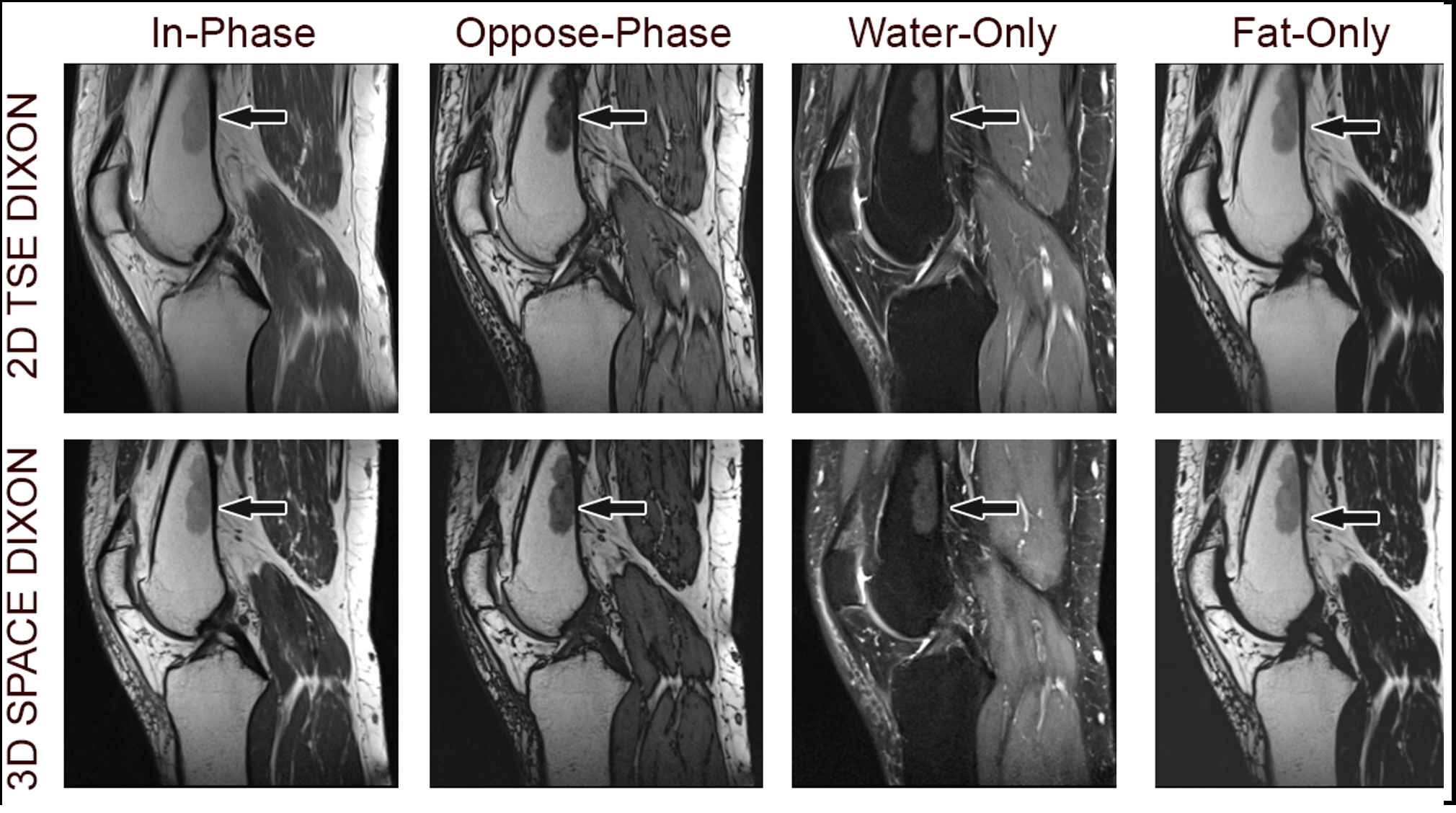
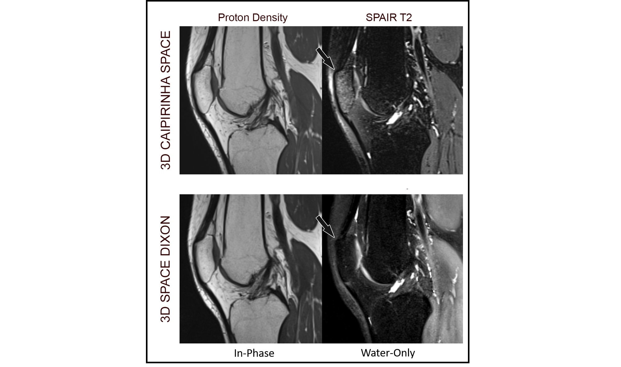
Figure 4 shows representative 3D SPACE-DIXON MR images from a volunteer along with the fat and water images from a 2D TSE DIXON acquisition. Note that the signal alterations observed in the SPACE-DIXON images are well correlated with the TSE DIXON water image. Figure 5 compares non-fat suppressed PD-weighted and SPAIR fat suppressed T2-weighted images from a 3D CAIPIRINHA SPACE sequence with the in-phase and water-only images from SPACE-DIXON. It can be observed that the SPACE-DIXON water-only image has more uniform fat suppression compared to fat suppressed SPACE.
Conclusions
A SPACE-DIXON technique was developed in this study and evaluated in phantom experiments and in vivo acquisitions. This technique demonstrated improved efficiency to provide water-only and in-phase images in a single acquisition, compared to the FS and non-FS images in two acquisitions. Uniform fat suppression was also demonstrated in 3D in vivo musculoskeletal imaging. Preliminary in vivo results indicate the clinical utility of this technique for proton density-weighted 3D MRI.Acknowledgements
No acknowledgement found.References
1. Del Grande F, Santini F, Herzka DA, et al. Fat-suppression techniques for 3-T MR imaging of the musculoskeletal system. Radiographics. 2014;34(1):217-233. doi:10.1148/rg.341135130
2. Glover, Gary H. "Multipoint Dixon technique for water and fat proton and susceptibility imaging." Journal of Magnetic Resonance Imaging 1.5 (1991): 521-530.
3. Glover GH, Schneider E. Three-point Dixon technique for true water/fat decomposition with B0 inhomogeneity correction. Magn Reson Med 1991; 18: 371–383.
4. Glover GH. Multipoint Dixon technique for water and fat proton and susceptibility imaging. J Magn Reson Imaging 1991; 1: 521–530.
Figures