3932
A Free-Breathing Hybrid Technique for Simultaneous T2w, T1w, PDFF, and R2* Imaging at 0.55T1Siemens Medical Solutions USA, Malvern, PA, United States, 2Department of Radiology, New York University Grossman School of Medicine, New York, NY, United States, 3Department of Radiology, Weill Cornell Medical College, New York, NY, United States, 4Siemens Healthcare GmbH, Erlangen, Germany
Synopsis
Recently, there has been renewed interest in imaging at low field strengths. The increased T2* relaxation times at 0.55T and reduced chemical shift between fat and water requires the need for longer TEs for accurate fat fraction and R2* estimation, often resulting in longer acquisition times. In this work, we present a scan-efficient hybrid TSE-GRE technique for the simultaneous acquisition of T2- and T1-weighted images along with quantification of fat fraction and R2*. The use of a stack-of-stars trajectory allows SNR efficient free breathing imaging at 0.55T.
Background and Motivation
With ongoing advancements in MRI technology, there has been an increased interest in imaging at lower field strengths (<1T) [1-3]. Recently, the ability to perform diagnostic abdominal imaging at 0.55T was demonstrated [4]. Conventional abdominal imaging protocols include the acquisition of T2- and T1-weighted images. Optionally, chemical shift encoded sequences such as T1w-DIXON are also acquired for quantification of liver fat to enable improved diagnosis of fatty liver disease [5]. These techniques acquire gradient echo images at multiple echo times (TE) to separate fat and water signal while also estimating the R2* component corresponding to iron overload.The increased T2* relaxation times at 0.55T and reduced chemical shift between fat and water requires the need for longer TEs for accurate fat fraction (FF) and R2* estimation, often resulting in longer acquisition times. In this work, we present a scan-efficient hybrid TSE-GRE technique for the simultaneous acquisition of T2- and T1-weighted images along with quantification of fat fraction and R2*. We investigate the effect of TE on FF and R2* estimation performance using phantoms and in vivo data.
Theory
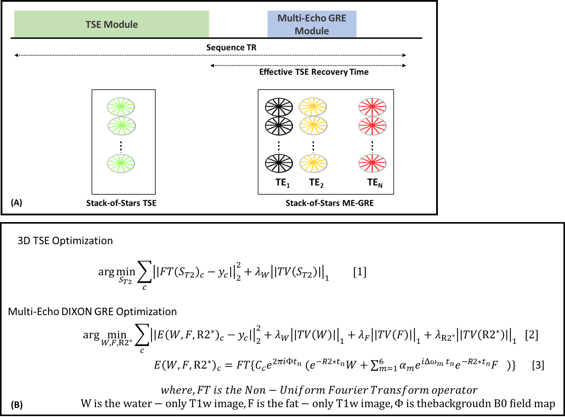
Pulse SequenceA previously proposed [6] Stack-of-Stars hybrid TSE-GRE pulse sequence was extended to acquired multiple echoes in the GRE echo train (Fig1A). By using a low flip angle for the gradient echo module, the impact on the TSE magnetization recovery is minimized. K-space is sampled using a radial stack-of-stars trajectory for both the TSE and GRE modules. Radial views are rotated by 2o between the GRE echoes using blipped gradients to improve k-space coverage.
Image Reconstruction
TSE and GRE data are reconstructed using a respiratory-compensated iterative reconstruction algorithm. The respiratory signal is extracted from the center of k-space of the GRE data and processed using principal component analysis and coil clustering [7]. The resultant signal is used in the reconstruction of both TSE and GRE data. The TSE and GRE data are reconstructed by solving the optimization problem formulated in Eq.1 and Eq.2 respectively (Fig1B) [6,8]. To better condition the fat/water separation, the acquired multi-echo data are first reconstructed using a non-iterative NUFFT operation and used to generate the background field map ($$$\Phi$$$) and an initial guess for fat and water signals.
Methods
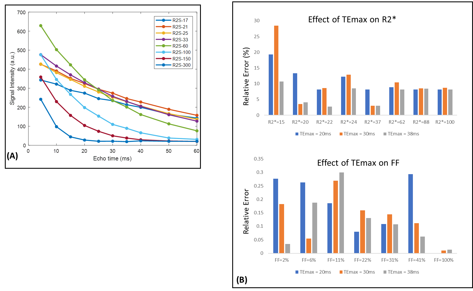
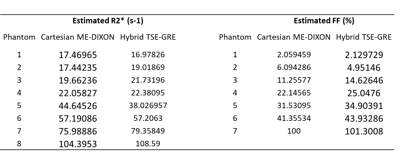
Phantom ImagingThe prototype sequence was tested on a commercial MRI system (MAGNETOM Aera 1.5T, Siemens Healthcare, Erlangen, Germany) modified to operate at a field strength of 0.55T. Quantification accuracy was evaluated using a set of fat fraction and R2* phantoms with FF values ranging from 0%-100% and R2* values from 15–93s-1. Reference estimates of R2* were acquired using a 2D Cartesian multi-echo GRE sequence with TEs=5ms–60ms, ∆TE=5ms and NEX=8. Reference FF estimate was obtained using a 3D Cartesian multi-echo GRE sequence. Data were acquired with the prototype hybrid sequence with the following parameters: resolution 1.56mmx1.56mmx3mm, radial views=400, TR=1.2sec, TSE ETL=42. To evaluate the effect of TE on the estimation accuracy, hybrid data was acquired with the following TEs for the GRE module: [6,12,20,30,38] ms, [6,12,18,25,30] ms, and [3,6,9,14,20] ms. A reproducibility study was performed by scanning the same set of phantoms in a two-week interval.
In vivo Imaging
All data were acquired after informed consent. A liver scan of a healthy volunteer was performed with the following parameters under free-breathing conditions: resolution=1.7mmx1.7mmx3mm, slices=36, TSE ETL=36, radial views=380. Other imaging parameters were identical to the phantom scans. Acquisition time=8min 30sec.
Results
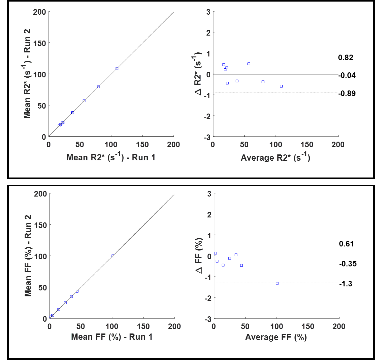
Impact of the TE time on R2* estimation is demonstrated in Figure 2. Figure 2A shows the signal evolution from the 2D multi-echo GRE reference acquisition for phantoms with varying R2*. Note that for R2* values of clinical interest at 0.55T (11.6–23.2s-1 [3]), sufficient temporal sampling of the T2* decay curve is needed to accurately estimate the R2*. This is also demonstrated in Figure 2B where the error in R2* estimation (for phantoms with longer T2*) increases at lower TEmax. However, error in estimated fat fraction is still within acceptable limits. Note that the R2* and FF values estimated from the hybrid technique are comparable to that from Cartesian multi-echo DIXON (Fig3).Figure 4 shows a Bland-Altman plot comparing the FF and R2* values obtained from two separate measurements. The FF and R2* estimates have coefficients of reproducibility of 0.86 and 0.95, respectively. The coefficient of variation was 0.96% and 1.5% for R2* and FF indicating very high agreement between measurements.
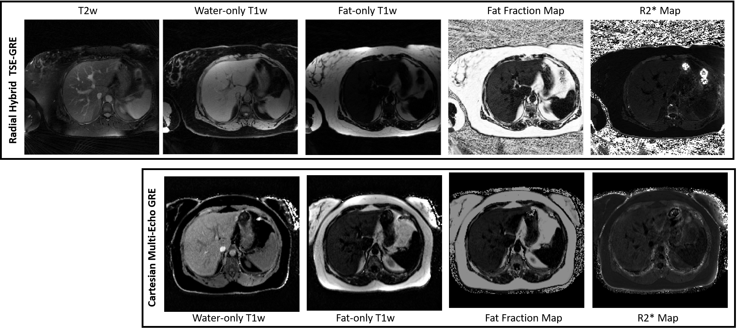
Figure 5 demonstrates the proposed technique on a volunteer. The hybrid sequence yields co-registered T2-weighted and T1-weighted images in addition to the quantitative maps. Further, due to the longer TR, number of slices in the Cartesian sequence had to be reduced to 20 to fit it into a 25 sec breath-hold.
Conclusions
We present a free-breathing hybrid TSE-multi-echo GRE technique for simultaneous T2w, T1w, PDFF, and R2* imaging in the liver at 0.55T. Phantom experiments were used to optimize protocol parameters for improved quantification accuracy. Preliminary in vivo results indicate clinical utility of the technique for FF and R2* quantification at low field strengths.Acknowledgements
No acknowledgement found.References
1. Runge VM, Heverhagen JT. Advocating the Development of Next-Generation, Advanced-Design Low-Field Magnetic Resonance Systems. Invest Radiol. 2020;55(12):747-753.
2. Sheth KN, Mazurek MH, Yuen MM, et al. Assessment of Brain Injury Using Portable, Low-Field Magnetic Resonance Imaging at the Bedside of Critically Ill Patients. JAMA Neurol. 2020.
3. Campbell-Washburn AE, Ramasawmy R, Restivo MC, et al. Opportunities in Interventional and Diagnostic Imaging by Using High-Performance Low-Field-Strength MRI. Radiology. 2019;293(2):384-393.
4. Chandarana, H., Bagga, B., Huang, C. et al. Diagnostic abdominal MR imaging on a prototype low-field 0.55 T scanner operating at two different gradient strengths. Abdom Radiol (2021)
5. Yu H, McKenzie CA, Shimakawa A, Vu AT, Brau ACS, Beatty PJ, Pineda AR, Brittain JH, Reeder SB. Multiecho reconstruction for simultaneous water-fat decomposition and T2* estimation. J Magn Reson Imaging. 2007;26:1153–61.
6. Benkert T, Mugler JP 3rd, Rigie DS, Sodickson DK, Chandarana H, Block KT. Hybrid T2 - and T1 -weighted radial acquisition for free-breathing abdominal examination. Magn Reson Med. 2018;80(5):1935-1948. doi:10.1002/mrm.27200
7. Feng L, Axel L, Chandarana H, Block KT, Sodickson DK, Otazo R. XD-GRASP: golden-angle radial MRI with reconstruction of extra motion-state dimensions using compressed sensing. Magn Reson Med. 2016;75:775-788
8. Schneider, M, Benkert, T, Solomon, E, et al. Free-breathing fat and R2* quantification in the liver using a stack-of-stars multi-echo acquisition with respiratory-resolved model-based reconstruction. Magn Reson Med. 2020; 84: 2592– 2605
Figures