3926
Bridging Structural MRI with Cognitive Function for Individual Level Classification of Early Psychosis via Deep Learning
Yang Wen1,2,3, Chuan Zhou2, Leiting Chen2, Yu Deng4, Martine Cleusix5, Raoul Jenni5, Philippe Conus6, Kim Q. Do5, and Lijing Xin1,3
1Animal imaging and technology core (AIT), Center for Biomedical Imaging (CIBM), Ecole Polytechnique Fédérale de Lausanne (EPFL), Lausanne, Switzerland, 2University of Electronic Science and Technology of China, Chengdu, China, 3Laboratory of Functional and Metabolic Imaging, Ecole Polytechnique Fédérale de Lausanne (EPFL), Lausanne, Switzerland, 4Department of Biomedical Engineering, King's College London, London, United Kingdom, 5Center for Psychiatric Neuroscience, Department of Psychiatry, Centre Hospitalier Universitaire Vaudois (CHUV) and University of Lausanne (UNIL), Lausanne, Switzerland, 6Service of General Psychiatry, Department of Psychiatry, Centre Hospitalier Universitaire Vaudois (CHUV) and University of Lausanne (UNIL), Lausanne, Switzerland
1Animal imaging and technology core (AIT), Center for Biomedical Imaging (CIBM), Ecole Polytechnique Fédérale de Lausanne (EPFL), Lausanne, Switzerland, 2University of Electronic Science and Technology of China, Chengdu, China, 3Laboratory of Functional and Metabolic Imaging, Ecole Polytechnique Fédérale de Lausanne (EPFL), Lausanne, Switzerland, 4Department of Biomedical Engineering, King's College London, London, United Kingdom, 5Center for Psychiatric Neuroscience, Department of Psychiatry, Centre Hospitalier Universitaire Vaudois (CHUV) and University of Lausanne (UNIL), Lausanne, Switzerland, 6Service of General Psychiatry, Department of Psychiatry, Centre Hospitalier Universitaire Vaudois (CHUV) and University of Lausanne (UNIL), Lausanne, Switzerland
Synopsis
The aims of this study were: (1) to test the feasibility of using a deep learning model with 7T sMRI as an input to predict cognition levels (CLs) at the single-subject level, and (2) to investigate whether the inclusion of CLs estimation could facilitate the classification for early psychosis (EP) patients and healthy controls (HCs). Promising accuracy was achieved in estimating CLs and the inclusion provides considerable classification improvement. Fivefold cross-validating experiments demonstrated higher classification AUC-ROC scores over published methods. Therefore, deep learning can be used to estimate CLs and CL estimation improves the classification performance of EP.
Introduction
Artificial intelligence (AI) approaches, particularly machine learning (ML) and deep learning (DL), have been extensively studied to accelerate medical data analysis and assist clinical interventions in many pathological contexts. Many applications have been conducted in psychiatric disorders using neuroimaging measures as input and incorporated with AI models to establish automated diagnostic workflows at a single subject level1. Previous machine learning works in schizophrenia have used handcrafted features extracted from sMRI data to distinguish patients from healthy individuals2, but such feature extraction process usually involves a long computational time. To reduce computational cost, recent efforts have focused on using directly sMRI images as input, and promising results have been achieved with the help of the latest AI models (e.g., convolutional neural networks, CNNs)3 4. However, these studies have mainly focused on patients at chronic stage, the classification of early psychosis (EP) patients from healthy controls (HCs), is more challenging5, because the brain structural changes in EP patients are mild and not evident, making computer-aided classification methods less robust and accurate.Furthermore, progressive cognitive deficit is one major feature of schizophrenia, inspiring the possibility of using individual cognition levels, in addition to sMRI images, to facilitate automated classification of EP patients from HCs. Several recent studies have used the DL framework to incorporate cognitive estimation into the workflow to facilitate the diagnosis of Alzheimer’s disease by explicitly including cognitive measures as secondary inputs6. However, this approach requires additional cognitive assessment that is not part of routine neuropsychiatric clinical examinations. Moreover, although several studies have been done using sMRI images to identify individual cognitive impairments via DL7, to the best of our knowledge, no study has been done to incorporate cognition estimation for classifying EP patients and controls. As shown in Fig.1A, given the sMRI image, we seek to estimate participant's cognitive level and classify EP patients from healthy individuals in a fully automated manner.
Methods
Input data-The GM and WM probability maps were considered as two different feature channels and make channel concatenations to generate a single 3D array as the input to our model. Unlike previous study4, where different segmentation components were used as multiple inputs and fed into a model in parallel, the multi-channel 3D array helps to reduce the training parameters and retain all the information from GM and WM. Besides the WM and GM maps, volumetric and surface analysis was also performed with CAT12 toolkit to calculate region of interest (ROI) volumes and cortical surface thickness as features for comparison. The analysis was adopted with default settings and obtained 388 ROI volume features and 219 cortical thickness features after filtering out the null values.Deep learning model: Three-dimensional multi-task learning framework-Given the sMRI array as input, a three-dimensional CNN model (3D-CNN) was used to encode visual features, similar in previous studies3 4. To predict both the cognitive level and the probability of EP for each participant, a multi-task learning framework is introduced. Based on the same visual features extracted by the 3D-CNN, three independent multilayer perceptron networks were used as individual subbranches for different tasks, including EP classification, cognitive level classification (CLC) and cognitive level regression (CLR). The complete architecture of our 3D-CNN encoder and multi-task learning framework is depicted in Fig.1B. Several popular classification models were selected for comparison, including random forest (RF), supported vector machine (SVM), gradient boost machine (GBM) and 2D-CNNs8.
Model optimization-Our framework is an end-to-end deep learning system and thus several loss functions were used to train the proposed model for parameter updating. Specifically, for classification tasks (i.e., EP and CLC), the cross-entropy loss is used. For the task of CLR, the mean square error loss is used. As an end-to-end framework, training losses are back-propagated from three multi-task subbranches to the 3D-CNN, updating the parameters of the entire network with an optimization algorithm.
Results
Our method, as demonstrated in Fig.2, achieved the best overall accuracy on CLs estimation, and as shown in Fig.3, obtained the best AUC-ROC score of 0.711 on EP/HC classification with the inclusion of CL estimation. The discriminative regions identified by our model as potential biomarkers for EP classification and CLC are shown in Fig. 4. Evidently, for the EP classification, the entire GM structure is emphasized, and saliency appears in the frontal, occipital, and temporal lobe regions, as well as putamen. Furthermore, by comparing the attentional maps between EP classification and CLC subtasks, we found that the thalamus contributed more to EP classification than CLC, suggesting that the thalamus plays an important role in the recognition of EP.Discussion and conclusion
This study demonstrated the feasibility of using a deep learning model with sMRI as an input to predict CLs at the single-subject level, as well as the feasibility of including CLs estimation to facilitate EP classification. Good accuracy and AUC-ROC score are achieved under a fivefold cross-validation. All of the regions recognized by our model in Fig.4 are highly consistent with the results in previous studies, such as putamen, occipital lobe and frontal lobe whose volume were considered associated to schizophrenia occurrence9 10 and cognition deficits11 in group-level analysis, indicating that the potential of identification of biomarkers from sMRI by DL methods.Acknowledgements
This work was supported by Sichuan Science and Technology Program (No.2019YJ0176 / 2019YJ0177 / 2019YFQ0005), China Scholarship Council, and National Center of Competence in Research (NCCR) ”SYNAPSY - The Synaptic Bases of Mental Diseases” from the Swiss National Science Foundation (n◦ 51AU40 185897 to KQD &PC). We acknowledge access to the facilities and expertise of the CIBM Center for Biomedical Imaging, a Swiss research center of excellence founded and supported by Lausanne University Hospital (CHUV), University of Lausanne (UNIL), EPFL, University of Geneva (UNIGE) and Geneva University Hospitals (HUG).References
- Noor M B T, Zenia N Z, Kaiser M S, et al. Detecting neurodegenerative disease from mri: A brief review on a deep learning perspective. International Conference on Brain Informatics. Springer, Cham, 2019: 115-125.2.
- Lu X, Yang Y, Wu F, et al. Discriminative analysis of schizophrenia using support vector machine and recursive feature elimination on structural MRI images. Medicine, 2016, 95(30).3.
- Oh J, Oh B L, Lee K U, et al. Identifying schizophrenia using structural MRI with a deep learning algorithm. Frontiers in psychiatry, 2020, 11: 16.4.
- Hu M, Sim K, Zhou J H, et al. Brain MRI-based 3D Convolutional Neural Networks for Classification of Schizophrenia and Controls. 2020 42nd Annual International Conference of the IEEE Engineering in Medicine & Biology Society (EMBC). IEEE, 2020: 1742-1745.5.
- Cortes-Briones J A, Tapia-Rivas N I, D'Souza D C, et al. Going deep into schizophrenia with artificial intelligence. Schizophrenia Research, 2021.6.
- Spasov S, Passamonti L, Duggento A, et al. A parameter-efficient deep learning approach to predict conversion from mild cognitive impairment to Alzheimer's disease. Neuroimage, 2019, 189: 276-287.7.
- Sui J, Jiang R, Bustillo J, et al. Neuroimaging-based individualized prediction of cognition and behavior for mental disorders and health: methods and promises. Biological psychiatry, 2020, 88(11): 818-828.8.
- Li Z, Li W, Wei Y, et al. Deep learning based automatic diagnosis of first-episode psychosis, bipolar disorder and healthy controls. Computerized Medical Imaging and Graphics, 2021, 89: 101882.9.
- Haijma S V, Van Haren N, Cahn W, et al. Brain volumes in schizophrenia: a meta-analysis in over 18 000 subjects. Schizophrenia bulletin, 2013, 39(5): 1129-1138.10.
- Wright I C, Rabe-Hesketh S, Woodruff P W R, et al. Meta-analysis of regional brain volumes in schizophrenia. American Journal of Psychiatry, 2000, 157(1): 16-25.11.
- Antonova E, Kumari V, Morris R, et al. The relationship of structural alterations to cognitive deficits in schizophrenia: a voxel-based morphometry study. Biological psychiatry, 2005, 58(6): 457-467.
Figures

Figure 1. Illustrations of (A) our workflow for classification of early psychosis patients from healthy controls and cognitive estimation on six dimensions, and (B) the deep learning architecture with a 3D-CNN feature encoder and three independent multilayer perceptron subbranches for different subtasks of early psychosis classification and cognitive estimations.

Figure 2. Accuracy of our model in two-categorized CLC task compared with different ML and DL counterparts in six cognitive estimation dimensions.

Figure 3. Performance of ROC curves for EP/HC classification with fivefold cross-validation. The proposed model used GM map as input.
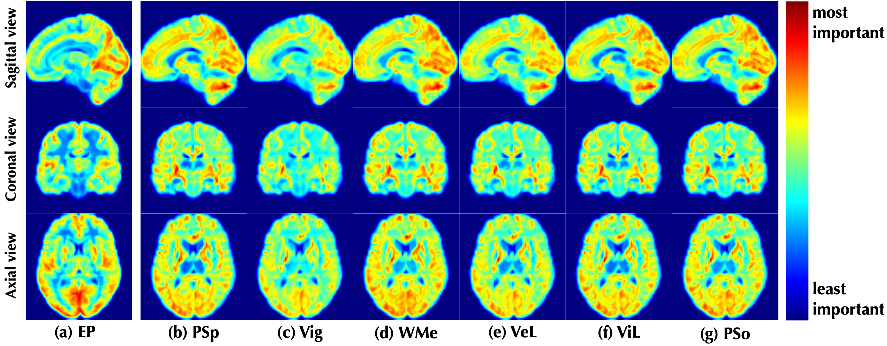
Figure 4. Visualization of the discriminative positions identified by the proposed model on (a) EP classification and (b-g) CLC tasks with attentional weights. The results were shown as the mean of all cases in the data set. We used both WM and GM images as input.
DOI: https://doi.org/10.58530/2022/3926