3920
Improving Motion-Robust Structural Imaging at 7T with Deep Learning-Based PROPELLER Reconstruction
Daniel V Litwiller1, Xinzeng Wang2, R Marc Lebel3, Baolian Yang4, Jeffrey McGovern4, Brian Burns5, and Suchandrima Banerjee6
1GE Healthcare, Denver, CO, United States, 2GE Healthcare, Houston, TX, United States, 3GE Healthcare, Calgary, AB, Canada, 4GE Healthcare, Waukesha, WI, United States, 5GE Healthcare, Olympia, WA, United States, 6GE Healthcare, Menlo Park, CA, United States
1GE Healthcare, Denver, CO, United States, 2GE Healthcare, Houston, TX, United States, 3GE Healthcare, Calgary, AB, Canada, 4GE Healthcare, Waukesha, WI, United States, 5GE Healthcare, Olympia, WA, United States, 6GE Healthcare, Menlo Park, CA, United States
Synopsis
The high sensitivity of MRI at 7T enables brain imaging with unprecedented spatial resolution, which can be important to the assessment of a variety of neurological disorders, such as multiple sclerosis, epilepsy, and neurodegenerative disease. With sub-millimeter voxel dimensions, and prolonged acquisition times, however, sensitivity to motion and pulsatility is increased dramatically. This increased sensitivity to motion can be managed with techniques like PROPELLER. Here, we present an initial assessment of a deep learning-based image reconstruction for high-resolution, 7T PROPELLER, and evaluate its ability to improve signal-to-noise ratio, and anatomical conspicuity, without increasing scan time.
Introduction
The increased signal-to-noise ratio available at 7T allows imaging at proportionally higher spatial resolutions, which can be important for visualizing finer structural detail and subtle lesions in a variety of neurological disorders, including multiple sclerosis, epilepsy, movement, neurodegenerative, and psychiatric disorders1. In the case of conventional, multi-shot, fast spin echo imaging, however, these higher spatial resolutions are often achieved at the expense of increased motion sensitivity, which can be further exacerbated by extended imaging times2. Among the existing methods for mitigating motion, PROPELLER (MultiVane, BLADE, etc.), is a well-established technique for compensating for rigid body motion, as found in the brain3. The purpose of this work was to evaluate a deep learning-based image reconstruction technique for increasing the signal-to-noise ratio (SNR) and anatomical conspicuity, without increasing scan time.Methods
In vivo imaging of consented volunteers was performed on a SIGNA™ 7.0T MRI scanner (GE Healthcare, Waukesha, WI), using a 32-channel head coil (Nova Medical, Wilmington, MA), and a T2-weighted PROPELLER acquisition with 200 um in-plane resolution, 1-mm slice thickness (40 nanoliter voxel), and other imaging parameters, summarized in Table 1. The PROPELLER images were reconstructed using both conventional methods, and a previously described deep learning-based image reconstruction (DL Recon), that was trained in a supervised fashion using thousands of paired training samples to increase both SNR and in-plane image resolution4. Reconstructions of the high-resolution, 7T PROPELLER images were inspected for reduced noise, improved anatomical conspicuity, and the overall absence of image artifacts.Results & Discussion
Figure 1 contains several axial T2-weighted images of the brain, acquired at 7T with 2D high-resolution fast spin echo (top), and PROPELLER (bottom). Even though both sequences provide excellent SNR and contrast, several sources of motion artifact are apparent in the FSE images, whereas the PROPELLER images show minimal motion contamination, and flow artifacts are suppressed. Figure 2 shows a comparison of conventional reconstruction and DL Recon for a high-resolution, coronal T2-weighted PROPELLER scan acquired with 200 um in-plane resolution. The SNR is considerably improved, and the finer structures in the amygdala, hippocampus, and cerebellum are much better delineated in the DL Recon image. In the zoomed inset, please note the improved visualization of the deep cerebellar nuclei, the cerebellar cortex, and the vermis. Figure 3 shows a sagittal acquisition with conventional reconstruction and DL Recon. Hippocampal layers are more distinct with DL Recon and the head, body, and tail of the hippocampus can be clearly seen. DL Recon also improves conspicuity of subtle vascular structures and Virchow-Robin spaces. Figure 4 depicts an enlarged portion of an axial slice through the midbrain, hippocampus, and amygdala. DL Recon improves overall SNR and anatomical conspicuity, enabling confident visualization of the hippocampal layers, as well as the substantia nigra, and complex vasculature branching from the Circle of Willis.Conclusion
The combination of ultra-high field (7T), a high-performance receiver array (32 channels), a motion robust acquisition (PROPELLER), and a state-of-the-art deep learning reconstruction, enables extremely high-resolution in-vivo imaging of the human brain with minimal artifacts. Motion and flow artifacts are well-suppressed by the acquisition, while SNR and overall anatomical conspicuity are improved by the deep learning-based image reconstruction. We demonstrate detailed visualization of deep brain structures, which are often difficult to detect due to SNR limitations, motion corruption, and/or flow artifacts.Acknowledgements
No acknowledgement found.References
- Balchandani P, Naidich TP. Ultra-high-field MR neuroimaging. American Journal of Neuroradiology. 2015 Jul 1;36(7):1204-15.
- Federau C, Gallichan D. Motion-correction enabled ultra-high resolution in-vivo 7T-MRI of the brain. PloS one. 2016 May 9;11(5):e0154974.
- Pipe JG. Motion correction with PROPELLER MRI: application to head motion and free-breathing cardiac imaging. Magn Reson Med. 1999 Nov;42(5):963-9.
- Lebel RM. Performance characterization of a novel deep learning-based MR image reconstruction pipeline. ArXiv 2020;abs/2008.06559.
Figures
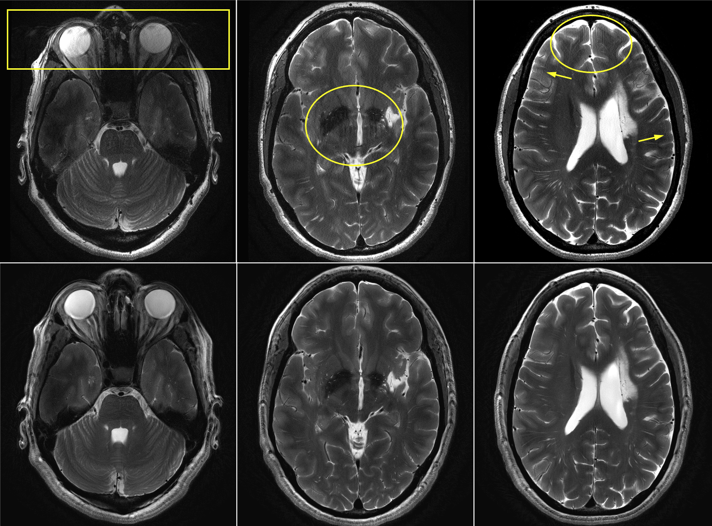
Figure 1. Axial T2-weighted images at three slice locations, acquired at 7T with high-resolution, multi-shot fast spin echo (top), and PROPELLER (bottom). Whereas the PROPELLER images are relatively free from detectable motion and/or flow artifacts, the conventional FSE images contain classic motion artifacts from a variety of sources, including motion of the orbits (top left), pulsation of the ventricles (top middle), and bulk motion of the head (top right).
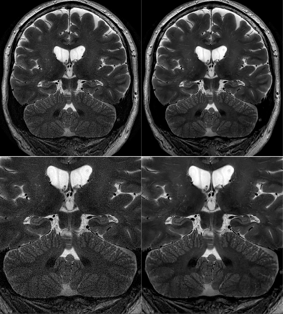
Figure 2. Coronal T2-weighted PROPELLER images with 200 um in-plane resolution, and 1-mm slice thickness (40 nanoliter voxel), reconstructed with conventional reconstruction (left), and deep learning-based reconstruction (right), demonstrating substantially improved signal-to-noise and anatomical conspicuity. Coronal section of the whole brain (top), and an enlarged view of the limbic system, including the hippocampus, and cerebellum (bottom).
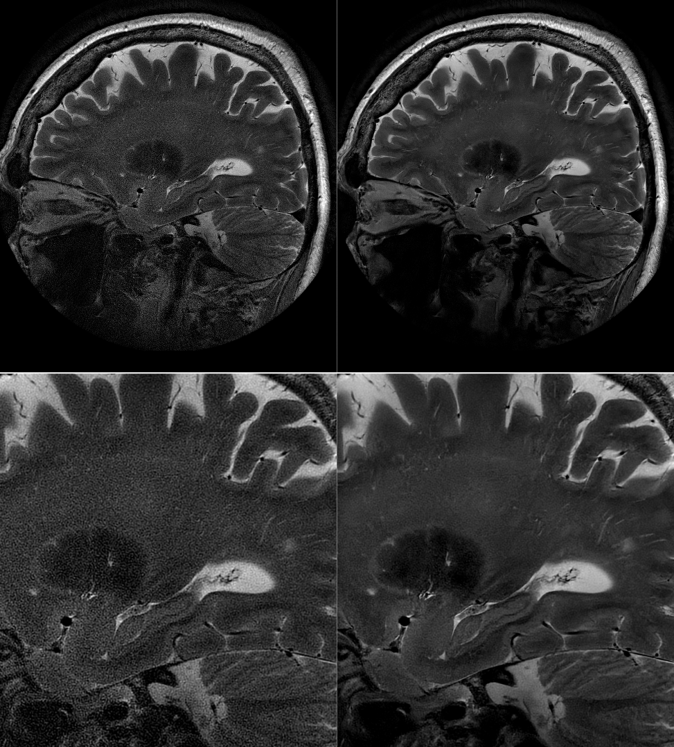
Figure 3. Sagittal T2-weighted PROPELLER images with 200 um in-plane resolution, and 1-mm slice thickness (40 nanoliter voxel), reconstructed with conventional reconstruction (left), and deep learning-based reconstruction (right). A substantial SNR improvement is evident with the DL reconstruction relative to the conventional, which facilitates detection of the hippocampal head, body, and tail.
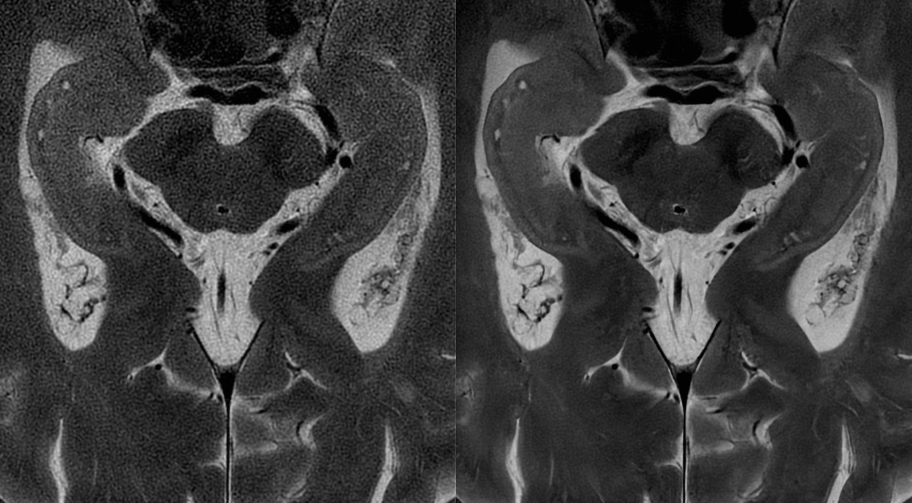
Figure 4. Enlarged axial T2-weighted PROPELLER images with 200 um in-plane resolution, and 1-mm slice thickness, reconstructed with conventional reconstruction (left), and deep learning-based reconstruction (right). A substantial SNR improvement is evident with the DL reconstruction relative to the conventional, which shows hippocampal layers and clearly depicts cerebrospinal fluid spaces within the hippocampus. Reduced ringing and improved SNR with DL Recon provides clear depiction of very small vascular structures, particularly within the ventricles.
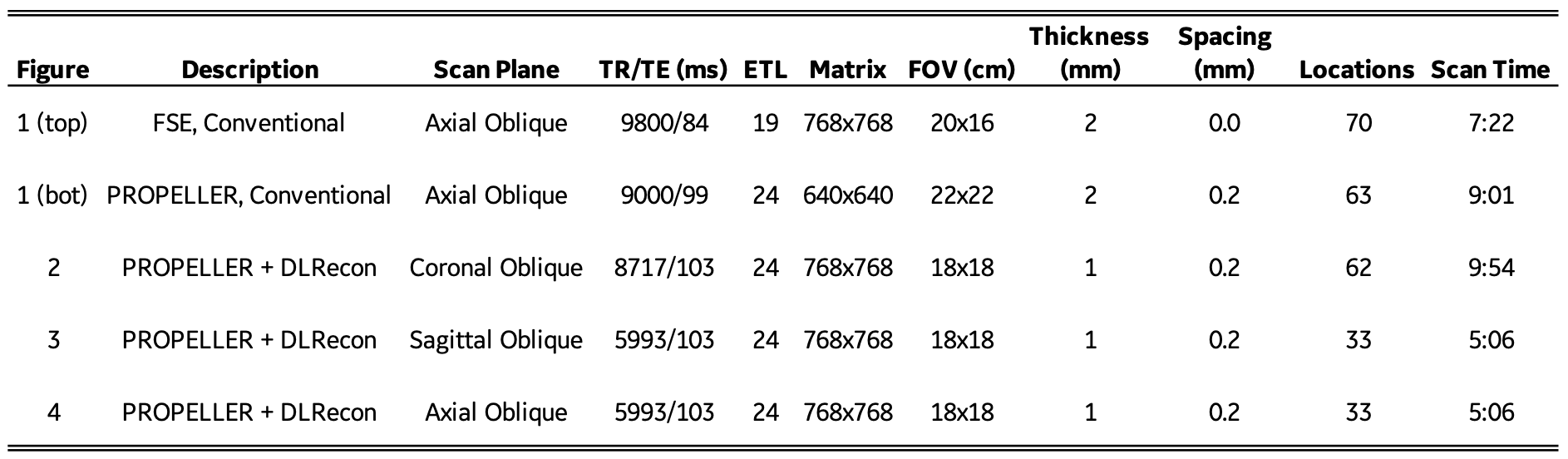
Table 1. Imaging parameters for Figures 1-4, including scan time.
DOI: https://doi.org/10.58530/2022/3920