3919
Robust Diffusion-Weighted Imaging with Deep Learning-Based DW PROPELLER Reconstruction1GE Healthcare, Houston, TX, United States, 2GE Healthcare, Waukesha, WI, United States, 3GE Healthcare, Denver, CO, United States, 4MD Anderson Cancer Center, Houston, TX, United States
Synopsis
Compared to DW-EPI, DW PROPELLER is less sensitive to susceptibility, chemical shift, and motion, and thus shows better image quality in areas, such as skull base, head-neck and pelvis. However, the SNR and in-plane resolution of DW PROPELLER images are often inferior to DW-EPI and often requires a longer scan time to compensate for this. In this work, we evaluated a deep-learning reconstruction method to improve the SNR of in-plane resolution of DW PROPELLER images without increasing acquisition time.
Introduction
Compared to DW-EPI, Diffusion Weighted PROPELLER (DW PROPELLER) is less sensitive to susceptibility, chemical shift, and motion, and thus shows better image quality in areas such as skull base, head-neck and pelvis (1-4). However, the SNR and in-plane resolution of DW PROPELLER images are often inferior to DW-EPI and often requires a longer scan time to achieve similar SNR and in-plane resolution. The purpose of this work is to evaluate a deep-learning reconstruction method to improve the SNR and in-plane resolution of DW PROPELLER images without increasing acquisition time.Methods
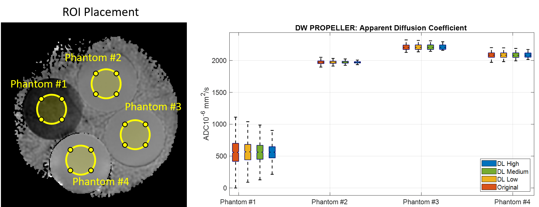
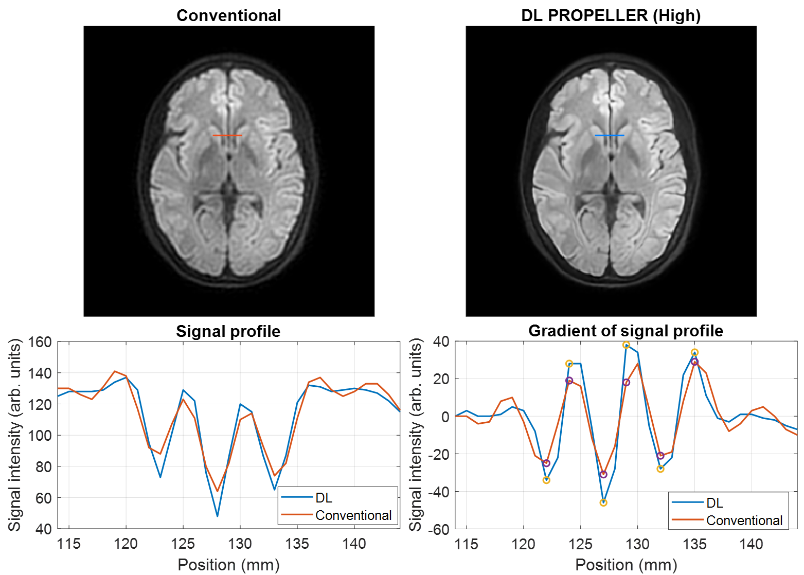
All of the diffusion weighted images were acquired on a 3T MRI scanner (SIGNA™ Premier, GE Healthcare, Waukesha, WI). A water-fat phantom was used to quantitatively compare the apparent diffusion coefficient (ADC) measurements in the images reconstructed using both the conventional and the deep learning-based reconstruction (DLRecon) methods (5). The DLRecon images were reconstructed at three denoising levels: low, medium, and high. The ADC maps were generated using the product software and algorithm. Four regions-of-interest (ROI#1 – #4) were placed in a uniform section of the four compartments to measure the ADC values (Figure 1). The water-fat phantom contains 4 total compartments (including 1 oil and 3 water-based) with different ADC values.The edge sharpness was evaluated in the human brain by drawing a line profile across a ventricle. Sharpness was quantified by comparing the maximum gradient in the line profile in the DLRecon images relative to the conventional image. This provided a relative measure of sharpness, independent of absolute intensity.
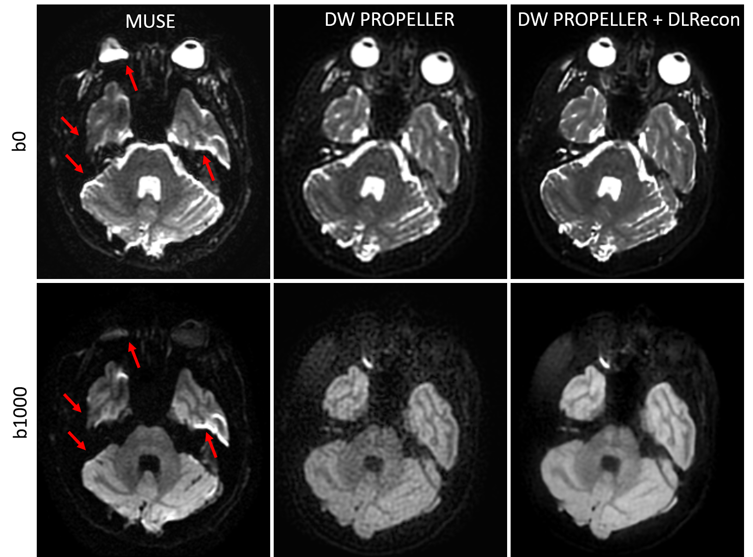
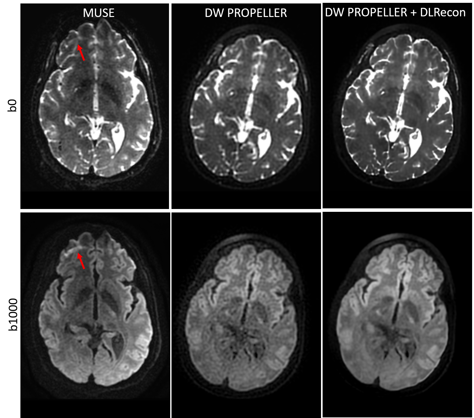
To evaluate the DW PROPELLER image quality, 3-shot EPI with multiplexed sensitivity encoding (MUSE) was used as the reference. Both the DW PROPELLER and DW EPI images were acquired at 1000 s/mm2 with 1.5-mm isotropic acquisition resolution using a 48-channel head coil. For optic nerve imaging, the DW PROPELLER images were acquired with 1.2x1.2 mm2 in-plane resolution, and 2.5 mm slice thickness, using a 21-channel head-and-neck coil. The coronal DW PROPELLER images were acquired with 1.5x1.5 mm2 in-plane resolution, and 4-mm slice thickness, using a 21-channel head-and-neck coil.
Results and Discussion
As shown in Figure 1, DLRecon doesn’t alter the measured mean ADC values, but reduces the standard deviation, and improves the precision of the ADC measurement at different denoising levels. The ADC values of the four ROIs are, Non-DL: 555.9 ± 210.3, 1972.5 ± 27.5, 2209.5 ± 40.6 and 2085.7 ± 46.3 10-6mm2/s; DLRecon Low: 558.8 ± 179.7, 1972.5 ± 22.3, 2211.2 ± 40.1 and 2085.0 ± 43.9 10-6mm2/s; DLRecon Medium: 559.8 ± 157.0, 1972.7 ± 18.6, 2211.2 ± 39.5, and 2085.2 ± 42.7 10-6mm2/s. DLRecon High: 563.1 ± 125.5, 1972.4 ± 13.9, 2211.8 ± 36.7 and 2086.3 ± 39.7 10-6mm2/s. Compared to the conventional image, there is no statistically significant change of the mean ADC, but the standard deviations of the ADC measurements are smaller with a higher denoising level, indicating that DL was effective in removing the noise and improving the precision of ADC measurement at different denoising levels (Fig. 1). This result bodes well for investigating the more general result of DL-recon aiding in decreasing bias and error of ADC values in low SNR or high b-value scenarios as well.As shown in Figure 2, DLRecon improves edge sharpness. A line profile across the ventricle was plotted to evaluate the edge sharpness. In the signal profile, DL showed a smaller full width half maximum (FWHM) than the conventional recon. The maximum gradient in the line profile in the DLRecon images is also higher than the conventional recon, indicating that DLRecon provides improved edge sharpness compared to the conventional recon (Fig. 2).
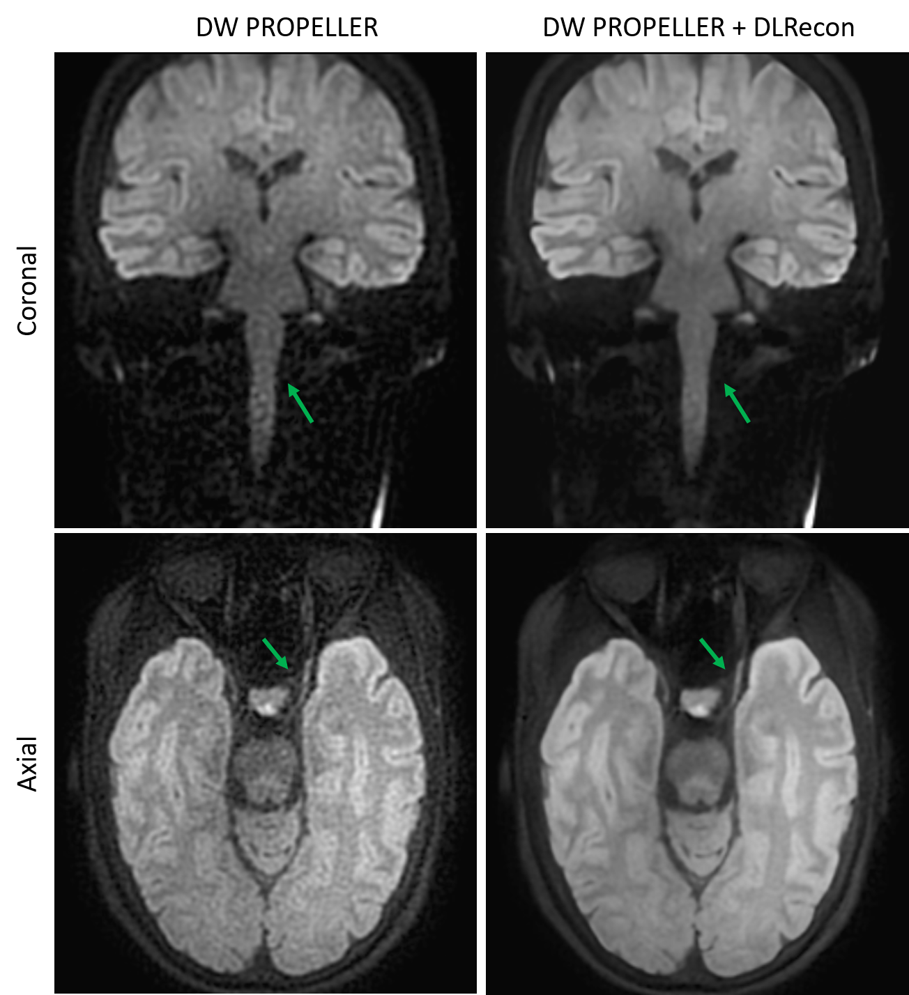
Compared to multi-shot DWEPI, DW PROPELLER is less sensitive to geometric distortion (Figure 3) and chemical shift artifacts (Figure 4). However, the conventional propeller DW images have a lower SNR and in-plane resolution compared to multi-shot EPI. With DLRecon, the SNR and in-plane resolution of DW PROPELLER images were significantly improved (Figures 3, 4), and are closer to multi-shot DWEPI, but with less geometric distortion and chemical shift artifact. With improved SNR and in-plane resolution, the DLRecon based DW PROPELLER achieved a better visualization of spinal cord in the coronal head neck imaging, and a better visualization of optical nerves in images of the skull base, as shown in Figure 5.
Conclusion
The deep learning-based DW PROPELLER reconstruction provides high-quality and more robust diffusion-weighted imaging by improving SNR and in-plane resolution, without increasing scan times. The deep learning-based reconstruction method can also be used to reduce the scan time of DW PROPELLER, without compromising SNR or resolution.Acknowledgements
No acknowledgement found.References
1. Juan CJ, Chang HC, Hsueh CJ, Liu HS, Huang YC, Chung HW, Chen CY, Kao HW, Huang GS. Salivary glands: echo-planar versus PROPELLER Diffusion-weighted MR imaging for assessment of ADCs. Radiology. 2009;253(1):144-52.
2. Mahmoud OM, Tominaga A, Amatya VJ, et al. Role of PROPELLER diffusion-weighted imaging and apparent diffusion coefficient in the evaluation of pituitary adenomas. European journal of radiology. 2011;80(2):412-7.
3. Mahmoud OM, Tominaga A, Amatya VJ, et al. Role of PROPELLER diffusion weighted imaging and apparent diffusion coefficient in the diagnosis of sellar and parasellar lesions. European journal of radiology. 2010;74(3):420-7
4. Fu Q, Kong XC, Liu DX, et al. Clinical comparison of single-shot EPI, readout-segmented EPI and TGSE-BLADE for diffusion-weighted imaging of cerebellopontine angle tumors on 3 tesla. Magnetic Resonance Imaging. 2021;84:76-83.
5. Wang X, Litwiller D, Lebel M, et al. High Resolution T2W imaging using Deep Learning Reconstruction and Reduced Field-of-View PROPELLER. ISMRM 2020;0820
Figures