3912
Real-time survey base estimation of gestational age to guide a fetal MRI scan1Centre for the developing Brain, King's College London, London, United Kingdom
Synopsis
Fetal MRI is an excellent tool for estimating the fetal gestational age using the head circumference measurements. This study measures the biometry information using fast 30 sec survey scans for the measurement of fetal head circumference using 3D UNet, Bi-parietal diameter, Frontal-occipital diameter for the estimation of the fetal gestational age in the second half of gestation. The results show high accuracy in gestational age measurements which correlates well with the clinical data.
Introduction
Fetal MRI scans provide high resolution information about the fetus and allows the estimation of their biological gestational age. MRI scans have demonstrated an improvement in diagnostic accuracy of 23% when the gestational age is between 18 and 24 weeks, and 29% at over 24 weeks, compared to ultrasound [1, 2]. Increasing emphasis is recently placed on maximizing the scanning efficiency and ensuring that the most pertinent data is acquired at the time of the scan. This study therefore uses the data acquired during the first 30 seconds of any fetal acquisition – the data from the pilot or survey to gain real-time information steering the remainder of the scan. Here, we demonstrate how the fetal gestational age between 20-38 weeks gestation can be determined using fetal head circumference from this 30-sec MRI survey scan and correlate our results with the data obtained by the paediatric radiologists. Deviations between the chronological GA and the thus determined growth-based GA can give an early indication that additional growth assessment, eg focusing on the placental oxygenation, is required.Methods
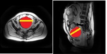
Pilot scans are fast 30-sec gradient echo fetal localiser sequences. In this study we have used 155 survey scans acquired between 20-38 weeks GA, containing 5 thick slices obtained from the maternal axial, coronal and sagittal planes. Fetal brain localisation is then performed using a 3D U-Net[6], which generated brain masks separately for the three planes. Once the segmentation results are obtained, smoothing is performed to obtain a contour of the fetal head. Fetal head circumference is then calculated as: HC = (FO+BP) * π/2 , where FO is greatest frontal-occipital diameter from the sagittal sequence and BP is the greatest bi-parietal diameter from the axial sequence [5] as shown in fig 1.Results
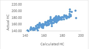
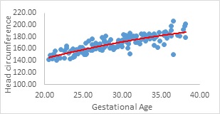
The graph in fig 2 shows plot of the calculated head circumference (HC) against the actual head circumference as measured by an experienced fetal radiologist with greater than 10 years of experience. We observe that there is a correlation of 0.93 between the two measurements. The graph in fig 3 shows the results of the polynomial regression model for the measurement of the head circumference against gestational age. We observe that the increase in gestational age increases the head circumference following a second order polynomial regression model, which matches previous studies [3,4]. We also performed a second experiment by evaluating the head circumference against gestational age divided into bins of 2 weeks. This improved the Person’s correlation coefficient of 0.79 to 0.86.Conclusion
The findings of this study demonstrate a first step towards real-time extraction of basic information from the first 30 sec of the fetal scan. These results support the use of MRI biometry charts to improve the fetal growth evaluation and offer an estimation model for fetal gestational age in the second half of the gestation. This in turn provides vital information in assessment of fetal development and neonatal care. Next steps include to use the thus obtained information in real-time to steer the focus of the scan to e.g. include placental measurements or detailed growth assessment if deviations between chronological and growth-based GA are found.Synopsis
Fetal MRI is an excellent tool for estimating the fetal gestational age using the head circumference measurements. This study measures the biometry information using fast 30 sec survey scans for the measurement of fetal head circumference using 3D UNet, Bi-parietal diameter, Frontal-occipital diameter for the estimation of the fetal gestational age in the second half of gestation. The results show high accuracy in gestational age measurements which correlates well with the clinical data.Acknowledgements
No acknowledgement found.References
1.Cai S et al. Childs Nerv Syst. 2020.
2.Grifths PD et al. The Lancet. 2017;389:538–46.
3.Kyriakopoulou V et al., Brain Struct Funct. 2017;222:2295-307.
4.Harreld JH et al., AJNR Am J Neuroradiol. 2011;32:490-4.
5.Smyth AE et al., (ASNR) 58th Annual Meeting, 2020.
6.Uus A et al., bioRxiv; 2021.
Figures