3881
Can a sub 2-minute MRI exam compete with CT for imaging sinus anatomy?1Physics and Atmospheric Science, Dalhousie University, Halifax, NS, Canada, 2Biomedical Translational Imaging Centre, QEII Health Sciences Centre, Halifax, NS, Canada, 3Medicine, Dalhousie University, Halifax, NS, Canada, 4Diagnostic Radiology, Dalhousie University, Halifax, NS, Canada, 5Diagnostic Imaging, Nova Scotia Health, Halifax, NS, Canada, 6School of Biomedical Engineering, Dalhousie University, Halifax, NS, Canada
Synopsis
MRI of the paranasal sinuses with bSSFP sequences is not typically performed due to prohibitive exam times and the prevalence of artifacts caused by off-resonance effects. We have shown that bSSFP images acquired in under 2 minutes using high-performance gradients at low field resist banding artifacts and demonstrate SNR and resolution sufficient to delineate relevant structures. This is supported by high SNR values in tissues and fluid-filled spaces, and low SNR in the air-filled maxillary sinus. 2-minute scan times compete with CT for clinical work flow and demonstrate the potential of MRI for high throughput sinus pathology evaluation.
Introduction
Computed tomography (CT) is typically employed when imaging the paranasal sinuses due to its high air/bone contrast, speed of acquisition and high in-plane resolution1. However, there are some crucial disadvantages to this modality including reduced resolution in the superior-inferior direction and exposure of the patient to harmful ionizing radiation.MRI is free of radiation, demonstrates superior soft tissue contrast, and is capable of high isotropic resolution, but it is difficult to obtain images of this anatomically complex region with both high resolution and signal-to-noise ratio (SNR) at clinical field strengths. This is due, primarily, to the multiple air/bone/tissue interfaces in this area which produce susceptibility-induced off-resonance artifacts.
Balanced steady-state free precession (bSSFP) sequences present the highest SNR efficiency2, and therefore enable high resolution images. However, bSSFP sequences demonstrate a particular sensitivity to off resonance effects, resulting in band-like signal loss in regions of decreased field uniformity such as the numerous interfaces in the paranasal region. These artifacts can be shifted out of relevant structures by a reduction in TR, or by phase-cycling techniques3, but this can be difficult to achieve with conventional clinical systems due to inadequate gradient performance and patient SAR limits4.
Recent advancements in gradient technology have broadened the range of possible values for TR when combined with a low-field, head-only system (EvryTM, Synaptive Medical)5. This 0.5-T MR uses an edge-gradient design that permits high slew rates (200 T/m/s) without peripheral nerve stimulation. Such a system provides a unique opportunity to take advantage of the benefits of imaging at low field, and it enables bSSFP imaging of the paranasal sinuses with high SNR and isotropic resolution comparable to clinical systems. We aim to describe the unique protocol developed and to demonstrate the quality of the images that can be obtained in a sub 2-minute exam.
Methods
Images were acquired under a Nova Scotia Health REB-approved protocol from patients (n=18) with clinical requisitions for suspected sinus abnormalities requiring CT imaging, and informed written consent was obtained.The protocol included an axial bSSFP (T2/T1 weight) sequence acquired at 0.5-T with gradients operated at a maximum strength of 50 mT/m and a slew rate of 200 T/m/s. Sequence parameters selected were 18cm FOV, 226x226 acquisition matrix, NEX=1, RBW=150kHz, TR/TE = 3.5ms/1.6ms, flip-angle=30°. Images were acquired with isotropic resolution of 0.8mm and interpolated onto a 0.4mm grid. These parameters resulted in a scan time of 1min52s without reaching specific absorption rate limits.
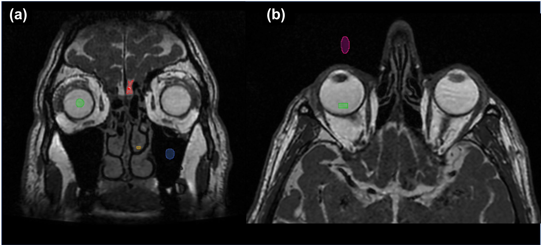
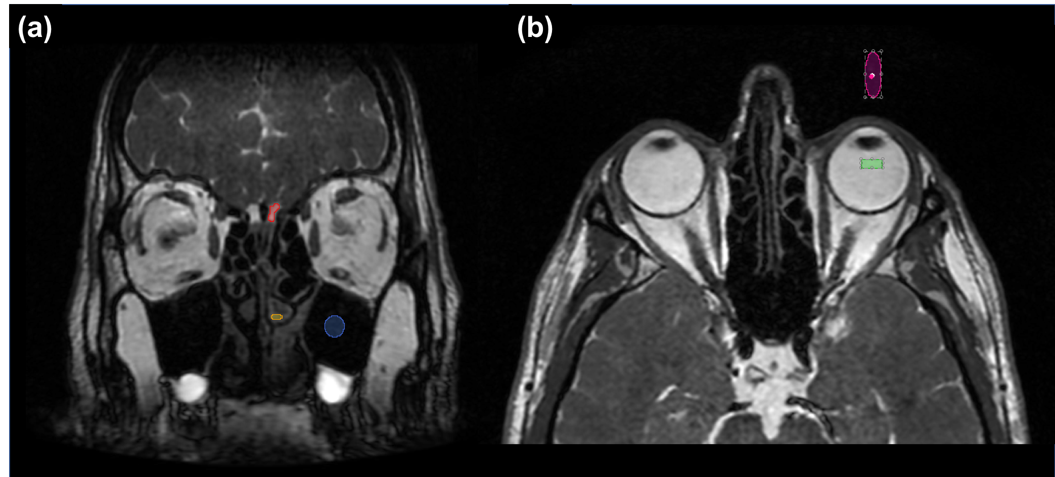
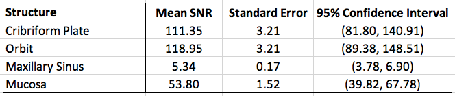
Regions of interest (ROIs) were drawn around the cribriform plate, orbit, maxillary sinus, and mucosa (Figures 1 and 2), and they each spanned 7 slices in the anterior-posterior direction. A noise region was drawn outside the head, anterior to the orbit. The SNR for each structure was calculated by taking the ratio of the signal in the ROI and the standard deviation of pixel intensities in the noise region. This was done for all subjects and the mean, standard deviation, and 95% confidence level across subjects were determined for each structure (Figure 3).
Results and Discussion
The sample acquisitions shown in Figures 1 and 2 demonstrate that the superior field homogeneity at 0.5-T, combined with a high slew rate, provides the ability to utilize a short TR without inducing banding artifacts typical of bSSFP sequences in this region.Figure 3 displays the resulting mean SNR, standard error, and 95% confidence intervals of the ROIs across patients. The high SNR values support the ability to delineate between air and relevant structures; of note is the SNR of the maxillary sinus, which has been shown to be low relative to that of the mucosa, allowing for contrast-to-noise amendable to visualizing the drainage pathways of the ostiomeatal complex.
The high SNR efficiency of the bSSFP sequence permitted isotropic resolution comparable to clinical systems. The result is demonstrated in Figure 4, where the edges of the cribriform plate and drainage pathways of the left middle turbinate are clearly visualized. Given the excess SNR obtained, future exploration of even higher resolution protocols could be considered.
Conclusions
We have shown that is it possible to perform bSSFP imaging of the paranasal sinuses at low field with high-performance gradients. The images obtained are resistant to banding artifacts typical of bSSFP acquisitions in this region, and the resolution and SNR achieved is sufficient to visualize relevant structures. This exam requires less than 2-minutes, making it competitive with CT exam times, and demonstrates the potential use of MRI for imaging the paranasal sinuses.Acknowledgements
Funding for this research was provided by the NSERC Discovery grant, the RNS Innovation grant, the Brain Repair Centre KT grant, and the Radiology Research Foundation grant.References
[1] Branstetter BF, Weissman JL. Role of MR and CT in the Paranasal Sinuses. Otolaryngologic Clinics of North America. 2005;38:1279-1299.
[2] Schleffer K, Lehnhardt S. Principles and Applications of Balanced SSFP Techniques. European Radiology. 2003;13:2409-2418.
[3] Nayak KS, Lee HL, Hargreaves BA. Wideband SSFP: alternating repetition time balanced steady state free precession with increased band spacing. Magnetic Resonance in Medicine. 2007;58.5:931-938.
[4] Huang SY, Seethamraju RT, Patel P, Hahn PF, Kirsch JE, Guimaraees AR. Body MR Imaging; Artifacts, k-Space, and Solutions. Radiographics. 2015;35(5):1439-1460.
[5] Stainsby JA, Bindseil GA, Connell IRO, Thevathasan G, Curtis AT, Beatty PJ, Harris CT, Wiens CN, and Panther A. Imaging at 0.5 T with high-performance system components. Proc. ISMRM 2019, no.1194.
Figures