3880
Folate Intake during Pregnancy is Associated with Greater White Matter Development in the Newborn Brain
Natalie E. Phelan1, Rajikha Raja2, Aline Andres3,4, and Xiawei Ou2,3,4,5
1College of Medicine, University of Arkansas for Medical Sciences, Little Rock, AR, United States, 2Department of Radiology, University of Arkansas for Medical Sciences, Little Rock, AR, United States, 3Department of Pediatrics, University of Arkansas for Medical Sciences, Little Rock, AR, United States, 4Arkansas Children's Nutrition Center, Little Rock, AR, United States, 5Arkansas Children's Research Institute, Little Rock, AR, United States
1College of Medicine, University of Arkansas for Medical Sciences, Little Rock, AR, United States, 2Department of Radiology, University of Arkansas for Medical Sciences, Little Rock, AR, United States, 3Department of Pediatrics, University of Arkansas for Medical Sciences, Little Rock, AR, United States, 4Arkansas Children's Nutrition Center, Little Rock, AR, United States, 5Arkansas Children's Research Institute, Little Rock, AR, United States
Synopsis
This study examined potential relationships between maternal folate intake during pregnancy and neonatal brain white matter development. Healthy pregnant women were recruited at early pregnancy and their newborns underwent a brain MRI examination at ~2 weeks of age, including diffusion tensor imaging to assess white matter development. We found that folate intake during the second trimester of pregnancy positively correlated with fractional anisotropy (FA) values in brain white matter regions including the genu of corpus callosum and the external capsule. Our findings suggest positive impact of maternal folate intake during pregnancy on offspring brain white matter development.
Introduction
Folate supplementation before conception and during early pregnancy has well-documented neonatal benefit, most notably the prevention of neural tube defects. It is less known the potential effects of maternal folate intake throughout pregnancy on subsequent white matter development in offspring, however recent research has indicated that maternal folate status is implicated in key neurodevelopmental processes in the fetus. Imaging the healthy newborn brain using sensitive methods allows us to assess early white matter development in relation to maternal dietary exposures including folate intake. In this study we evaluated folate intake level in healthy pregnant women at different trimesters, and correlated that with their newborn’s brain white matter development assessed by diffusion tensor imaging.Methods
Healthy pregnant women and their newborns were recruited for this prospective study. Inclusion criteria for the pregnant women were singleton pregnancy; ≥21 years of age; conceived without assisted fertility treatments. Exclusion criteria for the pregnant women were: pre-existing medical conditions such as diabetes mellitus, seizure disorder, and serious psychiatric disorders; drug or alcohol abuse; sexually transmitted diseases; medical complications during pregnancy such as gestational diabetes and pre-eclampsia. In addition, newborns born pre-term (<37 weeks of gestation) or with medical conditions or medications known to influence growth and development, or unable to complete MRI without sedation were also excluded. In total, 44 pregnant women and their newborns were included in this study. Folate intake levels of the pregnant women were assessed using 3 day food records obtained twice for each semester which were analyzed with the Nutrition Data System for Research (NDSR, University of Minnesota, http://www.ncc.umn.edu/products/). At ~2 weeks of postnatal age, the newborns underwent an MRI examination of the brain during natural sleep without sedation which included a short diffusion tensor imaging sequence with diffusion-weighting gradients in 15 uniformly distributed directions and a maximum b-value of 700 s/mm2. The diffusion weighted images were exported to a workstation with the open source FMRIB Software Library (FSL Version 5.0, created by the Oxford Center for Functional MRI of the Brain, Oxford, United Kingdom) installed on a VMware Linux virtual machine (VMware, Inc., Palo Alto, CA) for post-processing. Eigenvalues for the diffusion tensors were computed and diffusion tensor MRI parameter maps including fractional anisotropy (FA), mean diffusivity (MD), axial diffusivity (AD), and radial diffusivity (RD) were generated using the FDT toolbox in FSL. Tract-based spatial statistics (TBSS) methods were used for diffusion tensor MRI data analysis. Potential relationships between maternal folate intake levels at different trimesters of pregnancy and diffusion parameters in the newborn brain were evaluated using randomization tools in FSL with 5000 permutations. Postmenstrual age at MRI and sex of the newborns, maternal age at birth, and maternal education level were included as covariates in the permutation analysis. Threshold Free Cluster Enhancement (TFCE) method was used for multiple comparison correction, and correlations with P<0.05 after family-wise error (FWE) correction were regarded as significant.Results
TBSS analysis showed significant correlations between maternal folate intake level during the second trimester of pregnancy and diffusion imaging parameters in the newborn brain white matter. Specifically, higher folate intake at the second trimester of pregnancy is significantly correlated (P<0.05, FWE corrected) with higher FA values in multiple brain white matter regions, mainly involving the genu of the corpus callosum and the external capsule (Figure 1). There were no negative correlations between folate intake at second trimester of pregnancy and FA values in the newborn brain. Also there were no other significant relationships (P<0.05, FWE corrected) between folate levels at different trimesters and diffusion parameters.Conclusions
Maternal folate intake during the second trimester is associated with higher FA values in the newborn brain, indicating better brain white matter development.Acknowledgements
This project was supported by NIH 1R01HD099099 and USDA-ARS 6026-51000-012-06S.References
No reference found.Figures
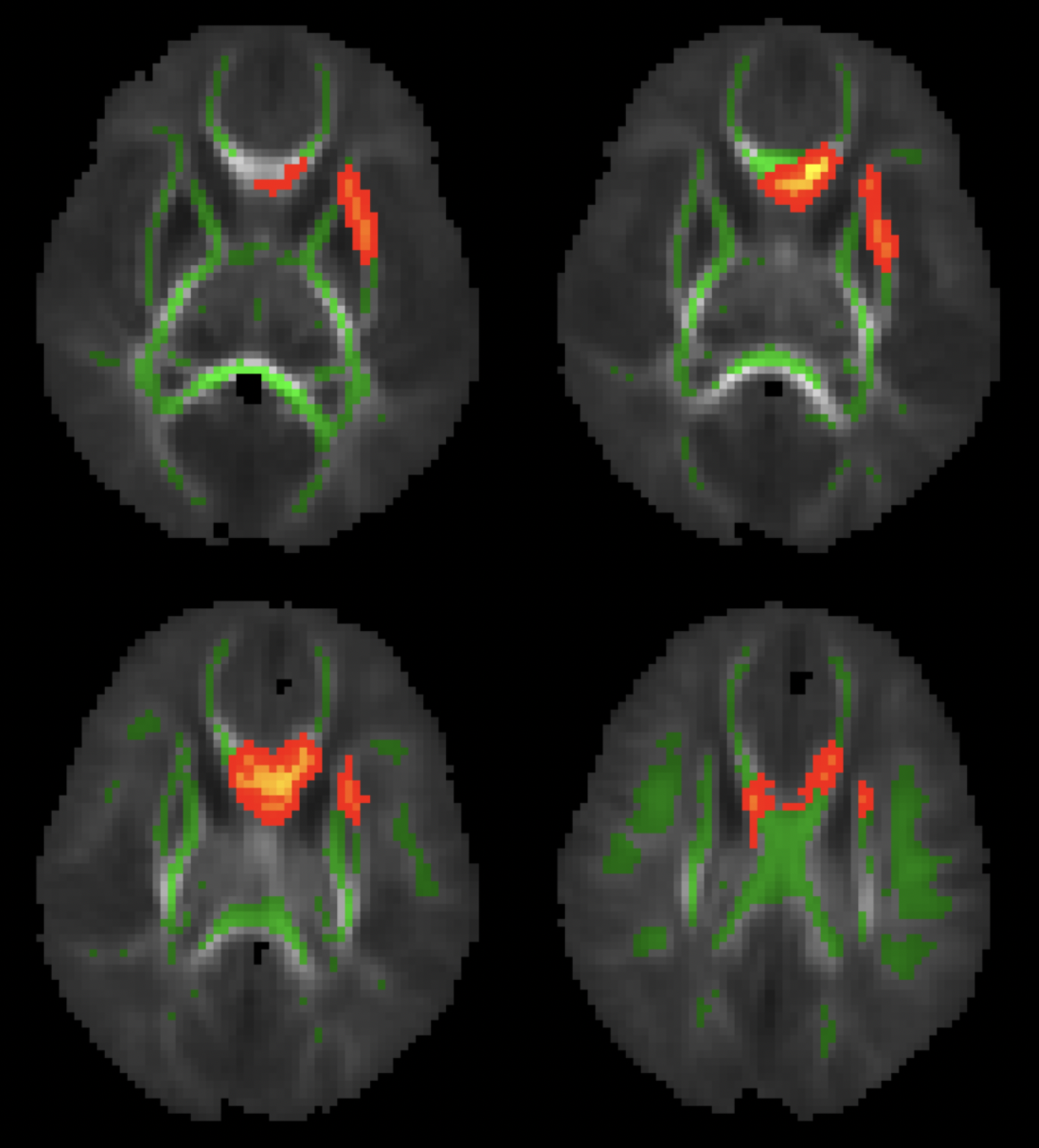
Figure 1: White matter regions (red/orange) showing significant (P<0.05, FWE corrected) and positive correlations between maternal folate intake level at second trimester of pregnancy and neonatal brain white matter FA values.
DOI: https://doi.org/10.58530/2022/3880