3865
Design of a Metamaterial-inspired Flexible Structure for MRI Field Enhancement at 3T: a Numerical Approach1Institute of Biomedical Engineering, Boğaziçi University, Istanbul, Turkey, 2Department of Electric and Electronics Engineering, Boğaziçi University, Istanbul, Turkey
Synopsis
MR imaging has a significant role in medical diagnostics. Imaging quality depends on SNR levels. Higher B0 field and consequently higher RF-field intensity increase SNR but it also elevates the total body SAR. This can be alleviated by local magnetic field enhancement. Metamaterial based structures have shown promising applications in this regard. Here we report a new metamaterial inspired resonant structure for local field enhancement in 3T MRI machine. We show field enhancement capabilities of our design in planar and cylindrical forms, each of which can be used based on the anatomical site under investigation.
INTRODUCTION
MR is an important imaging modality mainly because it can offer high tissue contrast ratio and high resolution at the same time without using any form of ionizing radiation. Signal to noise ratio (SNR) is one of the most important parameters used for characterizing the image quality. Higher SNR can be achieved through increase in the B0 field and consequently elevated RF field intensity. But high RF fields cause heating problems in patients with implants1. Thus, lower RF fields are usually utilized for these patients to reduce the total body specific absorption rate (SAR). This takes a toll on the diagnostic power of the image. An emerging approach to address this problem is the local field enhancement via resonant structures. In recent years, Metamaterial based resonant structures have shown promising applications in local field enhancement 2-7. These devices offer higher SNR without increasing the B0 field or the total body SAR. The dimensions of these structures depend on their resonant frequencies. Thus, the structures introduced so far were either bulky structures or studied in 7T MRI machines where the resonant structures can be much smaller and easier for use in the MRI bore. However, 1.5T or 3T MRI machines are much more abundant in clinical setups. Here we report a novel metamaterial-inspired flexible resonant structure design that we developed for 3T MRI machine. We investigated the local field enhancement behavior of the design in planar and cylindrical forms, both of which can be used based on the anatomical site under investigation.METHODS
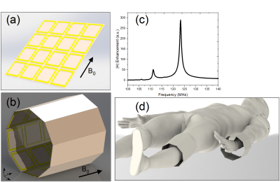
Figure 1 (a) and (b) show the rendered geometrical structures of the planar and cylindrical resonant structures. The planar structure is a 21.4x21.4x0.1 cm3 thin square. The cylindrical structure is nonagon with a diameter of 15 cm and length of 16.2 cm. Both structures are designed with thin polyimide substrate and copper as the conductive layer. Finite Difference Time Domain (FDTD) simulations were conducted first to find the resonant modes of the device and then to study the excitation of the device further on using a left circularly polarized plane wave. We also included a mineral oil phantom (ε = 5) in our simulations. Magnetic field and B1+ enhancement results are calculated based on the ratios of magnetic fields with and without resonant structure under the oil phantom ( $$$\frac{|H|_{with RS}}{|H|_{phantom}}$$$).RESULTS
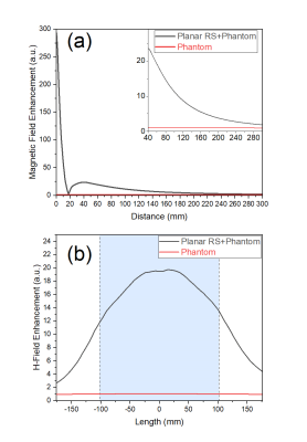
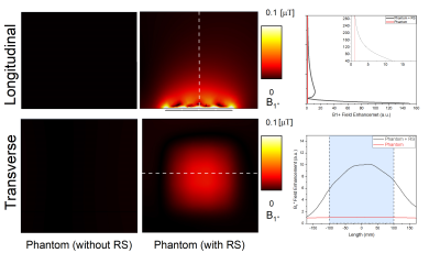
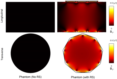
Figure 1(c) shows the resonance frequency of the planar structure that was tuned to 123.6 MHz. Figure 2 summarized the penetration depth of the magnetic field enhancement (a) and its transverse profile 6 cm inside the phantom (b). Figure 3 shows the B1+ field contours in the oil phantom with and without the resonant structure under it. Figure 4 demonstrates B1+ field distribution for the cylindrical phantom and corresponding cylindrical resonant structure placed around it.DISCUSSION
Magnetic field enhancement profile towards the depth of the phantom, depicted in Figure 2 (a), shows that even though there is a decaying performance, magnetic field enhancement is still significantly high (~3.8) even at 20cm above the structure. As it is seen in figure 2(b), field enhancement inside the phantom has a sine shaped transverse profile with the maximum in the middle. The same behavior can be seen in B1+ field distribution for planar structure (Figure 3). While planar structure provides a field enhancement closer to the surface (Figure 3), the cylindrical structure generates a more homogeneous field inside the structure (Figure 4). The B1+ field in the middle of the cylindrical phantom increased up to 55-folds with the cylindrical resonant structure around it. Placing the region of interest (e.g., Wrist, knee (Figure 1(d)) in the middle of the structure would generate a homogeneous signal enhancement.CONCLUSION
We have reported a novel design of a metamaterial inspired resonant structure for magnetic field manipulation in 3T MRI machine. We have shown that our planar and cylindrical structure can significantly enhance the local magnetic field and can be chosen based on the anatomical site under investigation.
Acknowledgements
There is no acknowledgment.References
1. S. Nazarian, R. Beinart, H. Halperin, Magnetic resonance imaging and implantable devices, Circ. Arrhythm. Electrophysiol. 6 (2013) 419–428.
2. Shchelokova, Alena V., et al. "Experimental investigation of a metasurface resonator for in vivo imaging at 1.5 T." Journal of Magnetic Resonance 286 (2018): 78-81.
3. Schmidt, Rita, et al. "Flexible and compact hybrid metasurfaces for enhanced ultra high field in vivo magnetic resonance imaging." Scientific reports 7.1 (2017): 1-7.
4. Zhao, Xiaoguang, et al. "Intelligent metamaterials based on nonlinearity for magnetic resonance imaging." Advanced Materials 31.49 (2019): 1905461.
5. Stoja, Endri, et al. "Improving Magnetic Resonance Imaging with Smart and Thin Metasurfaces." arXiv preprint arXiv:2105.09634 (2021).
6. Chi, Zhonghai, et al. "Adaptive Cylindrical Wireless Metasurfaces in Clinical Magnetic Resonance Imaging." Advanced Materials 33.40 (2021): 2102469.
7. Schmidt, Rita, and Andrew Webb. "Metamaterial combining electric-and magnetic-dipole-based configurations for unique dual-band signal enhancement in ultrahigh-field magnetic resonance imaging." ACS applied materials & interfaces 9.40 (2017): 34618-34624.
Figures


Figure 2: Penetration depth in the middle of the planar resonant structure located under the phantom (a). The transverse profile of the H-field enhancement 6cm deep inside the phantom(b). The area that was directly above the resonant structure is indicated as the blue region in the figure.

