3858
rNOE imaging detects distinctive neuropathology in Intracerebral Hemorrhage (ICH) under deferoxamine treatment
Ho Chi Joseph Lai1, Tian Yang2, Jianpan Huang1, Yang Liu1, Youngjin Lee2, and Wai Yan Kannie Chan1,3,4,5
1Department of Biomedical Engineering, City University of Hong Kong, Hong Kong, Hong Kong, 2Department of Neuroscience, City University of Hong Kong, Hong Kong, Hong Kong, 3Russell H. Morgan Department of Radiology and Radiological Science, The Johns Hopkins University School of Medicine, Baltimore, MD, United States, 4City University of Hong Kong Shenzhen Research Institute, Shenzhen, China, 5Hong Kong Centre for Cerebro-Cardiovascular Health Engineering, Hong Kong, Hong Kong
1Department of Biomedical Engineering, City University of Hong Kong, Hong Kong, Hong Kong, 2Department of Neuroscience, City University of Hong Kong, Hong Kong, Hong Kong, 3Russell H. Morgan Department of Radiology and Radiological Science, The Johns Hopkins University School of Medicine, Baltimore, MD, United States, 4City University of Hong Kong Shenzhen Research Institute, Shenzhen, China, 5Hong Kong Centre for Cerebro-Cardiovascular Health Engineering, Hong Kong, Hong Kong
Synopsis
We have shown that CEST (APTw and rNOE) contrast demonstrated distinctive changes of ICH mouse brains longitudinally1. As demonstrated by our published work2, the rNOE changes could be primarily associated with changes in myelin. Here, we studied the APTw and rNOE contrast after ICH up to 14 days, and under DFX treatment. We observed regional changes of APTw and rNOE contrast in the core and peri-hematoma regions, especially the significant difference (P<0.05) on day 3 with and without DFX treatment. Our immunohistochemistry data indicated that rNOE contrast correlated with myelin, which supports that rNOE could detect myelin pathology during ICH.
Introduction
We demonstrated that APTw and rNOE contrast of ICH could indicate distinctive pathology1. Since the molecular mechanism of injury in ICH is not fully revealed, here we would like to further study the underlying neuropathology indicated by CEST MRI at 3T. Lipid plays an important role during the evolvement of ICH, especially lipid peroxidation, which could cause white matter injury in the brain3. In our recent published work2, we demonstrated that rNOE could detect myelin-related neuropathology. We hypothesized that rNOE could detect lipid-related pathology or lipid peroxidation in ICH to identify the injury in the brain during ICH.In this study, we imaged the ICH mice with and without DFX treatment using CEST MRI, and we analyzed the changes of APTw and rNOE contrast over two weeks after ICH induction. Since the iron content in hematoma could attenuate the CEST contrast, we studied the effect of iron on CEST contrast in vitro and compare CEST contrast in ICH mice with DFX, i.e. iron chelation treatment. This is to study the feasibility of imaging changes in hematoma using CEST and the detection of related neuropathology. After that, representative mice from each time point were sacrificed for immunohistochemical studies, including the protein and myelin pathology3.
Methods
C57BL/6 mice (8-10 weeks, Jackson Laboratory) were induced with ICH by following a reported protocol4. Half of the ICH mice were randomly selected for iron chelation treatment5. Deferoxamine (DFX, Sigma) was delivered by intraperitoneal injection 6hr after the stroke induction and repeated every 12hr for 7 consecutive days, with 200mg/kg per dose. The control mice were injected with the same volume of saline.The ICH mice were imaged by a horizontal bore 3T Bruker BioSpec scanner over 2weeks. The scanning parameters were based on the protocol from our group6. The imaging parameters were as follows: TR=6000ms; TE=6ms; FOV=20×20mm; matrix=96×96; thickness=1.5mm; RARE factor=32; B1=0.8μT and saturation time=3s. Frequency offsets of the Z-spectrum included ±15ppm, ±10ppm, ±9ppm, and distributed between ±8ppm with 0.25ppm step size. ROIs for the core and peri-hematoma were drawn based on the corresponding T2-weighted images.
Representative mice were sacrificed at specific time points for the immunohistochemical study. Two immunohistochemical staining methods, Myelin Basic Protein (MBP)7 and FluoroMyelin™ (FM, ThermoFisher)8, were co-stained and the brain sections were imaged with fluorescence microscopy to reveal the pathological changes of myelin after ICH.
All statistical analysis was done by GraphPad Prism software, version 8.0 (GraphPad Software Inc.), and considered statistically significant for P<0.05 by two-tailed Student’s t-test.
Results and Discussion
In Fig. 1A, we observed distinctive changes in rNOE and APTw contrast within hematoma over two weeks. Since the iron content could have an effect on the CEST contrast, we applied DFX treatment to further study the CEST contrast in hematoma5. We observed a significant decrease in the rNOE signal on day 3, while rNOE in the DFX group is significantly lower than that in the control group (P<0.05) (Fig. 1B). Interestingly, the changes in the APTw (Fig. 1C) were not obvious. In addition, we found that there is an observable difference between the core and periphery of the hematoma, hence we performed a separate ROI analysis on these two regions (Fig. 2C, D). The DFX-treated group showed a significant decrease in rNOE contrast on day 3 for both core and peri-hematoma, which corresponds to a 26.8% and 27.3% drop, respectively. Yet, no significant difference was observed at other time points. rNOE is sensitive to lipids and proteins, which are the major components of myelin. During hematoma evolution3, the observed decrease on day 3 could be due to the loss of myelin, while the noticeable difference in rNOE between the core and peri-hematoma on day 7 in DFX group could indicate the change in myelin-related lipids or protein during regeneration. This is also supported by our immunohistochemistry (Fig. 3).Both lipid (FM) and protein (MBP) components of myelin showed a decrease on day 3 in DFX, while a distinctive contrast between the core and peri-hematoma was observed on day 7 (Fig. 3B).
We further studied the correlation between the rNOE contrast and FM (Fig. 4). We observed a strong correlation (R=0.816, ***P<0.0001), which could indicate the rNOE signal change came from the myelin lipid. In contrast, the MBP intensities in Fig. 4B did not show a significant correlation (R=0.285, P=0.2852). This validated our rNOE findings, which could detect ICH pathology related to myelin.
Conclusion
rNOE and APTw significantly decreased on day 3 after ICH. rNOE detected a 27% decrease in ICH mice after DFX treatment on day 3. As validated by the myelin-specific immunohistochemistry, rNOE correlated with the amount of myelin lipid in hematoma. Moreover, a slightly higher rNOE contrast in the core than in the peri-hematoma was observed on day 7 in the DFX group but not in the control group, which could indicate myelin-related changes during recovery. This supports that rNOE could detect myelin-related neuropathology in ICH at 3T and indicate recovery after ICH. Nevertheless, these findings demonstrated that rNOE and APTw could detect specific molecular changes during ICH and after DFX treatment.Acknowledgements
We are grateful to receive funding support from the Research Grants Council: 11102218; City University of Hong Kong: 7005210, 7005433, 9680247, 9667198 and 9609307; National Natural Science Foundation of China: 81871409.References
- Lai JHC, Liu J, Huang J, Liu Y, Chen Z, Xiao P et al. CEST MRI of temporal changes of hematoma in Intracerebral Hemorrhage ( ICH ) mouse at 3T. Int Soc Magn Reson Med 2021; : 2014–2016.
- Huang J, Xu J, Lai JHC, Chen Z, Yan C, Mak HKF et al. NeuroImage : Clinical Relayed nuclear Overhauser effect weighted ( rNOEw ) imaging identifies multiple sclerosis. NeuroImage Clin 2021; 32: 102867.
- Fu X, Zhou G, Zhuang J, Xu C, Zhou H, Peng Y et al. White Matter Injury After Intracerebral Hemorrhage. Front Neurol 2021; 12. doi:10.3389/fneur.2021.562090.
- Li M, Li Z, Ren H, Jin WN, Wood K, Liu Q et al. Colony stimulating factor 1 receptor inhibition eliminates microglia and attenuates brain injury after intracerebral hemorrhage. J Cereb Blood Flow Metab 2017; 37: 2383–2395.
- Wu H, Wu T, Xu X, Wang J, Wang J. Iron toxicity in mice with collagenase-induced intracerebral hemorrhage. J Cereb Blood Flow Metab 2011; 31: 1243–1250.
- Han X, Huang J, To AKW, Lai HCJ, Xiao P, Wu EX et al. CEST MRI detectable liposomal hydrogels for multiparametric monitoring in the brain at 3T. Theranostics 2020. doi:10.7150/thno.40146.
- Tse KH, Cheng A, Ma F, Herrup K. DNA damage-associated oligodendrocyte degeneration precedes amyloid pathology and contributes to Alzheimer’s disease and dementia. Alzheimer’s Dement 2018; 14: 664–679.
- Monsma PC, Brown A. FluoroMyelinTM Red is a bright, photostable and non-toxic fluorescent stain for live imaging of myelin. J Neurosci Methods 2012; 209: 344–350.
Figures
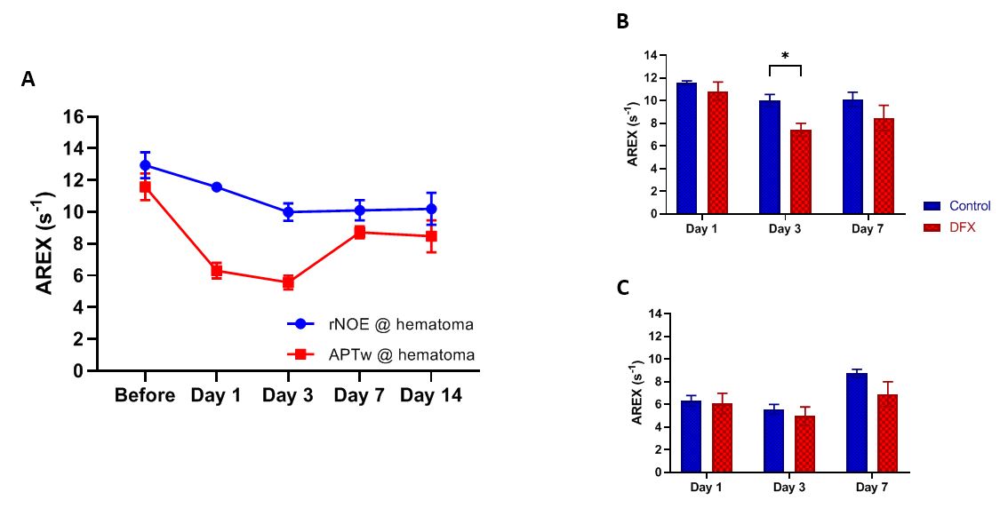
rNOE
and APTw
changes in hematoma, without (control) and with DFX treatment.
(A)
shows the average AREX signal at hematoma from the ICH mice over 2 weeks.
(B)
and (C) show the rNOE and APTw changes, respectively, after DFX
treatment up to day 7. Significant reduction of rNOE was observed in the DFX treatment
group on day 3 compared to the control group (*P<0.05, n=5). Values
are means ± SEM.
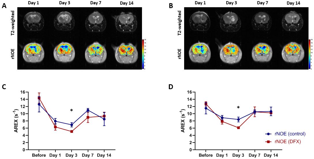
rNOE
contrast from the ICH mice, while (A) is the control group, and (B) is the DFX
group. The average rNOE signal change at (C) core hematoma
and (D) peri-hematoma
showed a significant difference on day 3 (*P<0.05, n=5). Values are means ±
SEM.
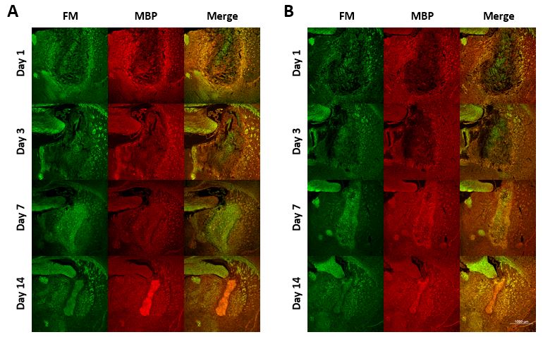
Immunohistochemistry by
FM (green) and MBP (red) staining to study myelin and related protein changes
at day 1, 3, 7, and 14. (A) represents the ICH mice; while (B) is the ICH mice
with DFX treatment. Distinctive difference in FM was observed on day 3.
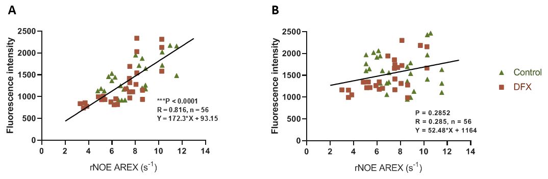
Figure 4. The correlation between
the fluorescence intensities (FM and MBP) with the rNOE
signal are plotted into (A) and (B) respectively. Only FM shows a good
correlation with rNOE.
DOI: https://doi.org/10.58530/2022/3858