3829
The location reliability of the functional connectivity of deep emotion-related regions towards functional connectivity guided rTMS therapy1Center for Cognition and Brain Disorders, The Affiliated Hospital of Hangzhou Normal University, Hangzhou, China, Hangzhou, China, 2Institute of Psychological Sciences, Hangzhou Normal University, Hangzhou, China, Hangzhou, China, 3Zhejiang Key Laboratory for Research in Assessment of Cognitive Impairments, Hangzhou, China, Hangzhou, China, 4Unit of Psychiatry, Department of Public Health and Medicinal Administration, & Institute of Translational Medicine, Faculty of Health Sciences, University of Macau, Macao SAR, China, Taipa, Macau, 5Centre for Cognitive and Brain Sciences, University of Macau, Macao SAR, China, Taipa, Macau, 6Faculty of Health Sciences, University of Macau, Macao SAR, China, Taipa, Macau, 7Institute of sports medicine and health, Chengdu Sport University, Chengdu, China, Chengdu, China, 8MR Research, GE Healthcare, Shanghai, China, Shanghai, China, 9Institute of Advanced Studies in Humanities and Social Sciences, University of Macau, Macao SAR, China, Taipa, Macau
Synopsis
The current study systematically investigated multiple factors that affect the reliability of the FC location and strength of the DLPFC with a few emotional related deep regions. The group-level voxel-wise FC strength reliability was low to moderate, indicating its little significance for guiding individualized rTMS treatment. In terms of the FC location reliability, the intra-individual distances of the center of gravity (COG) were 3.8-7.3 mm across different conditions, which suggest that the COG of the seed-based FC might be potential stimulation target for individualized precise rTMS treatment of affective brain disorders.
INTRODUCTION
Retrospective resting-state magnetic resonance imaging (RS-fMRI) studies indicated that the anti-depression efficacy of repetitive transcranial magnetic stimulation (rTMS) was closely associated with the functional connectivity (FC) strength of the DLPFC with the subgenual anterior cingulate cortex (sgACC), pregneual ACC (pgACC), or nucleus accumbens (NAc) 1-3. It is proposed that the FC-guided individualized precise rTMS on the DLPFC would be more effective 1,4. Nevertheless, the stability of the individualized FC-guided stimulation target is greatly dependent on the FC reliability. Thus, the current study systematically investigated the reliability of the RS-fMRI FC location as well as the FC strength with factors including different deep seed regions of interest (ROIs) (i.e., the sgACC, pgACC, pgACC, NAc), eyes open (EO) and closed (EC), different scanners, FC algorithms (Pearson vs. Spearman), scanning length (half a session vs. whole session), frequency bands, and location method (FC peak vs. center of gravity (COG)). We aimed to provide a stable individualized stimulation target for future FC-guided TMS therapy for emotion related disorders.METHODS
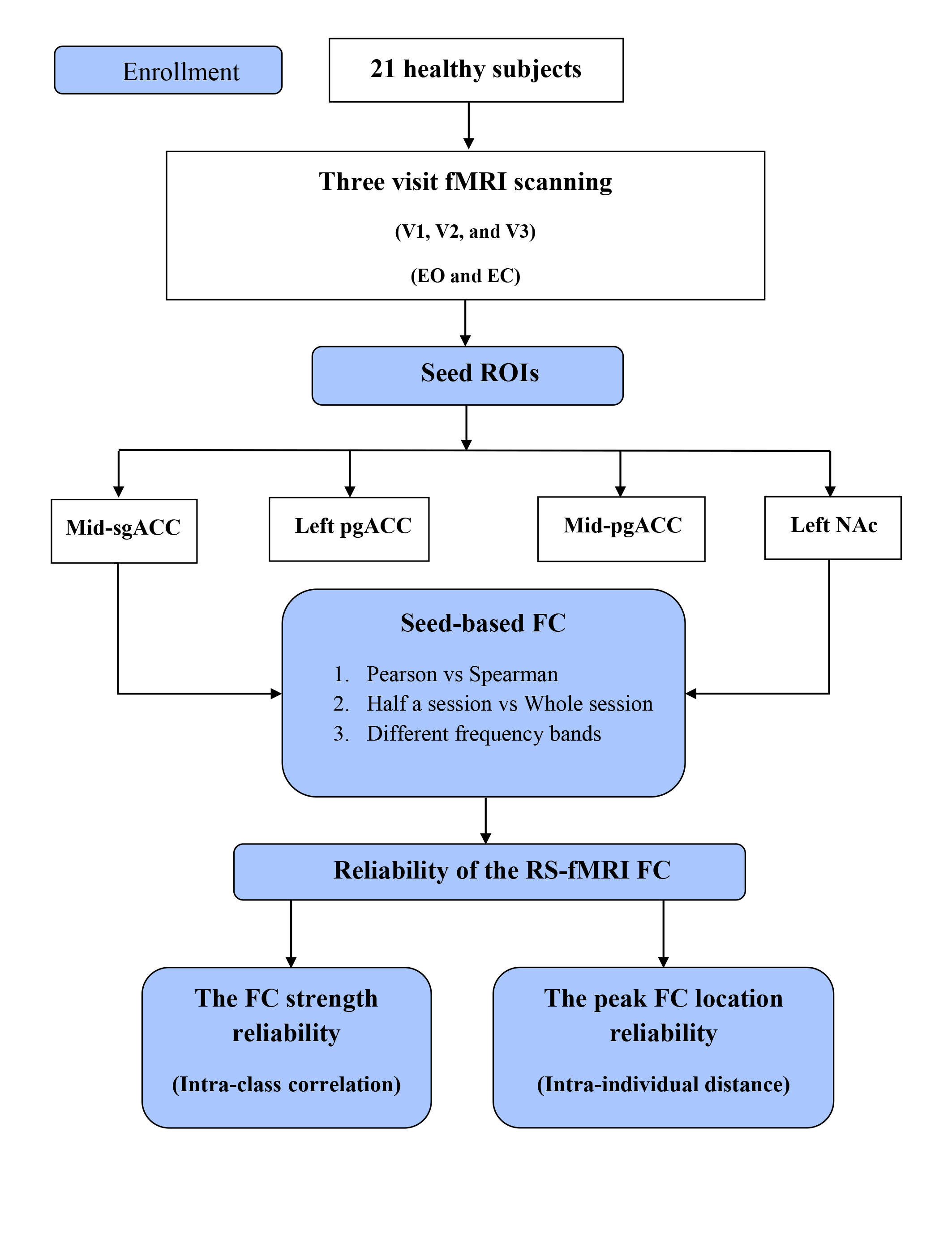
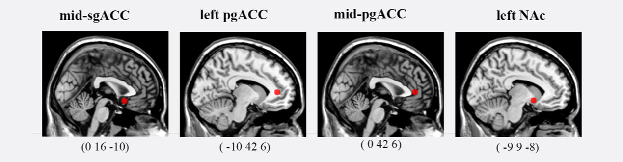
The overall study procedure was displayed in Figure 1 and based on open access data 5,6 (https://www.nitrc.org/projects/reliability/). Twenty-one subjects under RS-fMRI conditions with the first two times (V1, V2) scanned on a GE 3T scanner and the third visit (V3) on a Siemens 3T scanner.Four deep brain regions were selected as seed ROIs (spheres with 5 mm radius) centered at the mid-sgACC (0, 16, -10), left pgACC (-10, 42, 6), mid-pgACC (0, 42 6), and left NAc (-9, 9, -8) (Figure 2). We selected the mid-sgACC (0, 16, -10) as a deep effective region because our pervious study found that the negative correlation of sgACC-DLPFC was largely contributed by the mid-sgACC but not the right sgACC 7. For the pgACC and NAc, the coordinates were selected according to previous studies 2,8. The DLPFC mask was also defined following previous study 9.
For each seed ROI, the Pearson linear correlation (i.e., seed-based FC) between the seed time series and that of each voxel across the whole brain of different frequency bands was calculated. Spearman rank correlation were also calculated. In addition, to explore the reliability of scanning length, we split each session into two halves (with 115 time points for each half session, i.e., Half-1 and Half-2), and then the FC analysis was performed for each half time series. Finally, both the reliability of the FC’s strength and the location of the FC-guided stimulation target was estimated based on these different FC maps.
The reliability of the FC strength was evaluated with intra-class correlation (ICC). Regarding to the FC location reliability, intra-individual Euclidean distances of the potential individualized stimulation target between two visits (V1 vs. V2, V1 vs. V3) and two half sessions (Half-1 vs. Half-2) were used. Two methods were used to define the location of the potential individualized stimulation target, i.e., the peak FC location and the center of gravity (COG).
RESULTS
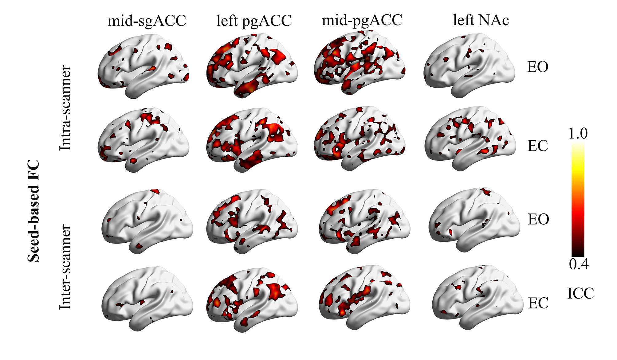
The overall FC strength reliability were displayed as follows (Figure 3):1) EO > EC
2) Intra-scanner reliability > inter-scanner reliability
3) Pearson Correlation » Spearman Correlation
4) Half session (Half-1 vs. Half-2) > Whole session (V1 vs. V2)
5) The left pgACC and mid-pgACC > other ROIs
6) Similar results across different frequency bands
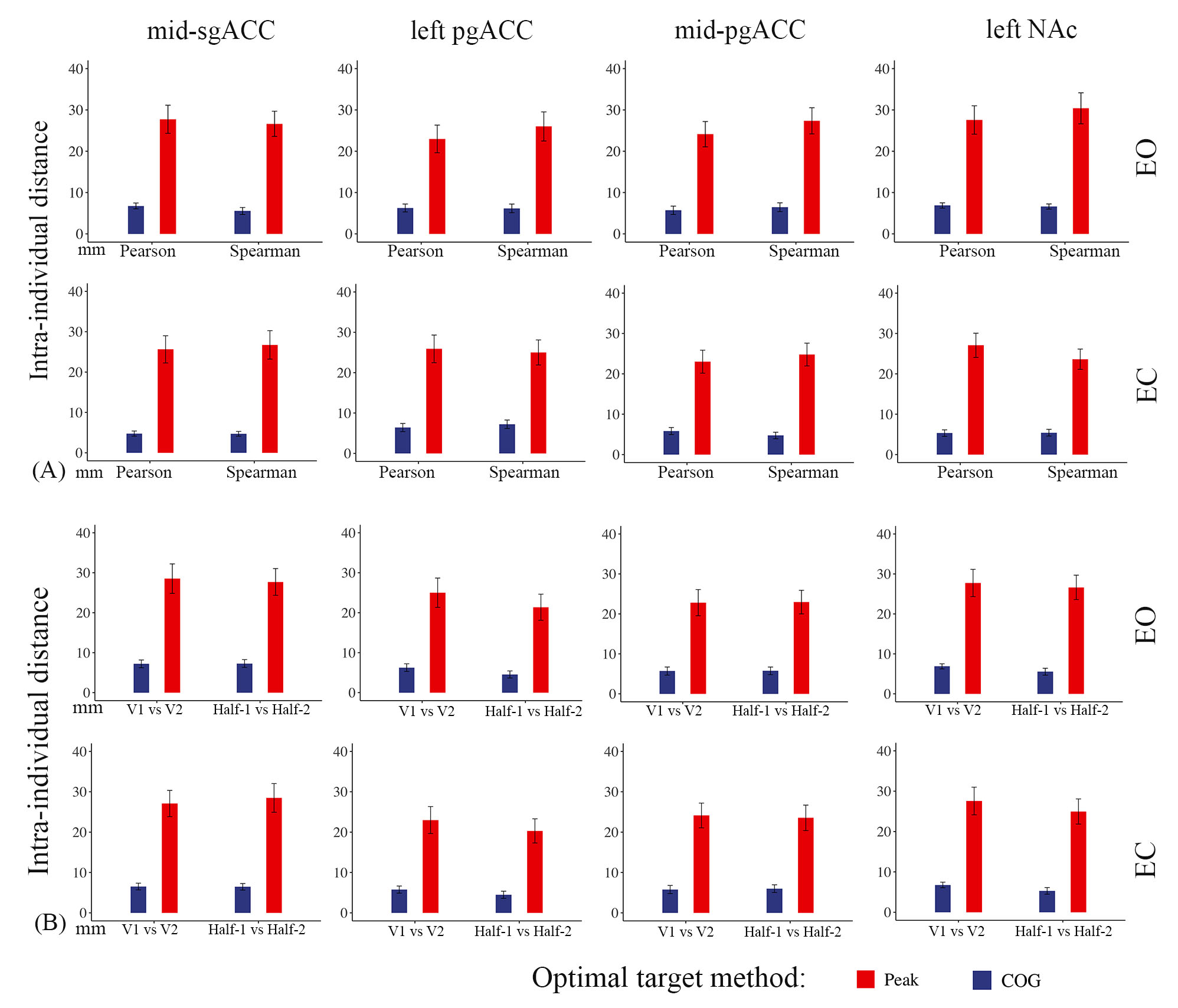
The location reliability of the potential stimulation targets were displayed as follows (Figure 4):
1) COG > peak
2) EO » EC
3) Intra-scanner distance » inter-scanner distance
4) Pearson Correlation » Spearman Correlation
5) Half session (Half-1 vs Half-2) » Whole session (V1 vs V2)
6) Mid-sgACC » Left pgACC » Mid-pgACC » NAc
7) Similar across different frequency bands
DISCUSSION
Generally, the overall results showed that the voxel-wise FC strength reliability was low to moderate across different conditions, including seed ROIs, EO vs EC, FC algorithms (Pearson vs Spearman), different scanners, frequency band, and scanning length. From a perspective of clinical rTMS treatment, individualized stimulation target (i.e., FC peak and COG in the current study), other than the above group-level voxel-wise FC, is helpful to guide precise localization of rTMS target. Consistent with previous studies 9,10, we found that the intra-individual distances of the COG were 3.8-7.3 mm across all conditions, which were smaller than that of FC peak with approximately 30 mm. These results indicate that the COG of seed-based FC could be a potential rTMS stimulation target.CONCLUSION
The COG of seed-based FC might be a potential rTMS stimulation target. Anyway, all potential stimulation targets should be tested in future rTMS treatment studies.Acknowledgements
None.References
1. Fox MD, Buckner RL, White MP, Greicius MD, Pascual-Leone A. Efficacy of transcranial magnetic stimulation targets for depression is related to intrinsic functional connectivity with the subgenual cingulate. Biol Psychiatry. 2012;72(7):595-603.
2. Du L, Liu H, Du W, et al. Stimulated left DLPFC-nucleus accumbens functional connectivity predicts the anti-depression and anti-anxiety effects of rTMS for depression. Transl Psychiatry. 2018;7(11):3.
3. Long Z, Du L, Zhao J, Wu S, Zheng Q, Lei X. Prediction on treatment improvement in depression with resting state connectivity: A coordinate-based meta-analysis. J Affect Disord. 2020;276:62-68.
4. Blumberger DM, Vila-Rodriguez F, Thorpe KE, et al. Effectiveness of theta burst versus high-frequency repetitive transcranial magnetic stimulation in patients with depression (THREE-D): a randomised non-inferiority trial. The Lancet. 2018;391(10131):1683-1692.
5. Yuan LX, Wang JB, Zhao N, et al. Intra- and Inter-scanner Reliability of Scaled Subprofile Model of Principal Component Analysis on ALFF in Resting-State fMRI Under Eyes Open and Closed Conditions. Front Neurosci. 2018;12:311.
6. Zhao N, Yuan LX, Jia XZ, et al. Intra- and Inter-Scanner Reliability of Voxel-Wise Whole-Brain Analytic Metrics for Resting State fMRI. Front Neuroinform. 2018;12:54.
7. Jing Y, Zhao N, Deng XP, et al. Pregenual or subgenual anterior cingulate cortex as potential effective region for brain stimulation of depression. Brain Behav. 2020;10(4):e01591.
8. Zhou M, Hu X, Lu L, et al. Intrinsic cerebral activity at resting state in adults with major depressive disorder: A meta-analysis. Prog Neuropsychopharmacol Biol Psychiatry. 2017;75:157-164.
9. Cash RFH, Cocchi L, Lv J, Wu Y, Fitzgerald PB, Zalesky A. Personalized connectivity-guided DLPFC-TMS for depression: Advancing computational feasibility, precision and reproducibility. Hum Brain Mapp. 2021.
10. Ning L, Makris N, Camprodon JA, Rathi Y. Limits and reproducibility of resting-state functional MRI definition of DLPFC targets for neuromodulation. Brain Stimul. 2019;12(1):129-138.
Figures